. 2008 Nov 1;68(21):8788-95.
doi: 10.1158/0008-5472.CAN-08-2135.

Insulin-like growth factor 2 is required for progression to advanced medulloblastoma in patched1 heterozygous mice

Affiliations

Insulin-like growth factor 2 is required for progression to advanced medulloblastoma in patched1 heterozygous mice

Ryan B Corcoran et al. Cancer Res. .

Abstract

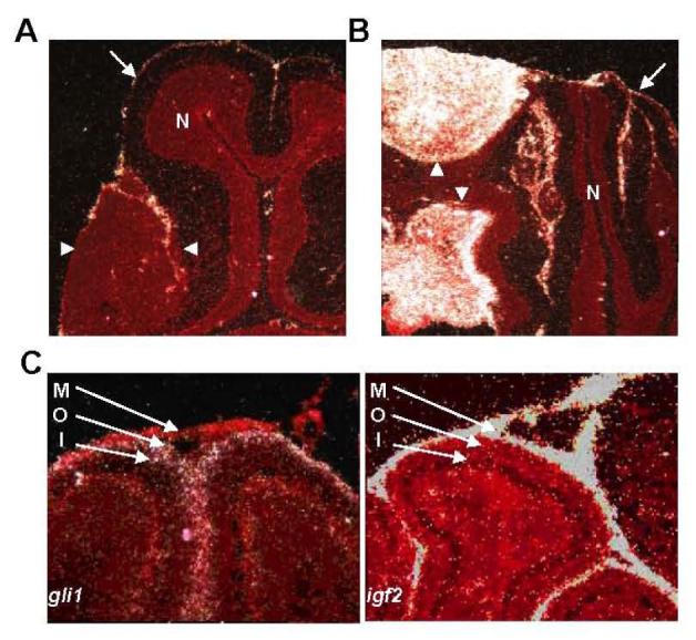
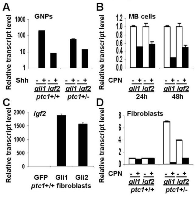
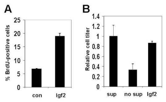
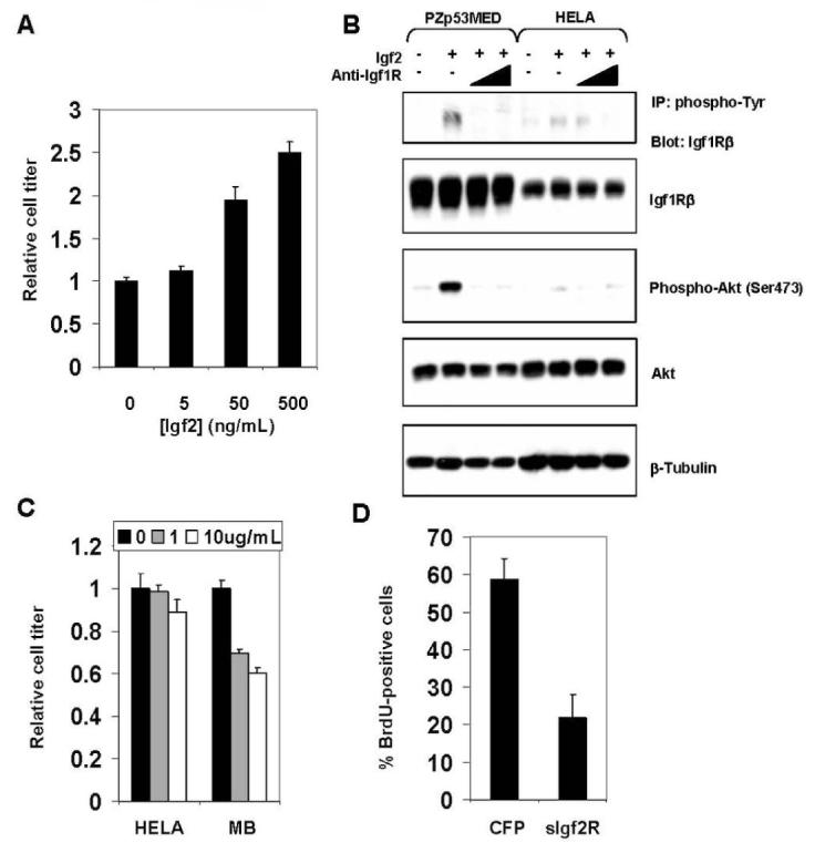
Medulloblastoma (MB) can arise in the cerebellum due to genetic activation of the Sonic Hedgehog (Shh) signaling pathway. During normal cerebellum development, Shh spurs the proliferation of granule neuron precursors (GNP), the precursor cells of MB. Mutations in the Shh receptor gene patched1 (ptc1+/-) lead to increased MB incidence in humans and mice. MB tumorigenesis in mice heterozygous for ptc1+/- shows distinct steps of progression. Most ptc1+/- mice form clusters of preneoplastic cells on the surface of the mature cerebellum that actively transcribe Shh target genes. In approximately 15% of mice, these preneoplastic cells will become fast-growing, lethal tumors. It was previously shown that the loss of function of insulin-like growth factor 2 (igf2) suppresses MB formation in ptc1+/- mice. We found that igf2 is not expressed in preneoplastic lesions but is induced as these lesions progress to more advanced MB tumors. Igf2 is not required for formation of preneoplastic lesions but is necessary for progression to advanced tumors. Exogenous Igf2 protein promoted proliferation of MB precursor cells (GNP) and a MB cell line, PZp53(MED). Blocking igf2 signaling inhibited growth of PZp53(MED) cells, implicating igf2 as a potential clinical target.


Figures

Figure 1
Figure 1
Igf2 transcription increases at a late stage of MB tumorigenesis. (A) Multiple stages of MB tumorigenesis in the ptc1+/- mouse. GNPs (green) in the normal developing postnatal cerebellum proliferate in response to Shh produced by underlying Purkinje cell neurons (red). Throughout the first 3 weeks of postnatal life, GNPs stop responding to Shh, differentiate, and migrate into the internal granule cell layer to become mature granule neurons (blue). GNPs express Math1, while mature granule neurons do not. From 3 weeks of age to adulthood, no GNPs remain in the normal cerebellum, and Math1 expression is lost. However, in the majority of ptc1+/- mice (>85%) at 3 weeks of age, small clusters of Math1-expressing GNP-like cells persist as early, pre-neoplastic MB lesions. The majority of these lesions regress spontaneously by 6 weeks of age. A subset of these lesions (∼15% of total) progress to form more aggressive-appearing intermediate, asymptomatic MBs by 10 weeks and eventually advanced, symptomatic MBs by ∼20 weeks of age. (B) Pooled averages of igf2 transcript levels by microarray analysis of pre-neoplastic early MBs (early), intermediate MBs (inter), and advanced MBs (MB). Values are relative to normal P7 GNPs. Note that igf2 transcript levels are not increased until late in tumorigenesis. Asterisk denotes p<0.01. (C) Fold-enrichment of igf2 transcript in individual MB lesions relative to normal GNPs. Each measurement is the average of five different igf2 probes. Asterisk denotes p<0.05. (D) Quantification of igf2 transcript levels by quantitiative real-time PCR from individual early MBs (early), intermediate MBs (inter), and advanced MBs (MB). Values are relative to normal P7 GNPs. Asterisk denotes p<0.01. In (B-D) error bars denote standard deviation. P values were calculated using student’s T-test relative to normal P7 GNP controls.
Figure 2
Figure 2
Expression of igf2 in developing cerebella and MB. (A, B) In situ hybridization studies of early MB (A) and advanced MB (B) using a probe specific to igf2. Signal from hybridized probe appears white. Tissue is counterstained in red. In each panel, tumor is outlined by arrowheads. Igf2 transcript is not seen in early MB (A), but is seen at high levels in the advanced MB (B). Igf2 transcript is not seen in normal cerebellum (N), but is observed in the meninges (arrows) surrounding the cerebellum. (C) In situ hybridization studies of P7 cerebella using probes specific to igf2 or gli1. Igf2 transcript is observed in the meninges (M), but not in the outer (O) or inner (I) EGL. Conversely, the Shh target transcript gli1 is absent from the meninges, but is seen in the outer, but not inner EGL.
Figure 3
Figure 3
Igf2 transcription can be regulated by the Shh pathway. (A) Levels of gli1 and igf2 transcript are increased in ptc1+/+ or ptc1+/- P7 GNPs treated with Shh (3μg/mL) for 48h. (B) PZp53MED cells (MB cells) show decreased levels of gli1 and igf2 transcript when treated with the Shh pathway inhibitor cyclopamine (CPN) for 24 or 48h. (C) ptc1+/+ fibroblasts transfected with CMV promoter-driven Gli1 or Gli2 expression constructs showed increased levels of igf2 transcript after 24h compared with cells transfected with a GFP control. (D) ptc1+/- fibroblasts have increased levels of gli1 and igf2 transcript at baseline. In CPN-treated ptc1+/- fibroblasts, levels of both transcripts are reduced to levels comparable to those seen in ptc1+/+ fibroblasts. Error bars represent standard deviation.
Figure 4
Figure 4
Igf2 promotes GNP proliferation and survival. (A) The percentage of GNPs which are proliferating (measured by BrdU staining) is increased when cells are cultured in the presence of Igf2 versus control for 48h. (B) Igf2 increases GNP survival under conditions which normally lead to apoptosis. If P7 GNPs are cultured in media without supplement (no sup) for 48h, a majority undergo apoptosis compared to those cultured in supplemented media (sup). When Igf2 is added to unsupplemented media, GNP survival is restored. Error bars represent standard deviation.
Figure 5
Figure 5
Inhibition of Igf2 signaling reduces MB cell proliferation. (A) Proliferation of PZp53MED cells is increased in a dose-dependent manner by exogenous Igf2 protein. Cell titer was measured after 48h in culture. (B) Levels of Igf1R and Akt phosphorylation in PZp53MED and HELA cell controls treated for 30 min with Igf2 with or without increasing concentrations of an antibody that blocks the Igf1R (anti-Igf1R, 1μg/mL, 10μg/mL). Levels of Igf1R and Akt phosphorylation increase dramatically in PZp53MED cells and only minimally in HELA cells when treated with Igf2 protein. Co-treatment with anti-Igf1R antibody reduced Igf1R and Akt phosphorylation to levels equal or below baseline in PZp53MED cells. Phospho-Igf1R was detected by immunoprecipitation with anti-phosphotyrosine antibody and immunoblotting with anti-Igf1Rβ antibody. (C) Anti-Igf1R antibody inhibits MB cell, but not control cell (HELA), proliferation in a dose-dependent manner. Antibody was added for 48h at the specified concentrations, and cell titer was determined. (D) PZp53MED cells were co-transfected with YFP and either a CFP control or sIgf2R. Cells were assessed for BrdU staining after 36h (BrdU was added for the final 6h). The percentage of YFP-positive cells that were also BrdU-positive was calculated for each sample. Error bars represent standard deviation.

References

    1. Hahn H, Wojnowski L, Specht K, et al. Patched target Igf2 is indispensable for the formation of medulloblastoma and rhabdomyosarcoma. J Biol Chem. 2000;275:28341–4. - PubMed
    1. Goussia AC, Bruner JM, Kyritsis AP, Agnantis NJ, Fuller GN. Cytogenetic and molecular genetic abnormalities in primitive neuroectodermal tumors of the central nervous system. Anticancer Res. 2000;20:65–73. - PubMed
    1. Raffel C, Jenkins RB, Frederick L, et al. Sporadic medulloblstomas contain PTCH mutations. Cancer Res. 1997;57:842–5. - PubMed
    1. Johnson RL, Rothman AL, Xie J, et al. Human homolog of patched, a candidate gene for the basal cell nevus syndrome. Science. 1996;272:1668–71. - PubMed
    1. Ingham PW, Taylor AM, Nakano Y. Role of the Drosophila patched gene in positional signaling. Nature. 1991;353:184–7. - PubMed

Publication types

MeSH terms