. 2008 Nov 1;68(21):8871-80.
doi: 10.1158/0008-5472.CAN-08-1972.

Modulation of orphan nuclear receptor Nur77-mediated apoptotic pathway by acetylshikonin and analogues

Affiliations

Modulation of orphan nuclear receptor Nur77-mediated apoptotic pathway by acetylshikonin and analogues

Jie Liu et al. Cancer Res. .

Abstract

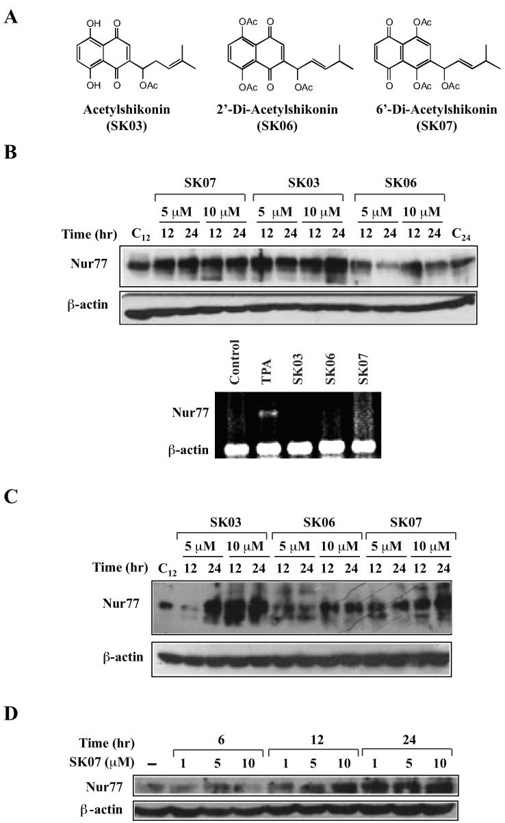
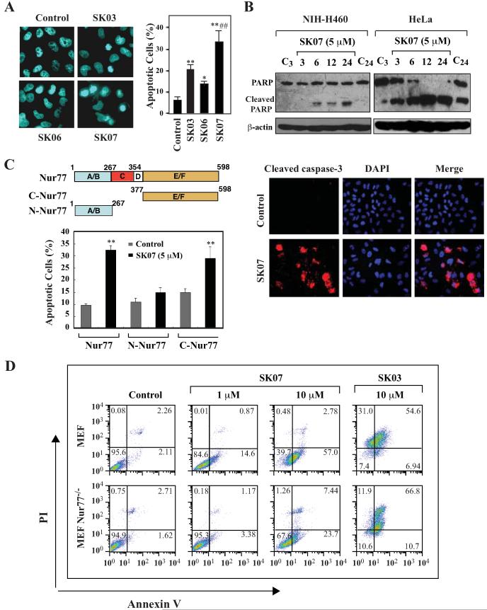
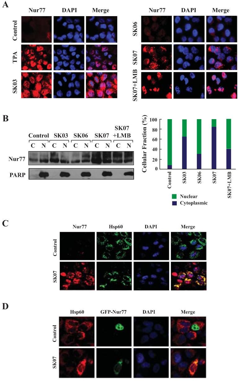
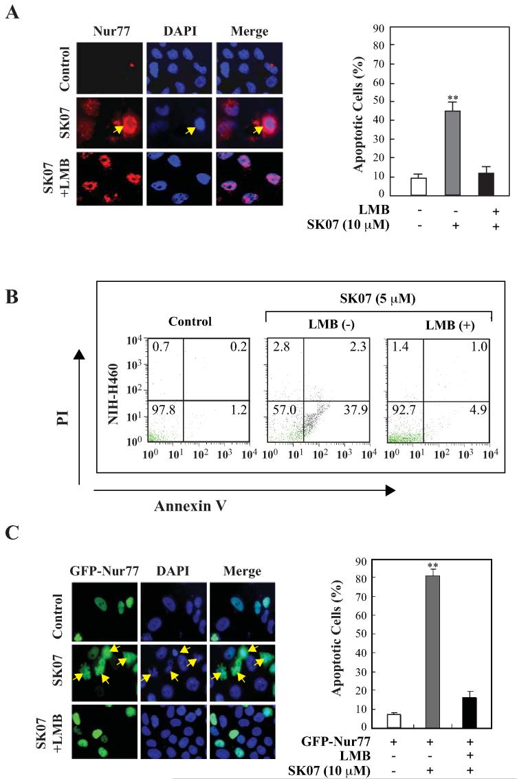
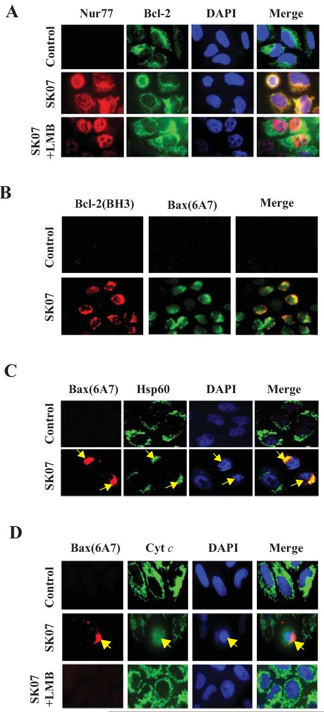
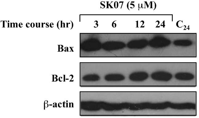
Shikonin derivatives, which are the active components of the medicinal plant Lithospermum erythrorhizon, exhibit many biological effects including apoptosis induction through undefined mechanisms. We recently discovered that orphan nuclear receptor Nur77 migrates from the nucleus to the mitochondria, where it binds to Bcl-2 to induce apoptosis. Here, we report that certain shikonin derivatives could modulate the Nur77/Bcl-2 apoptotic pathway by increasing levels of Nur77 protein and promoting its mitochondrial targeting in cancer cells. Structural modification of acetylshikonin resulted in the identification of a derivative 5,8-diacetoxyl-6-(1'-acetoxyl-4'-methyl-3'-pentenyl)-1,4-naphthaquinones (SK07) that exhibited improved efficacy and specificity in activating the pathway. Unlike other Nur77 modulators, shikonins increased the levels of Nur77 protein through their posttranscriptional regulation. The apoptotic effect of SK07 was impaired in Nur77 knockout cells and suppressed by cotreatment with leptomycin B that inhibited Nur77 cytoplasmic localization. Furthermore, SK07 induced apoptosis in cells expressing the COOH-terminal half of Nur77 protein but not its NH(2)-terminal region. Our data also showed that SK07-induced apoptosis was associated with a Bcl-2 conformational change and Bax activation. Together, our results show that certain shikonin derivatives act as modulators of the Nur77-mediated apoptotic pathway and identify a new shikonin-based lead that targets Nur77 for apoptosis induction.


Figures

Figure 1
Figure 1. Induction of Nur77 protein levels by acetylshikonin and analogs
A, structures of acytylshikonin and analogs. B, induction of Nur77 expression by shikonins in NIH-H460 lung cancer cells. Western blotting analysis was employed to evaluate the effect of shikonins on Nur77 protein levels (upper panel). NIH-H460 cells were treated with SK03, SK06, SK07 (5 or 10 µM), or vehicle for 12 or 24 hr. Nur77 protein expression was detected by using anti-Nur77 antibody. The blots were reprobed with anti-β-actin antibody for loading control. As for RT-PCR analysis (lower panel) of Nur77 mRNA expression, the same cells were treated with SK03, SK06 or SK07 (5 µM) for 3 hr, while TPA (100 ng/ml) was used as positive control. Total RNA were purified and subjected to RT-PCR analysis. Nur77 and β-actin products were simultaneously amplified by including primers for both Nur77 and β-actin in the same reaction system, in which β-actin expression level served as an internal control. C, induction of Nur77 expression by shikonins in HeLa cervical cancer cells. HeLa cells were treated with shikonins as indicated. Anti-Nur77 antibody was utilized to detect the expression of Nur77 protein by Western blotting analysis. D, dose- and time-dependent induction of Nur77. Cells were treated with vehicle or with SK07 at 1, 5 or 10 µM in serum-free medium for the indicated period of time. Nur77 expression was determined by Western blotting analysis using anti-Nur77 antibody.
Figure 2
Figure 2. Induction of apoptosis by SK07 and the role of Nur77
A, DAPI staining. NIH-H460 cells cultured in serum-free medium were treated with SK03, SK06, SK07 (5 μM), or vehicle for 24 hr and subjected to DAPI staining. Apoptotic cells were compared between different treatments. *p<0.05 (vs control); **p<0.01 (vs control); ##p<0.01 (vs SK03 or SK06). B, PARP cleavage and caspase-3 activation. NIH-H460 or HeLa cells were treated with 5 μM SK07 in serum-free medium for the indicated time. Total cell lysates were subjected to Western blotting assay for PARP cleavage using anti-PARP antibody (upper panel). As for analysis of caspase-3 activation (lower panel), NIH-H460 cells were treated with shikonins as described above and stained with an antibody recognizing the cleaved caspase-3. Nuclei were visualized by co-staining with DAPI. C, transfection of Nur77 mediates the apoptotic effect of SK07. HEK293T cells were transfected with full-length Nur77, N-Nur77 (A/B domain) or C-Nur77 (E/F domain) and subjected to SK07 treatment (5 μM) or vehicle in serum-free medium for 8 hr. Apoptotic cells examined by DAPI staining were compared between transfected cells carried different mutants. **p<0.01 (vs control). D, the apoptotic effect of SK07 is impaired in Nur77 knockout MEFs. MEF cells or MEF Nur77−/− cells were treated with vehicle or the indicated concentration of SK07 or SK03 for 24 hr and stained with PI/annexin V. Apoptosis was analyzed by FACS analysis.
Figure 3
Figure 3. Acetylshikonins induce cytoplasmic localization of Nur77
A, Nur77 translocation. NIH-H460 cells treated with SK03, SK06, or SK07 (5 μM) for 24 hr were subjected to immunostaining with anti-Nur77 antibody. Cells treated with 100 ng/mL TPA (3 hr) were used as positive control. Subcellular localization of Nur77 in cells treated with SK07 alone was compared to that in cells with cotreatment of SK07 and LMB (10 ng/mL). Cells were co-stained with DAPI to visualize the nuclei. B, cellular fractionation. Nuclear and cytoplasmic fractions were prepared from NIH-H460 cells with similar treatments as indicated in A, and subjected to Western blotting analysis. The purity of cellular fraction was confirmed by using anti-PARP antibody. Ratio of cytoplasmic Nur77 (C) to nuclear Nur77 (N) expression was quantified by BandScan 5.0. C and D, Nur77 mitochondrial targeting. NIH-H460 cells transfected with or without GFP-Nur77 were treated with 5 μM SK07 for 24 hr. Endogenous Nur77 expression was immunostained with anti-Nur77 antibody. Cells were co-stained with anti-Hsp60 antibody to recognize the mitochondria and with DAPI to visualize the nuclei. Fluorescent microscopy was used to determine the overlap of Nur77 and Hsp60.
Figure 4
Figure 4. SK07-induced apoptosis is dependent on Nur77 expression and Nur77 nuclear export
A, Nur77 cytoplasmic localization and apoptosis. NIH-H460 cells were treated with 10 μM SK07 in serum-free medium in the presence or absence of LMB (10 ng/mL) for 24 hr. Subcellular localization of endogenous Nur77 was examined by immunostaining using anti-Nur77 antibody (R&D) and apoptosis was determined by DAPI staining. B, analysis of the effect of LMB on SK07-induced apoptosis by PI/annexin V staining. NIH-H460 cancer cells were treated with SK07 (5 μM) in the presence or absence of LMB (10 ng/mL) for 24 hr. Apoptosis was determined by PI/annexin V staining. C, cytoplasmic localization of transfected Nur77 and apoptosis. NIH-H460 cells transfected with GFP-Nur77 expression vector were treated with 10 μM SK07 in serum-free medium in the presence or absence of LMB (10 ng/mL) for 24 hr. Subcellular localization of GFP-Nur77 was examined by fluorescence microscopy and apoptosis was examined by DAPI staining. Quantification was conducted in nontransfected and transfected cells respectively. **p<0.01 [vs control or LMB (+)].
Figure 5
Figure 5. SK07 induces Bcl-2 conformational change and Bax activation
A, colocalization of Nur77 with Bcl-2. NIH-H460 cells treated with vehicle or 5 μM SK07 in the presence or absence of LMB (10 ng/mL) for 24 hr were subjected to co-staining with anti-Nur77 and anti-Bcl-2 antibodies. Fluorescent microscopy was used to analyze the subcellular localization of Nur77 and Bcl-2. B, Bcl-2 conformational change and Bax activation. NIH-H460 cells treated with 5 μM SK07 were examined for Bcl-2 conformational change by anti-Bcl-2(BH3) antibody. Its association with Bax activation was analyzed by co-staining with anti-Bax (6A7) antibody. Cells were also stained with DAPI to visualize the nucleus. C, translocation of Bax to mitochondria. NIH-H460 cells treated with 5 μM SK07 were examined for Bax activation by antibody against Bax (6A7). Cells were co-stained with anti-Hsp60 antibody to reveal Bax colocalization with mitochondria. DAPI staining was employed to visualize the nuclei. The colocalization of active Bax with mitochondria and their association with apoptotic nuclear morphology were compared. D, Bax activation and cytochrome c release. NIH-H460 cells treated with 5 μM SK07 were stained with anti-Bax (6A7) and anti-cytochrome c (cyt c) antibodies. Bax activation was compared to its induction of cytochrome c release as indicated by diffusive cyt c staining. LMB was used to determine its effect on both Bax activation and cyt c release.
Figure 6
Figure 6. SK07 does not affect Bax and Bcl-2 expression levels
Cells were treated with vehicle or 5 μM SK07 for the indicated period of time. Total cell lysates were prepared and examined for Bcl-2 and Bax expression by Western blotting analysis. β-actin expression was used as a loading control.

References

    1. Zhang XK. Targeting Nur77 translocation. Expert Opin Ther Tar. 2007;11:69–79. - PubMed
    1. Moll UM, Marchenko N, Zhang XK. p53 and Nur77/TR3 - transcription factors that directly target mitochondria for cell death induction. Oncogene. 2006;25:4725–43. - PubMed
    1. Chen GQ, Lin B, Dawson MI, Zhang XK. Nicotine modulates the effects of retinoids on growth inhibition and RAR beta expression in lung cancer cells. Int J Cancer. 2002;99:171–8. - PubMed
    1. Wu Q, Liu S, Ye XF, Huang ZW, Su WJ. Dual roles of Nur77 in selective regulation of apoptosis and cell cycle by TPA and ATRA in gastric cancer cells. Carcinogenesis. 2002;23:1583–92. - PubMed
    1. Stasik I, Rapak A, Kalas W, Ziolo E, Strzadala L. Ionomycin-induced apoptosis of thymocytes is independent of Nur77 NBRE or NurRE binding, but is accompanied by Nur77 mitochondrial targeting. BBA-Mol Cell Res. 2007;1773:1483–90. - PubMed

Publication types

MeSH terms