. 2008 Nov 1;68(21):8998-9004.
doi: 10.1158/0008-5472.CAN-08-2658.

Aurora kinase A inhibition leads to p73-dependent apoptosis in p53-deficient cancer cells

Affiliations

Aurora kinase A inhibition leads to p73-dependent apoptosis in p53-deficient cancer cells

Altaf A Dar et al. Cancer Res. .

Abstract

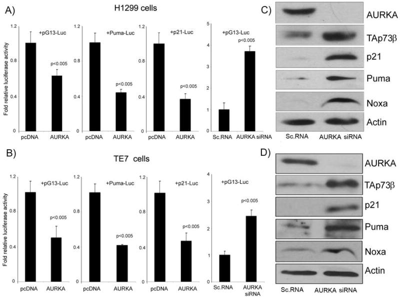
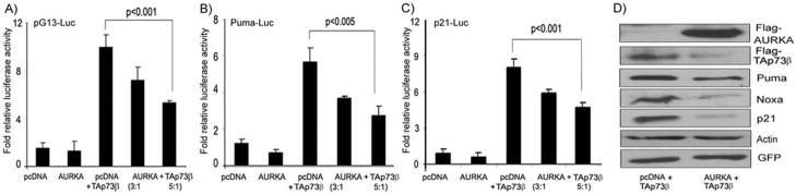
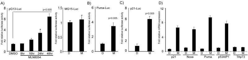
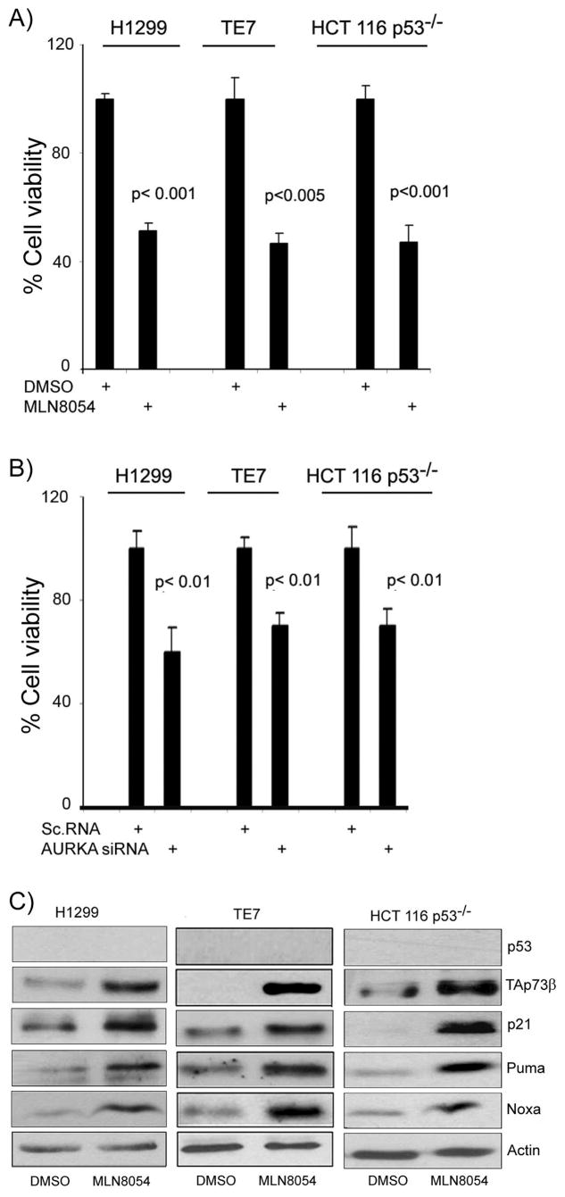
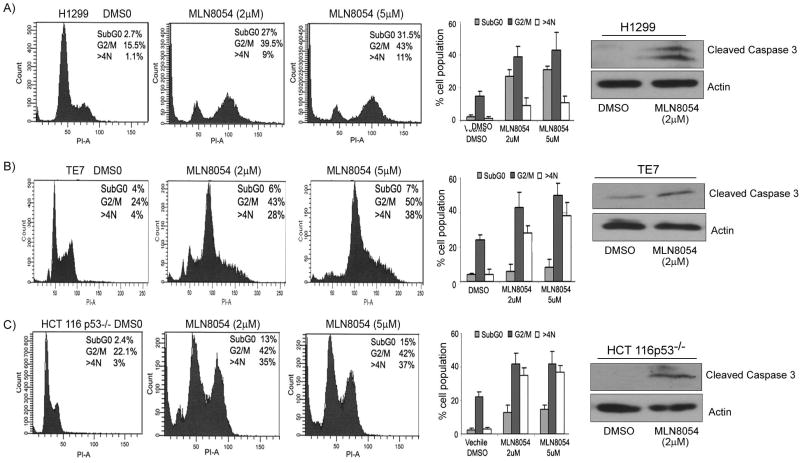
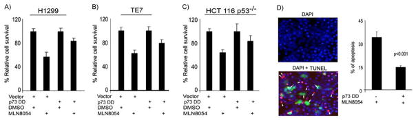
We investigated the role of Aurora kinase A (AURKA) in regulating p73-dependent apoptosis using the p53-deficient cancer cell lines H1299, TE7, and HCT116p53(-/-). Overexpression of AURKA led to down-regulation of the TAp73-induced activation of the p53/p73-dependent luciferase reporter plasmid (pG13-luc). The reduction in the TAp73 transcription activity was confirmed by measuring the activity of luciferase reporters for p21/WAF1, and PUMA. The siRNA knockdown of endogenous AURKA reversed these effects and Western blot analysis showed a significant increase in the protein level of TAp73 and its downstream transcription targets, PUMA, NOXA, and p21/WAF1. The coexpression of AURKA together with TAp73 inhibited the activation of the pG13-luc, PUMA-luc, and p21/WAF1-luc reporter plasmids with reduction in the protein levels of TAp73 transcription targets. Treatment with AURKA-selective small molecule inhibitor MLN8054 led to a significant increase in the activities of pG13-luc, PUMA-luc, and p21/WAF1-luc reporter plasmids. This effect was accompanied by a significant increase in the mRNA and protein levels of several TAp73 transcription targets: p21/WAF1, PUMA, and NOXA. Flow cytometry cell cycle analysis, after MLN8054 treatment, showed more than a 2-fold increase in cell death. The apoptotic outcome was corroborated by showing an increase in cleaved caspase-3 protein levels by Western blot. Using terminal deoxynucleotidyl-transferase-mediated dUTP nick-end labeling assay, we showed that the expression of dominant-negative mutant TAp73 expression plasmid (p73DD) counteracted the MLN8054-induced cell death. Taken together, our results indicate that AURKA regulates TAp73-dependent apoptosis and highlight the potential of the AURKA inhibitor MLN8054 in treating cancers that are defective in p53 signaling.


Figures

Figure 1
Figure 1. AURKA expression interferes with the transcriptional activity TAp73
A–B) Luciferase assays using pG13-luc, Puma-luc and p21/WAF1-luc reporter vectors were used to study the effect of AURKA expression on TAp73 in H1299 (A) and TE7 (B) p53-deficient cancer cell lines. The cells were transfected with pcDNA3-AURKA (AURKA), empty vector control (pcDNA3), AURKA-siRNA, or scrambled siRNA (control). Overexpression of AURKA led to reduction in the luciferase activity of the three reporters whereas AURKA-siRNA led to an induction of the pG13-luc reporter. C–D) H1299 (C) and TE7 (D) cells were transiently transfected with scrambled siRNA or AURKA siRNA oligonucleotides. Cell lysates were analyzed by Western blotting for AURKA, TAp73 p21/WAF1, Puma, Noxa, and β–actin. The knockdown of AURKA with siRNA up-regulated the protein expression of endogenous TAp73β and its downstream targets; p21/WAF1, Puma, and Noxa.
Figure 2
Figure 2. AURKA regulates exogenous TAp73 and its targets
A–C) The luciferase assays were performed on HCT116p53−/− cells that were transfected with pcDNA3-AURKA at different ratios (3:1 or 5:1) with TAp73β–pcDNA3, or empty vector control (pcDNA3) in combination with pG13-luc (A), Puma-luc (B), and p21/WAF1-luc (C) reporter plasmids. While TAp73β expression activated all three reporters, the co-expression of AURKA together with TAp73 reduced the promoter activities of the pG13-luc and the TAp73 downstream targets (Puma and p21/WAF1). D) HCT116p53−/− cells were co-transfected with GFP and TAp73β in combination with either empty vector (pcDNA3) or AURKA-pcDNA3 expression plasmid. Cell lysates were subjected to Western blot analysis using antibodies against Flag, for detection of the exogenous AURKA, TAp73, Puma, Noxa, p21/WAF1, β–Actin, and GFP. The transfection and gel loading were normalized for equal GFP and actin. In agreement with the reporter assays (A–C), the co-transfection with AURKA and TAp73β plasmids led to a significant decrease of the TAp73β, Puma, Noxa, and p21/WAF1 protein levels.
Figure 3
Figure 3. MLN8054 up-regulates promoter activity and mRNA levels of TAp73 target genes
A) Luciferase assay of HCT116p53−/− cells transfected with pG13-luc reporter vector and treated with AURKA inhibitor MLN8054 (2μM) for different time periods as indicated. The pG13 promoter activity was significantly induced by MLN8054 (2μM in a time-dependent manner. The MG15-luc has mutated binding sites for p53 family members and was used as a negative control. The treatment with MLN8054 (2μM did not induce promoter activity of the MG15-luc (mutant of pG13-luc). B–C) The HCT116p53−/− cells were transfected with Puma-luc and p21/WAF1-luc reporter vectors, respectively, and treated with 2μM MLN8054 (M) or DMSO (D) for 24 hours. These cells exhibited induction of the Puma and p21/WAF1 promoter activities. D) Quantitative real time RT-PCR analysis demonstrated more than five fold increase in the expression of various downstream pro-apoptotic targets (p21/WAF1, NOXA, PUMA, and p53AIP1), after treatment with MLN8054 (2μM for 24 hrs). However, no effect on the mRNA level of TAp73 was observed.
Figure 4
Figure 4. The MLN8054 treatment suppresses cell survival and induces endogenous TAp73 and its target genes at the protein level in p53-deficient cells
A) Cell viability assay of three p53-deficient cells (H1299, TE7, and HCT116p53−/−) was carried out by Cell Titer Glo assay after treating them with MLN8054 (2μM for 72 hours. These cells showed approximately 50% reduction in cell viability. B) The knockdown of AURKA by specific validated siRNA in H1299, TE7 and HCT116p53−/− showed a similar effect on cell viability. C) The H1299, TE7, and HCT116p53−/− cells were treated with vehicle (DMSO) or MLN8054 (2μM) for 24 hrs. Cell lysates were subjected to immunoblotting for p53, TAp73, p21/WAF1, Puma, Noxa, and β–Actin. Treatment with MLN8054 up-regulated TAp73β and its downstream apoptotic targets p21/WAF1, Puma and Noxa at protein levels. The TAp63 was neither expressed nor induced by MLN8054 treatment (data not shown). The results indicate that MLN8054 can specifically induce the expression of TAp73, but not TAp63, in these p53-deficient cancer cell lines.
Figure 5
Figure 5. Cell cycle analysis following MLN8054 treatment in p53-deficient cancer cell lines
The H1299 (A), TE7 (B), and HCT116p53−/− (C) were treated with two different concentrations of MLN8054 (2μM and (5μM) for 24 hours. A significant increase in the percentage of cells in the SubG1 peak was seen in H1299 and HCT116p53−/− cells, summarized in the bar graphs. In addition, the inhibition of AURKA by MLN8054 led to accumulation of cells at G2/M phase with a subsequent increase in the polyploidy of cells. The cell lysates from panels A–C were subjected to immunoblotting for cleaved caspase-3. Gel loading was normalized for equal β–actin. Cleaved caspase-3 protein was up-regulated in MLN8054- treated cells, confirming the induction of apoptosis as suggested by the increase of cells in the SubG1 peak.
Figure 6
Figure 6. The dominant-negative TAp73 mutant abrogates the MLN8054-induced cell death in p53-deficient cells
A–C)The H1299 (A), TE7 (B), and HCT116p53−/− (C) cells were transfected with control vector (pcDNA3) or TAp73 dominant-negative (p73DD) expression plasmid and treated with vehicle (DMSO) or MLN8054 (2μM) for 24 hrs. Cell survival was determined by CellTiter Glo assay. While the control cells showed an approximately 50% reduction in cell viability, following ML8054 treatment, the p73DD- transfected cells had only 20% reduction in their cell viability. D) The HCT116p53−/− cells were transfected with control vector or p73DD-IRES-GFP plasmid, and then treated with vehicle (DMSO) or MLN8054 (2 μM) for 24 hrs. Apoptotic cells were detected by TUNEL assay (red fluorescence). The DAPI (blue fluorescence) was used as nuclear counterstain to count all cells. The majority of p73DD- expressing cells (green fluorescence, white arrows) resisted MLN8054 induced apoptosis as indicated by negative TUNEL staining. The bar graph summarizes the results and demonstrates a 50% reduction in the MLN8054-induced apoptosis level in p73DD- expressing cells as compared to control cells (P<.001).

References

    1. Giet R, McLean D, Descamps S, et al. Drosophila Aurora A kinase is required to localize D-TACC to centrosomes and to regulate astral microtubules. J Cell Biol. 2002;156:437–51. - PMC - PubMed
    1. Zhou H, Kuang J, Zhong L, et al. Tumour amplified kinase STK15/BTAK induces centrosome amplification, aneuploidy and transformation. Nat Genet. 1998;20:189–93. - PubMed
    1. Glover DM, Leibowitz MH, McLean DA, Parry H. Mutations in aurora prevent centrosome separation leading to the formation of monopolar spindles. Cell. 1995;81:95–105. - PubMed
    1. Kallioniemi A, Kallioniemi OP, Piper J, et al. Detection and mapping of amplified DNA sequences in breast cancer by comparative genomic hybridization. Proc Natl Acad Sci (USA) 1994;91:2156–60. - PMC - PubMed
    1. Dar AA, Zaika A, Piazuelo MB, et al. Frequent overexpression of Aurora Kinase A in upper gastrointestinal adenocarcinomas correlates with potent antiapoptotic functions. Cancer. 2008;112:1688–98. - PMC - PubMed

Publication types

MeSH terms