. 2008 Nov 1;68(21):9024-32.
doi: 10.1158/0008-5472.CAN-08-2494.

Plumbagin, a medicinal plant-derived naphthoquinone, is a novel inhibitor of the growth and invasion of hormone-refractory prostate cancer

Affiliations

Plumbagin, a medicinal plant-derived naphthoquinone, is a novel inhibitor of the growth and invasion of hormone-refractory prostate cancer

Moammir H Aziz et al. Cancer Res. .

Abstract

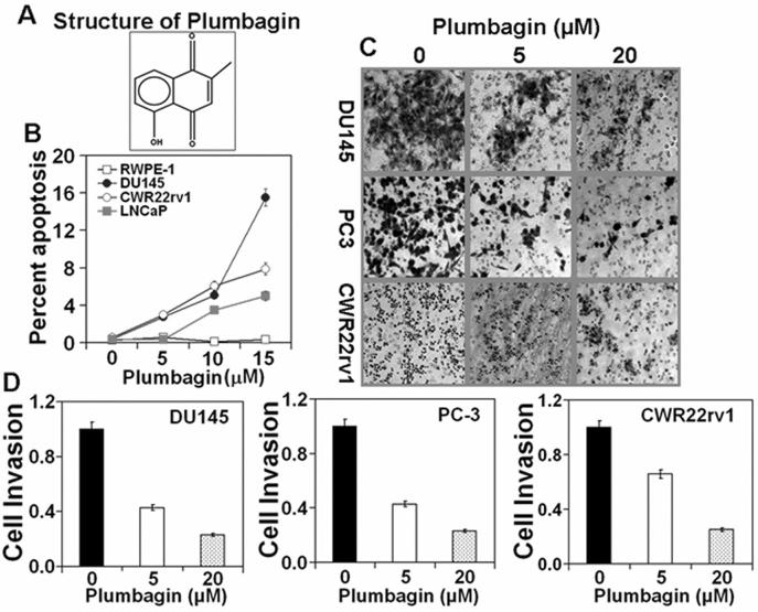
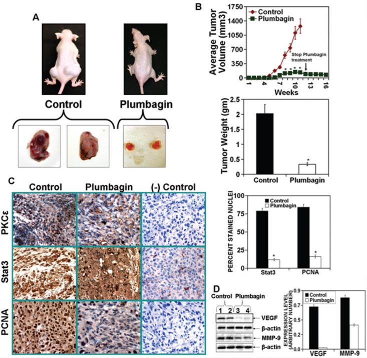
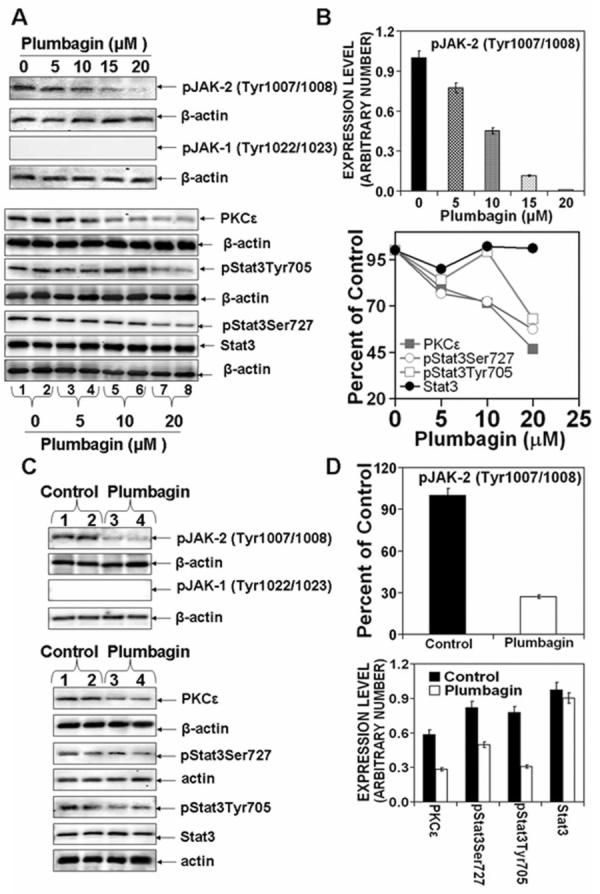
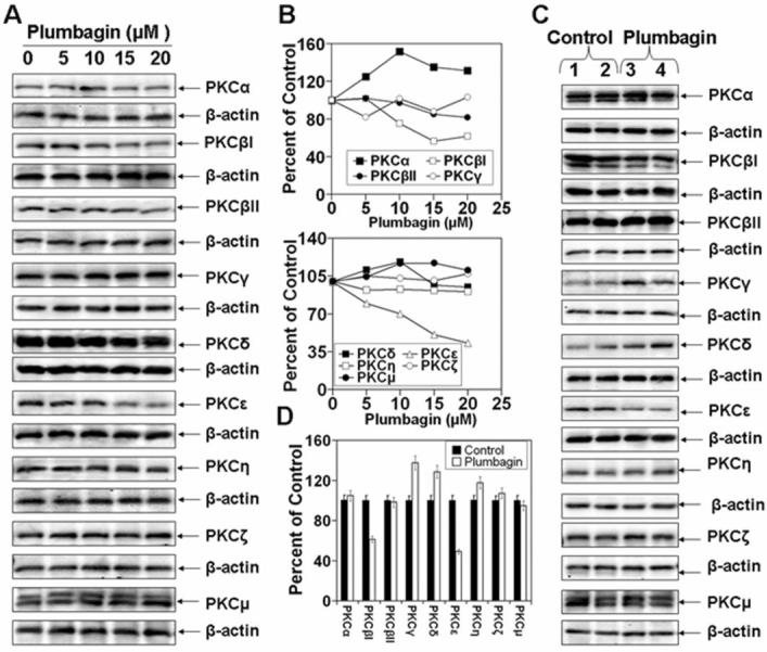
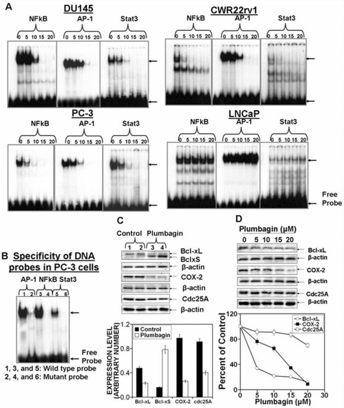
Prostate cancer (PCa) is the second leading cause of cancer-related deaths in men. Hormone-refractory invasive PCa is the end stage and accounts for the majority of PCa patient deaths. We present here that plumbagin (PL), a quinoid constituent isolated from the root of the medicinal plant Plumbago zeylanica L., may be a potential novel agent in the control of hormone-refractory PCa. Specific observations are the findings that PL inhibited PCa cell invasion and selectively induced apoptosis in PCa cells but not in immortalized nontumorigenic prostate epithelial RWPE-1 cells. In addition, i.p. administration of PL (2 mg/kg body weight), beginning 3 days after ectopic implantation of hormone-refractory DU145 PCa cells, delayed tumor growth by 3 weeks and reduced both tumor weight and volume by 90%. Discontinuation of PL treatment in PL-treated mice for as long as 4 weeks did not result in progression of tumor growth. PL, at concentrations as low as 5 micromol/L, inhibited in both cultured PCa cells and DU145 xenografts (a) the expression of protein kinase Cepsilon (PKCepsilon), phosphatidylinositol 3-kinase, phosphorylated AKT, phosphorylated Janus-activated kinase-2, and phosphorylated signal transducer and activator of transcription 3 (Stat3); (b) the DNA-binding activity of transcription factors activator protein-1, nuclear factor-kappaB, and Stat3; and (c) Bcl-xL, cdc25A, and cyclooxygenase-2 expression. The results indicate for the first time, using both in vitro and in vivo preclinical models, that PL inhibits the growth and invasion of PCa. PL inhibits multiple molecular targets including PKCepsilon, a predictive biomarker of PCa aggressiveness. PL may be a novel agent for therapy of hormone-refractory PCa.


Figures

Figure 1
Figure 1. Plumbagin induces apoptosis and inhibits cells invasion in prostate cancer cells
PCa cell lines (DU145, CWR22rv1, LNCaP and RWPE-1) at 70-80% confluency were serum starved for 24 hrs and then treated with PL at various (0, 5, 10, and 15 μM) concentrations in DMSO (final concentration 0.1%). At 24hrs after treatment, cells were collected for apoptosis analysis. CWR22rv1, DU145, and PC-3 cells were treated with 5μM or 20μM PL in DMSO (final concentration 0.1%) for 48hr and assayed cell invasion as described before (14). A: Structure of PL; B: Induction of apoptosis. Each value in the graph is the mean ± S.E. from three separate dishes. C: PCa cell invasion. Cells were stained with crystal violet and photographed at 40X magnification. D: Number of invading cells was estimated by colorimetric measurements at 560 nm according to assay instructions (Chemicon International, Temecula, CA). Each value in the graph is the mean ± S.E. from three separate wells. Similar results were observed in a repeat experiment.
Figure 2
Figure 2. Plumbagin inhibits expression of PKCε, activated Stat3, PCNA, VEGF and MMP-9 in ectopically xenografted DU145 cells
The DU145 cells (2.5 × 10⁶ cells in 100 μl of a 1:1 mixture of media:Matrigel) were implanted on both flanks of athymic nude mice (N=10 mice per group). The animals were treated with PL (2mg/kg body weight in PBS or PBS only, five days a week) by intra-peritoneal injection beginning at three days post cell implantation. At the end of the study, mice were sacrificed and digital photographs were taken. A (top): photographs of representative mice A (bottom): photographs of excised tumors. B (top): tumor growth kinetics. Tumor growth was measured weekly using digital calipers and the average tumor volume was graphed as a function of time. After 11 weeks, PL treatment was stopped and tumor growth was measured through 16 weeks post cell implantation. *, p<0.05 from the control group. B (bottom): Tumor weight at 11 weeks post cell implantation. C (left): Immunohistochemistry of tumor tissue for PKCε, Stat3 and PCNA with negative controls for specificity. Magnification, x40 (left). C (right): Quantitation of Stat3 and PCNA-positive stained nuclei (right). Columns: mean from ten different views; bars: SE. *, p<0.000 from the control group. D (left): Expression of VEGF and MMP-9 in tumors from PL treated and control mice. D (right): Quantitation of VEGF and MMP-9 expression.
Figure 3
Figure 3. Plumbagin inhibits PKCε expression as well as JAK-2 and Stat3 phosphorylation in DU145 cells in vitro and in vivo
A and B: DU145 cells at 70-80% confluency were serum starved for 24 hrs. Cells were treated with 0, 5, 10, 15 or 20 μM PL in DMSO (final concentration 0.1%) for 6hrs. Whole cell lysates were prepared and used for Western blot analysis of the indicated proteins. C and D: DU145 cells (2.5 × 10⁶ cells in 100 μl in a 1:1 of media:Matrigel) were implanted on both flanks of nude mice. Animals were treated with PL (2 mg/kg body weight in PBS or PBS only, five days a week) by intra-peritoneal injection beginning three days post implantation. At the end of the study, tumors from PL treated or control mice were excised and whole-cell lysates were prepared. Protein extracts (25 μg protein) were immunoblotted and indicated proteins detected with the appropriate antibodies. Protein levels were normalized to β-actin. Western blots (A and C) were quantitated (B and D) by densitometric analysis using Total lab Nonlinear Dynamic Image analysis software (Nonlinear USA Inc., Durham, NC).
Figure 4
Figure 4. Effects of plumbagin on the expression of PKC isoforms
DU145 cells at 70-80% confluency were serum starved for 24 hrs. Cells were treated with 0, 5, 10, 15 or 20 μM PL in DMSO (final concentration 0.1%) for 6hrs. Whole cell lysates were prepared and used for Western blot analysis of PKC isoforms (A). Tumors from PL treated or control mice were excised and whole-cell lysates were prepared to analyze the expression of PKC isoforms (C). Quantitation of Western blots (B and D).
Figure 5
Figure 5. Effects of plumbagin on the expression PI3K, AKT, p21 and p27
DU145 cells at 70-80% confluency were serum starved for 24 hrs. Cells were treated with 0, 5, 10, 15 or 20 μM PL DMSO (final concentration 0.1%) for 6hrs. Whole cell lysates were prepared and used for Western blot analysis of PI3K, AKT, p21 and p27 (A and C). Tumors from PL treated or control mice were excised and whole-cell lysates were prepared to analyze the expression PI3K, AKT, p21 and p27 (B and D).
Figure 6
Figure 6. Plumbagin inhibits DNA binding of transcription factors (TF) Stat3, NFkB and AP-1 and TF-regulated gene expression
PCa cells (DU145, PC-3, CWR22rv1 and LNCaP) at 70-80% confluency were serum starved for 24 hrs. Cells were treated with 0, 5, 10, 15, or 20 μM PL in DMSO (final concentration 0.1%) for 3hrs. Nuclear protein extracts were prepared by lysing cells in a hypotonic solution (10 mM HEPES, pH 7.5; 10 mM KCl; 0.1 mM EDTA, pH 8.0; 0.1 mM EGTA pH 8.0; 1 mM DTT; 0.5 mM PMSF; 0.5 mg/ml benzamide; 2 μg/ml aprotinin; 2 μg/ml leupeptin), with detergent (NP-40 at 6.25% (v/v)) followed by low speed (1500 × g for 30 secs) to collect nuclei. Nuclear proteins were extracted in a high-salt buffer (20 mM HEPES, pH 7.5; .4 M NaCl; 1 mM EDTA, pH 8.0; 1 mM EGTA pH 8.0; 1 mM DTT; 1 mM PMSF; 0.5 mg/ml benzamide; 2 μg/ml aprotinin; 2 μg/ml leupeptin) and nuclear membranes and genomic DNA removed by high-speed centrifugation. Nuclear protein extracts were stored at −70°C until used. A: EMSA of NFkB, AP-1 and Stat3 DNA binding; B: Specificity of AP-1, NFkB and Stat3 DNA binding. C and D: TF-regulated gene expression. C: Tumors from PL treated or control mice were excised and whole-cell lysates were prepared to analyze the expression of indicated proteins. D: DU145 cells at 70-80% confluency were serum starved for 24 hrs. Cells were treated with 0, 5, 10, 15 or 20 μM PL for 6hrs. Whole cell lysates were prepared and used for Western blot analysis of indicated proteins. Quantitation of Western blots C and D (Bottom).

Similar articles

Cited by

References

    1. Jemal A, Murray T, Ward E, et al. Cancer statistics, 2005. CA Cancer J Clin. 2005;55:10–30. - PubMed
    1. Edwards J, Bartlett JM. The androgen receptor and signal-transduction pathways in hormone-refractory prostate cancer. Part 2: Androgen-receptor cofactors and bypass pathways. BJU Int. 2005;95:1327–35. - PubMed
    1. Zhou J, Scholes J, Hsieh JT. Signal transduction targets in androgen-independent prostate cancer. Cancer Metastasis Rev. 2001;20:351–62. - PubMed
    1. Silvestris N, Leone B, Numico G, et al. Present status and perspectives in the treatment of hormone-refractory prostate cancer. Oncology. 2005;69:273–82. - PubMed
    1. Chau CH, Figg WD. Molecular and phenotypic heterogeneity of metastatic prostate cancer. Cancer Biol Ther. 2005;4:166–7. - PubMed

Publication types