

Moderate expression of prostate-specific membrane antigen, a tissue differentiation antigen and folate hydrolase, facilitates prostate carcinogenesis
- PMID: 18974153
- PMCID: PMC2748916
- DOI: 10.1158/0008-5472.CAN-08-2328
Moderate expression of prostate-specific membrane antigen, a tissue differentiation antigen and folate hydrolase, facilitates prostate carcinogenesis
Abstract
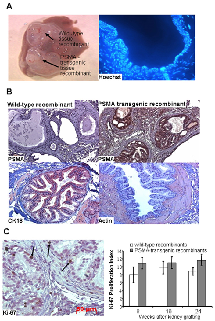
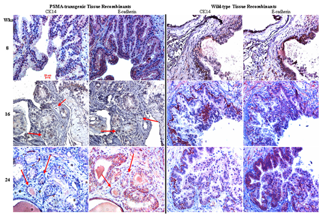
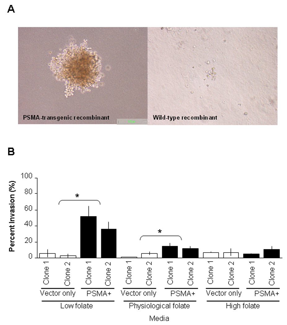
Increased expression of PSMA, a differentiation antigen with folate hydrolase activity, is an independent marker of prostate cancer progression. Mice expressing moderate levels of human PSMA in their prostate develop PIN-like lesions by 9 months. The aim of this study was to determine whether PSMA is involved in prostate carcinogenesis and progression and, if so, the possible mechanism by which PSMA may exert its effects. Using prostates from PSMA-transgenic mice, we developed a tissue recombinant model that exhibits small atypical glands with features of adenocarcinoma. This was not observed in tissue recombinants that were composed of prostate tissues from the wild-type siblings. Cells from PSMA-transgenic tissue recombinants have the ability to form colonies in semisolid agar. PSMA may facilitate this phenotype by increasing the invasive ability of cells. Ectopic PSMA expression on PC-3 cells increased the invasive capacity of cells in in vitro invasion assays, which could be competed out by folic acid. These results suggest PSMA facilitates the development of prostate cancer, and the invasive ability of these cells may be modulated by folate levels. These findings show a novel mechanism that may contribute to the known role of folate in cancer prevention, and may lead to the use of PSMA inhibitors as novel chemopreventive agents for prostate cancer. Moreover, our model should prove useful for further dissecting pathways involved in prostate carcinogenesis and progression.
Conflict of interest statement
Disclosure of potential conflicts of interest: No potential conflicts of interest to disclose.
Figures
Similar articles
-
Novel role of prostate-specific membrane antigen in suppressing prostate cancer invasiveness.Cancer Res. 2005 Feb 1;65(3):727-31. Cancer Res. 2005. PMID: 15705868
-
Expression of prostate-specific membrane antigen (PSMA), increases cell folate uptake and proliferation and suggests a novel role for PSMA in the uptake of the non-polyglutamated folate, folic acid.Prostate. 2010 Feb 15;70(3):305-16. doi: 10.1002/pros.21065. Prostate. 2010. PMID: 19830782
-
Targeting prostate-specific membrane antigen for personalized therapies in prostate cancer: morphologic and molecular backgrounds and future promises.J Biol Regul Homeost Agents. 2014 Oct-Dec;28(4):555-63. J Biol Regul Homeost Agents. 2014. PMID: 25620167 Review.
-
Prostate specific membrane antigen (PSMA) expression gives prostate cancer cells a growth advantage in a physiologically relevant folate environment in vitro.Prostate. 2006 Jun 1;66(8):867-75. doi: 10.1002/pros.20361. Prostate. 2006. PMID: 16496414
-
Tumor target prostate specific membrane antigen (PSMA) and its regulation in prostate cancer.J Cell Biochem. 2004 Feb 15;91(3):528-39. doi: 10.1002/jcb.10661. J Cell Biochem. 2004. PMID: 14755683 Review.
Cited by
-
Deficiency of DNA repair nuclease ERCC1-XPF promotes prostate cancer progression in a tissue recombination model.Prostate. 2012 Aug 1;72(11):1214-22. doi: 10.1002/pros.22472. Epub 2011 Dec 27. Prostate. 2012. PMID: 22212909 Free PMC article.
-
Elevated expression of prostate cancer-associated genes is linked to down-regulation of microRNAs.BMC Cancer. 2014 Feb 11;14:82. doi: 10.1186/1471-2407-14-82. BMC Cancer. 2014. PMID: 24517338 Free PMC article.
-
Prodrugs Targeting Prostate-Specific Membrane Antigen against Prostate Cancer.J Med Chem. 2025 Jun 26;68(12):12296-12330. doi: 10.1021/acs.jmedchem.4c02626. Epub 2025 Jun 12. J Med Chem. 2025. PMID: 40503743 Free PMC article. Review.
-
Prostate-specific membrane antigen cleavage of vitamin B9 stimulates oncogenic signaling through metabotropic glutamate receptors.J Exp Med. 2018 Jan 2;215(1):159-175. doi: 10.1084/jem.20171052. Epub 2017 Nov 15. J Exp Med. 2018. PMID: 29141866 Free PMC article.
-
Prostate-specific membrane antigen as a target for cancer imaging and therapy.Q J Nucl Med Mol Imaging. 2015 Sep;59(3):241-68. Epub 2015 Jul 24. Q J Nucl Med Mol Imaging. 2015. PMID: 26213140 Free PMC article. Review.
References
-
- Jemal A, Siegel R, Ward E, et al. Cancer statistics, 2008. CA Cancer J Clin. 2008;58:71–96. - PubMed
-
- Coffey DS. Prostate cancer: an overview of an increasing dilemma. Cancer. 1993;71:880–886. - PubMed
-
- Walczak JR, Carducci MA. Prostate cancer: a practical approach to current management of recurrent disease. Mayo Clin Proc. 2007;82:243–249. - PubMed
-
- O’Keefe DS, Bacich DJ, Heston WD. Comparative analysis of prostate-specific membrane antigen (PSMA) versus a prostate-specific membrane antigen-like gene. Prostate. 2004;58:200–210. - PubMed
-
- Bostwick DG, Pacelli A, Blute M, Roche P, Murphy GP. Prostate Specific Membrane Antigen expression in prostatic Intraepithelial Neoplasia and adneocarcinoma: a study of 184 cases. Cancer. 1998;82:2256–2261. - PubMed
Publication types
MeSH terms
Substances
Grants and funding
LinkOut - more resources
Full Text Sources
Medical
Molecular Biology Databases
Miscellaneous
