. 2008 Nov 4;105(44):16843-8.
doi: 10.1073/pnas.0807916105. Epub 2008 Oct 30.

The chemistrode: a droplet-based microfluidic device for stimulation and recording with high temporal, spatial, and chemical resolution

Affiliations

The chemistrode: a droplet-based microfluidic device for stimulation and recording with high temporal, spatial, and chemical resolution

Delai Chen et al. Proc Natl Acad Sci U S A. .

Abstract

Microelectrodes enable localized electrical stimulation and recording, and they have revolutionized our understanding of the spatiotemporal dynamics of systems that generate or respond to electrical signals. However, such comprehensive understanding of systems that rely on molecular signals-e.g., chemical communication in multicellular neural, developmental, or immune systems-remains elusive because of the inability to deliver, capture, and interpret complex chemical information. To overcome this challenge, we developed the "chemistrode," a plug-based microfluidic device that enables stimulation, recording, and analysis of molecular signals with high spatial and temporal resolution. Stimulation with and recording of pulses as short as 50 ms was demonstrated. A pair of chemistrodes fabricated by multilayer soft lithography recorded independent signals from 2 locations separated by 15 mum. Like an electrode, the chemistrode does not need to be built into an experimental system-it is simply brought into contact with a chemical or biological substrate, and, instead of electrical signals, molecular signals are exchanged. Recorded molecular signals can be injected with additional reagents and analyzed off-line by multiple, independent techniques in parallel (e.g., fluorescence correlation spectroscopy, MALDI-MS, and fluorescence microscopy). When recombined, these analyses provide a time-resolved chemical record of a system's response to stimulation. Insulin secretion from a single murine islet of Langerhans was measured at a frequency of 0.67 Hz by using the chemistrode. This article characterizes and tests the physical principles that govern the operation of the chemistrode to enable its application to probing local dynamics of chemically responsive matter in chemistry and biology.


Conflict of interest statement

The authors declare no conflict of interest.

Figures

Fig. 1.
Fig. 1.
The chemistrode delivers and records multiple molecular signals with high temporal and spatial resolution for off-line analysis by multiple analytical methods in parallel. (A) A conceptual schematic drawing of stimulation, recording, and analysis. See text for details. (B) Schematic of the chemistrode brought into contact with a hydrophilic substrate. (C) Time-lapse bright-field images (side view) of an incoming stimulus plug merging with the wetting layer above a hydrophilic glass surface and the formation of a response plug as the fluid exits the wetting layer (see Movie S1).
Fig. 2.
Fig. 2.
The chemistrode provides stimulation and recording with high temporal resolution. (A) Time-lapse fluorescence images of the delivery and capture of fluorescein within the wetting layer by 3 buffer plugs. See Movie S2 for more details. (B) Removal of fluorescein from the wetting layer by buffer plugs as a function of flow velocity. Data indicate rapid mass transport between the wetting layer and the plugs. Error bars are standard deviation (n = 5) (see SI Text). (C) Stimulation with a preformed array of fluorescent plugs containing fluorescein (green), sulforhodamine 101 (red), and buffer (gray) detected at the wetting layer with a confocal microscope (schematic on the left, experimental data on the right). A.U., arbitrary units. (D) Intensity of recorded 40-ms pulses of fluorescein measured at the tip of the chemistrode (site 1) and 10 cm downstream (site 2) (schematic on the left). Experimental data (right, green graphs of fluorescence intensity) show that pulses (shown as dashed gray lines) are reliably captured at site 1 and transported 10 cm to site 2 by response plugs in the chemistrode (upper graph and Movie S3) but not by the single-phase laminar flow in the same device (lower graph and Movie S4).
Fig. 3.
Fig. 3.
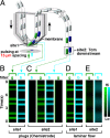
An array of chemistrodes operates at high spatial resolution. (A) A schematic drawing of the 2-layer chemistrode device used for sampling 2 signals, 8-methoxypyrene-1,3,6 trisulfonic acid (MPTS, blue) or fluorescein (green), released through 2 orifices separated by 15 μm (see SI Text). (B–E) A plot of fluorescence intensity of the 2 fluorescent signals, observed through green (g) and blue (b) filters, captured and transported by plugs of the chemistrode (B and C) and laminar flow (D and E) in the same geometry.
Fig. 4.
Fig. 4.
The chemistrode is compatible with parallel off-line analysis by independent analytical techniques and is compatible with living cells as a substrate. (A and B): Use of the chemistrode to record and analyze pulses of sample solution containing CaCl2, insulin, glucose, and a fluorescent dye, MPTS. (A) A schematic of the experiment. Pulses were generated at the surface. After recording with the chemistrode, recorded plugs were split into 4 identical daughter arrays for off-line analysis via fluorescence microscopy, FCS competitive immunoassay, and MALDI-MS. (B) Experimental data of Ca2+, insulin, glucose, and MPTS analyses combined and aligned to reveal the complete release profile of the 4 species. (C–E) Use of the chemistrode to stimulate a mouse islet of Langerhans and record insulin secretion every 1.5 s. A.U., arbitrary units. (C) A schematic of the experiment. An islet was cultured on a glass-bottom dish, and the chemistrode was positioned over the islet. Stimulation and recording took place while the islet was imaged by fluorescence microscopy. (D) A fluorescent image of an islet showing an increase of fluorescence of fluo-4, corresponding to the rise in intracellular [Ca2+]i in the islet upon stimulation. (E) Graphs showing the [Ca2+]i response and insulin secretion of a stimulated mouse islet. (Upper) Traces measured by fluorescence microscopy during stimulation and recording, showing the fluorescence intensity of fluo-4 (green) as an indicator of [Ca2+]i and the intensity of Alexa Fluor 594 (red) as a marker of the stimulant solution. (Lower) Traces with results of off-line analysis of plugs collected during recording, showing the fluorescence intensity of Alexa Fluor 594 marker and the calculated insulin secretion rate.

References

    1. Hamill OP, Marty A, Neher E, Sakmann B, Sigworth FJ. Improved patch-clamp techniques for high-resolution current recording from cells and cell-free membrane patches. Pflugers Arch. 1981;391:85–100. - PubMed
    1. Hubel DH, Wiesel TN. Receptive fields of single neurones in the cats striate cortex. J Physiol (London) 1959;148:574–591. - PMC - PubMed
    1. Wightman RM. Probing cellular chemistry in biological systems with microelectrodes. Science. 2006;311:1570–1574. - PubMed
    1. Kennedy RT, Thompson JE, Vickroy TW. In vivo monitoring of amino acids by direct sampling of brain extracellular fluid at ultralow flow rates and capillary electrophoresis. J Neurosci Methods. 2002;114:39–49. - PubMed
    1. Cellar NA, Burns ST, Meiners JC, Chen H, Kennedy RT. Microfluidic chip for low-flow push–pull perfusion sampling in vivo with on-line analysis of amino acids. Anal Chem. 2005;77:7067–7073. - PubMed

Publication types

LinkOut - more resources