. 2009 Mar;150(3):1321-9.
doi: 10.1210/en.2008-1090. Epub 2008 Oct 30.

Glucose-oxidized low-density lipoproteins enhance insulin-like growth factor I-stimulated smooth muscle cell proliferation by inhibiting integrin-associated protein cleavage

Affiliations

Glucose-oxidized low-density lipoproteins enhance insulin-like growth factor I-stimulated smooth muscle cell proliferation by inhibiting integrin-associated protein cleavage

Lee B Allen et al. Endocrinology. 2009 Mar.

Erratum in

  • Endocrinology. 2010 Jun;151(6):2967

Abstract

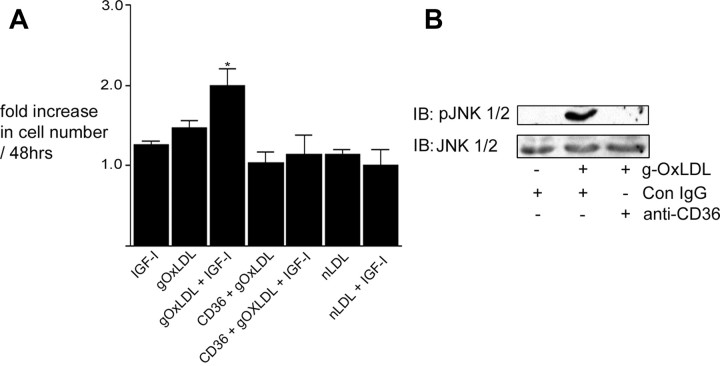
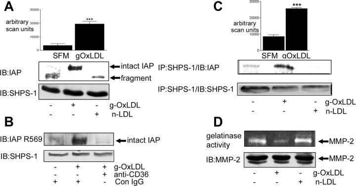
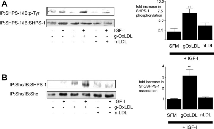
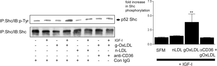
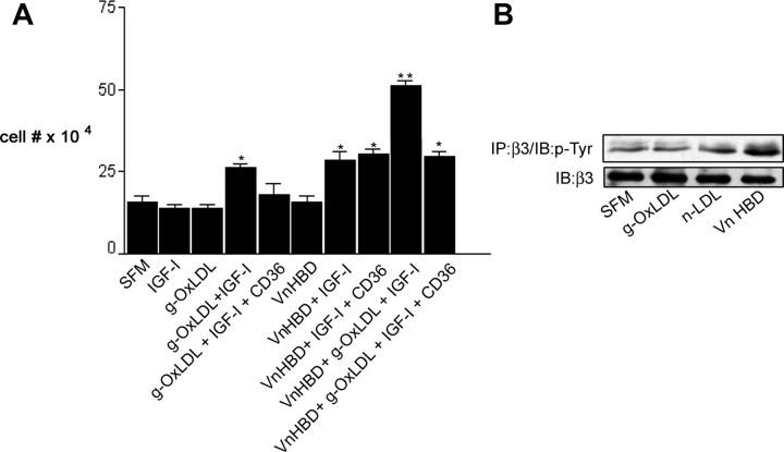
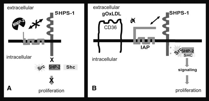
Prior published reports have demonstrated that glucose-oxidized low-density lipoproteins (g-OxLDL) enhance the proliferative response of vascular smooth muscle cells (SMC) to IGF-I. Our previous studies have determined that the regulation of cleavage of integrin-associated protein (IAP) by matrix-metalloprotease-2 (MMP-2) in diabetic mice in response to hyperglycemia is a key regulator of the response of SMC to IGF-I. Because chronic hyperglycemia enhances glucose-induced LDL oxidation, these studies were conducted to determine whether g-OxLDL modulates the response of SMC to IGF-I by regulating MMP-2-mediated cleavage of IAP. We determined that exposure of SMC to g-OxLDL, but not native LDL, was sufficient to facilitate an increase in cell proliferation in response to IGF-I. Exposure to an anti-CD36 antibody, which has been shown to inhibit g-OxLDL-mediated signaling, inhibited the effects of g-OxLDL on IGF-I-stimulated SMC proliferation. The effect of g-OxLDL could be attributed, in part, to an associated decrease in proteolytic cleavage of IAP leading to increase in the basal association between IAP and Src homology 2 domain-containing protein tyrosine phosphatase substrate-1, which is required for IGF-I-stimulated proliferation. The inhibitory effect of g-OxLDL on IAP cleavage appeared to be due to its ability to decrease the amount of activated MMP-2, the protease responsible for IAP cleavage. In conclusion, these data provide a molecular mechanism to explain previous studies that have reported an enhancing effect of g-OxLDL on IGF-I-stimulated SMC proliferation.


Figures

F<sc>ig</sc>. 1.
Fig. 1.
g-OxLDL increases IGF-I-stimulated SMC proliferation in a CD36-dependent manner. A, Cells (2 × 10⁴) were plated in each well of a 24-well plate before exposure to IGF-I (50 ng/ml), g-OxLDL in the presence of control antiserum (5 μg/ml), n-LDL (5 μg/ml), or g-OxLDL in the presence of the anti-CD36 (CD36) antiserum (all prepared in DMEM plus 0.2% platelet poor plasma). At 48 h after the addition of IGF-I (50 ng/ml), cell number was determined by trypan blue staining and counting. *, P < 0.05 when cell number in response to IGF-I in the presence of g-OxLDL is compared with the number of cells in the presence of IGF-I alone. B, SMC were grown to confluency in medium containing 5 mm glucose before overnight incubation in SFM. g-OxLDL (5 μg/ml) was added for 4 h. Where indicated, either control IgG or anti-CD36 antiserum was added for 30 min before the addition of the LDL preparation. Phosphorylation of JNK was determined by immunoblotting (IB) equal amounts of cell lysates with an antibody specific for the phosphorylated form (pJNK). To demonstrate that there as no significant difference in the amount of total protein, blots were reprobed with an antibody that recognizes total JNK protein.
F<sc>ig</sc>. 2.
Fig. 2.
g-OxLDL protects IAP from cleavage. SMC were grown to confluency in medium containing 5 mm glucose before overnight incubation in SFM. g-OxLDL (5 μg/ml) or n-LDL (5 μg/ml) was added for 4 h before collecting conditioned medium and lysing the cells. Where indicated, either control IgG or anti-CD36 antiserum was added for 30 min before the addition of the LDL preparations. A, Intact and fragmented IAP were visualized by immunoblotting (IB) cell lysates with the anti-IAP monoclonal antibody B6H12. Lysates were also immunoblotted with an anti-SHPS-1 antibody as a control for total protein. The graph shows the intensity of the intact IAP immunoreactive band expressed as arbitrary scanning units (mean ± sem, n = 3). ***, P < 0.005 when the values for IAP in the presence of g-OxLDL is compared with SFM alone. B, Intact IAP was visualized by immunoblotting (IB) cell lysates with the anti-IAP antibody that specifically recognizes intact IAP (R569). Lysates were also immunoblotted with an anti-SHPS-1 antibody as a control for total protein. C, IAP association with SHPS-1 was determined by immunoblotting cell lysates with an anti-SHPS-1 antibody and then immunoblotting with the anti-IAP monoclonal antibody B6H12. Lysates were also immunoprecipitated (IP) with an anti-SHPS-1 antibody and immunoblotted with the same antibody to demonstrate that the difference in the amount of IAP was not due to a significant change in SHPS-1 protein. The graph shows the intensity of the IAP immunoreactive band expressed as arbitrary scanning units (mean ± sem, n = 3). ***, P < 0.005 when the values for SHPS-1 association with IAP in the presence of g-OxLDL is compared with SFM alone. D, Conditioned medium was collected, concentrated, and then applied to a gelatin zymogram. After overnight incubation at 37 C (as described in Materials and Methods), the gel was stained with Coomassie blue, and cleared areas were visualized as an indicator of MMP-2 gelatinase activity (upper panel). Cell lysates were immunoblotted (IB) with an anti-MMP-2 antibody (lower panel). The results shown are representative of three independent experiments with similar results.
F<sc>ig</sc>. 3.
Fig. 3.
g-OxLDL enhances IGF-I-stimulated SHPS-1 phosphorylation and Shc recruitment to SHPS-1. SMC were grown to confluency in medium containing 5 mm glucose before overnight incubation in SFM. g-OxLDL (5 μg/ml) or n-LDL (5 μg/ml) was added for 4 h. IGF-I was added (100 ng/ml) for 5 min at the end of the 4-h incubation before lysing of the cells. A, SHPS-1 phosphorylation was determined by immunoprecipitating (IP) with an anti-SHPS-1 antibody and immunoblotting with an anti-phosphotyrosine antibody (p-Tyr). Equal amounts of cell lysates were also immunoprecipitated with the anti-SHPS-1 antibody and immunoblotted with the same antibody to demonstrate that the difference in response was not due to different amounts of SHPS-1 protein. The graph shows the increase in SHPS-1 phosphorylation in response to IGF-I (mean ± sem, n = 3). **, P < 0.01 when SHPS-1 phosphorylation in response to IGF-I in the presence of g-OxLDL is compared with the response to IGF-I alone. B, The extent of Shc association with SHPS-1 was determined by immunoprecipitating (IP) cell lysates with an anti-Shc antibody and then immunoblotting with an anti-SHPS-1 antibody. Equal amounts of cell lysates were also immunoprecipitated with the anti-Shc antibody and immunoblotted with the same antibody. The graph shows the increase in Shc association with SHPS-1 in response to IGF-I (mean ± sem, n = 3). **, P < 0.01 when Shc recruitment to SHPS-1 in response to IGF-I in the presence of g-OxLDL is compared with the response to IGF-I alone. The results shown are representative of three similar experiments performed independently.
F<sc>ig</sc>. 4.
Fig. 4.
g-OxLDL enhances IGF-I-stimulated Shc phosphorylation in a CD36-dependent manner. SMC were grown to confluency in medium containing 5 mm glucose before overnight incubation in SFM. g-OxLDL (5 μg/ml) or n-LDL (5 μg/ml) was added for 4 h. Where indicated, either control IgG or anti-CD36 antiserum (anti-CD36) was added for 30 min before the addition of the LDL preparations. IGF-I (100 ng/ml) was added for 5 min at the end of the 4-h incubation before lysing the cells. Shc phosphorylation was determined by immunoprecipitating (IP) with an anti-Shc antibody and immunoblotting with an anti-phosphotyrosine antibody (p-Tyr). Equal amounts of cell lysates were also immunoprecipitated with the anti-Shc antibody and immunoblotted with the same antibody. The graph shows the fold increase in Shc phosphorylation in response to IGF-I (mean ± sem, n = 3). **, P < 0.01 when Shc phosphorylation in the presence of g-OxLDL and IGF-I is compared with Shc phosphorylation in the presence of IGF-I alone. The results shown are representative of the results of three similar experiments performed independently.
F<sc>ig</sc>. 5.
Fig. 5.
CD36 blocks g-OxLDL but not Vn enhancement of IGF-I-stimulated SMC proliferation. A, Cells 2 × 10⁴) were plated in each well of a 24-well plate before exposure to g-OxLDL (5 μg/ml) with or without the anti-CD36 antiserum or control antiserum (Con IgG), a peptide homologous to the Vn HBD (10 μg/ml) or g-OxLDL and Vn HBD with or without the anti-CD36 antiserum (CD36). All treatments were prepared in DMEM plus 0.2% platelet-poor plasma. At 48 h after the addition of IGF-I (50 ng/ml), cell number was determined by trypan blue staining and counting. *, P < 0.05 when cell number in response to IGF-I is compared with SFM alone. **, P < 0.01 when cell number in response to IGF-I in the presence of g-OxLDL/Vn HBD is compared with the number of cells in the presence of IGF-I alone. B, SMC were grown to confluency in medium containing 5 mm glucose before overnight incubation in SFM. g-OxLDL (5 μg/ml), n-LDL (5 μg/ml), or the Vn HBD (10 μg/ml) was added for 4 h. After lysis, β3 phosphorylation was detected by immunoprecipitation (IP) with an anti-β3 antibody and immunoblotting with an anti-phosphotyrosine antibody (p-Tyr). To demonstrate that an equivalent amount of protein was loaded in each lane, samples were also immunoblotted with an anti-β3 antibody.
F<sc>ig</sc>. 6.
Fig. 6.
Diagrammatic representation of the regulation of IGF-I signaling by OxLDL increase in IAP association with SHPS-1. When SMC are grown in normal glucose-containing medium, the cleavage of IAP by MMP-2 prevents its interaction with SHPS-1. The consequence of this cleavage is an inability of IGF-I to stimulate SHPS-1 phosphorylation and therefore failure to form the SHPS-1-SHP-2-Src-Shc signaling complex that is required for signaling (A). In contrast, when SMC are exposed to g-OxLDL, the amount of MMP-2 activity in the conditioned medium is reduced, thus limiting the cleavage of IAP. Consequently, the association of IAP with SHPS-1 is enhanced, resulting in increased phosphorylation of SHPS-1 in response to IGF-I. The formation of this signaling complex then facilitates the proliferative response of SMC to IGF-I (B).

References

    1. Kannel WB, McGee DL 1979. Diabetes and cardiovascular risk factors: the Framingham study. Circulation 59:8–13 - PubMed
    1. Laing SP, Swerdlow AJ, Slater SD, Burden AC, Morris A, Waugh NR, Gatling W, Bingley PJ, Patterson CC 2003. Mortality from heart disease in a cohort of 23,000 patients with insulin-treated diabetes. Diabetologia 46:760–765 - PubMed
    1. Moss SE, Klein R, Klein BE 1991. Cause-specific mortality in a population-based study of diabetes. Am J Public Health 81:1158–1162 - PMC - PubMed
    1. Ross R 1993. The pathogenesis of atherosclerosis: a perspective for the 1990s. Nature 362:801–809 - PubMed
    1. Jones JI, Prevette T, Gockerman A, Clemmons DR 1996. Ligand occupancy of the α-V-β3 integrin is necessary for smooth muscle cells to migrate in response to insulin-like growth factor. Proc Natl Acad Sci USA 93:2482–2487 - PMC - PubMed

Publication types

MeSH terms