. 2008 Dec;173(6):1902-10.
doi: 10.2353/ajpath.2008.080201. Epub 2008 Oct 30.

Serum amyloid A, but not C-reactive protein, stimulates vascular proteoglycan synthesis in a pro-atherogenic manner

Affiliations

Serum amyloid A, but not C-reactive protein, stimulates vascular proteoglycan synthesis in a pro-atherogenic manner

Patricia G Wilson et al. Am J Pathol. 2008 Dec.

Abstract

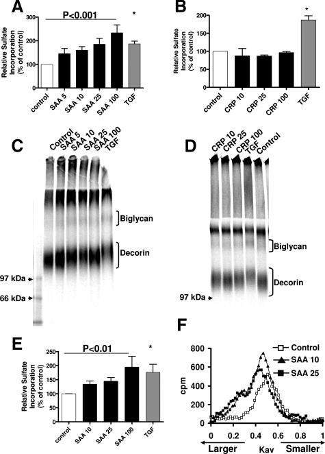
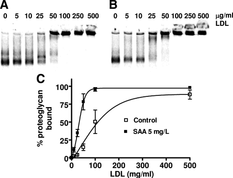
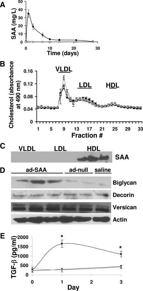
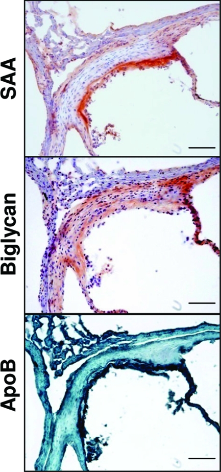
Inflammatory markers serum amyloid A (SAA) and C-reactive protein (CRP) are predictive of cardiac disease and are proposed to play causal roles in the development of atherosclerosis, in which the retention of lipoproteins by vascular wall proteoglycans is critical. The purpose of this study was to determine whether SAA and/or CRP alters vascular proteoglycan synthesis and lipoprotein retention in a pro-atherogenic manner. Vascular smooth muscle cells were stimulated with either SAA or CRP (1 to 100 mg/L) and proteoglycans were then isolated and characterized. SAA, but not CRP, increased proteoglycan sulfate incorporation by 50 to 100% in a dose-dependent manner (P < 0.0001), increased glycosaminoglycan chain length, and increased low-density lipoprotein (LDL) binding affinity (K(d), 29 microg/ml LDL versus 90 microg/ml LDL for SAA versus control proteoglycans; P < 0.005). Furthermore, SAA up-regulated biglycan via the induction of endogenous transforming growth factor (TGF)-beta. To determine whether SAA stimulated proteoglycan synthesis in vivo, ApoE(-/-) mice were injected with an adenovirus expressing human SAA-1, a null virus, or saline. Mice that received adenovirus expressing SAA had increased TGF-beta concentrations in plasma and increased aortic biglycan content compared with mice that received either null virus or saline. Thus, SAA alters vascular proteoglycans in a pro-atherogenic manner via the stimulation of TGF-beta and may play a causal role in the development of atherosclerosis.


Figures

Figure 1
Figure 1
SAA, but not CRP, increases vascular proteoglycan and glycosaminoglycan synthesis. A and B: Vascular smooth muscle cells were stimulated with SAA (A) or CRP (B) at the indicated concentrations (mg/L) or with TGF-β (gray bars, 2 ng/ml) for 24 hours. Sulfate incorporation was determined by cetyl pyridinium chloride precipitation as described in the Materials and Methods. Data shown are mean ± SEM from eight separate experiments (A) and three separate experiments (B). C and D: Proteoglycan size was estimated using sodium dodecyl sulfate-polyacrylamide gel electrophoresis (3.5% stacking gel with 4 to 12% gradient resolving gel). Lanes were loaded with equal counts; gels shown are representative of eight independent experiments (C) or three separate experiments (D). E and F: Vascular smooth muscle cells were stimulated with SAA at the indicated concentrations (mg/L) or with TGF-β (gray bars, 2 ng/ml) for 24 hours in the presence of 0.5 mmol/L xyloside. E: Sulfate incorporation was determined by cetyl pyridinium chloride precipitation as described in the Materials and Methods. Data shown are mean ± SEM from five separate experiments. F: Glycosaminoglycans were applied to Sepharose CL-6B columns as described in the Materials and Methods for analysis of hydrodynamic size. The curves shown are representative of five separate experiments. *P < 0.01 versus control.
Figure 2
Figure 2
Proteoglycans secreted by cells stimulated with SAA have higher LDL binding affinity than proteoglycans secreted by unstimulated cells. Shown are representative gels showing the binding of proteoglycans synthesized by unstimulated cells (A) and proteoglycans synthesized by cells stimulated with 5 mg/L SAA (B) to native human LDL. C: Binding curves show mean ± SEM from four separate experiments.
Figure 3
Figure 3
SAA stimulates biglycan via TGF-β and the FPRL1 receptor. A: Cells were stimulated with SAA at 25 or 100 mg/L, or with TGF-β (2 ng/ml). B: Cells were stimulated with CRP at 25 or 100 mg/L, or with TGF-β (2 ng/ml). C: Cells were stimulated with TGF-β (2 ng/ml), SAA 100 mg/L alone or in combination with TGF-β neutralizing antibody 1D11 (10 μg/ml), or with irrelevant control antibody 13C4 (10 μg/ml). D: Cells were stimulated with TGF-β (2 ng/ml), SAA 100 mg/L alone or in combination with Lipoxin A4 (LXA4, 5μmol/L), or PTX (0.5 μg/ml) for 24 hours. Proteoglycan core protein synthesis was analyzed by Western blot. Lanes were loaded with an equal amount of protein, and blotted for biglycan, versican, decorin, and actin. Blots shown are representative of three independent experiments (A) or two independent experiments (B--D).
Figure 4
Figure 4
Elevated human SAA stimulates proteoglycan synthesis in vivo. A: Mice were injected with an adenovirus expressing SAA (ad-SAA, black squares), a null virus (ad-null, open squares), or saline (gray squares, not visible because of overlap with open squares) on day 0, then samples were collected on the indicated days and human SAA quantified by enzyme-linked immunosorbent assay. Shown are mean ± SEM for n = 22 (ad-SAA), n = 11 (ad-null), and n = 9 (saline). B: Plasma aliquots from individual mice drawn 1 day after adenovirus or saline injection were analyzed by fast performance liquid chromatography for lipoprotein cholesterol distribution. Shown are mean ± SEM for n = 5 (ad-SAA and ad-null) and n = 4 (saline). C: Fractions from the VLDL, LDL, and HDL peaks (indicated by the horizontal bars) were collected and SAA content analyzed by Western blot. Blot shown is from a mouse that received ad-SAA, representative of five. D: Aortic biglycan content was determined by Western blot analysis of total aortic protein. Each lane represents individual mice, representative of n = 22 (ad-SAA), n = 11 (ad-null), and n = 9 (saline). E: Endogenously active TGF-β was measured in six mice per group from samples collected before, and 1 and 3 days after injections. Data shown are mean ± SEM. *P < 0.01 versus saline group.
Figure 5
Figure 5
SAA increased aortic biglycan content, which co-localizes with SAA and apoB. Mice were injected with ad-SAA, ad-null, or saline, and 28 days later vascular tissues were collected. Shown are representative photos from a mouse that received ad-SAA. In each photo the lumen is shown to the bottom right. Scale bars = 100 μm. Original magnifications, ×200.

Similar articles

Cited by

References

    1. McLaughlin T, Abbasi F, Lamendola C, Liang L, Reaven G, Schaaf P, Reaven P. Differentiation between obesity and insulin resistance in the association with C-reactive protein. Circulation. 2002;106:2908–2912. - PubMed
    1. Lemieux I, Pascot A, Prud'homme D, Almeras N, Bogaty P, Nadeau A, Bergeron J, Despres JP. Elevated C-reactive protein: another component of the atherothrombotic profile of abdominal obesity. Arterioscler Thromb Vasc Biol. 2001;21:961–967. - PubMed
    1. Fröhlich M, Imhof A, Berg G, Hutchinson WL, Pepys MB, Boeing H, Muche R, Brenner H, Koenig W. Association between C-reactive protein and features of the metabolic syndrome: a population-based study. Diabetes Care. 2000;23:1835–1839. - PubMed
    1. Visser M, Bouter LM, McQuillan GM, Wener MH, Harris TB. Elevated C-reactive protein levels in overweight and obese adults. JAMA. 1999;282:2131–2135. - PubMed
    1. Uhlar CM, Whitehead AS. Serum amyloid A, the major vertebrate acute-phase reactant. Eur J Biochem. 1999;265:501–523. - PubMed

Publication types

MeSH terms