

Genotype-correlated expression of lysyl oxidase-like 1 in ocular tissues of patients with pseudoexfoliation syndrome/glaucoma and normal patients
- PMID: 18974306
- PMCID: PMC2626384
- DOI: 10.2353/ajpath.2008.080535
Genotype-correlated expression of lysyl oxidase-like 1 in ocular tissues of patients with pseudoexfoliation syndrome/glaucoma and normal patients
Abstract
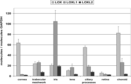
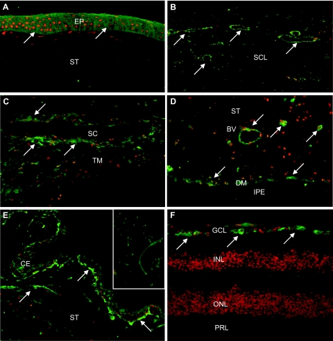
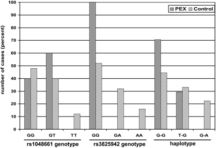
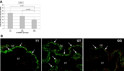
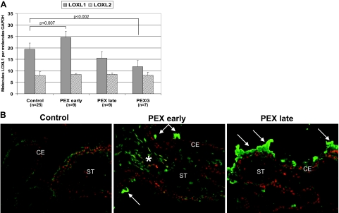
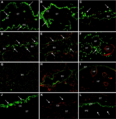
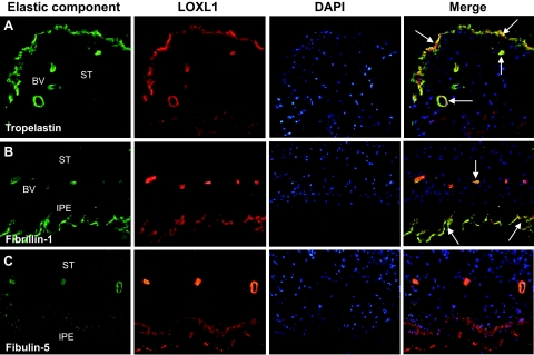
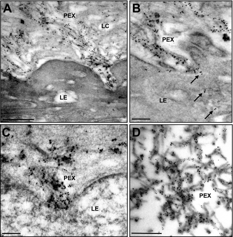
Pseudoexfoliation (PEX) syndrome is a generalized disease of the extracellular matrix and the most common identifiable cause of open-angle glaucoma. Two single nucleotide polymorphisms in the lysyl oxidase-like 1 (LOXL1) gene (rs1048661 and rs3825942) have been recently identified as strong genetic risk factors for both PEX syndrome and PEX glaucoma. Here we investigated the expression and localization of LOXL1, LOXL2, and lysyl oxidase (LOX) in tissues of PEX syndrome/glaucoma patients and controls in correlation with their individual single nucleotide polymorphism genotypes and stages of disease. LOXL1 ocular expression was reduced by approximately 20% per risk allele of rs1048661, whereas risk alleles of rs3825942, which were highly overrepresented in PEX cases, did not affect LOXL1 expression levels. Irrespective of the individual genotype, LOXL1 expression was significantly increased in early PEX stages but was decreased in advanced stages both with and without glaucoma compared with controls, whereas LOX and LOXL2 showed no differences between groups. LOXL1 was also found to be a major component of fibrillar PEX aggregates in both intra- and extraocular locations and to co-localize with various elastic fiber components. These findings provide evidence for LOXL1 involvement in the initial stages of abnormal fibrogenesis in PEX tissues. Alterations of LOXL1 activation, processing, and/or substrate specificity may contribute to the abnormal aggregation of elastic fiber components into characteristic PEX fibrils.
Figures
Similar articles
-
LOXL1 deficiency in the lamina cribrosa as candidate susceptibility factor for a pseudoexfoliation-specific risk of glaucoma.Ophthalmology. 2012 Sep;119(9):1832-43. doi: 10.1016/j.ophtha.2012.03.015. Epub 2012 May 24. Ophthalmology. 2012. PMID: 22633114
-
Molecular pathology of pseudoexfoliation syndrome/glaucoma--new insights from LOXL1 gene associations.Exp Eye Res. 2009 Apr;88(4):776-85. doi: 10.1016/j.exer.2008.08.012. Epub 2008 Sep 6. Exp Eye Res. 2009. PMID: 18809397 Review.
-
Correlation of Aqueous Humor Lysyl Oxidase Activity with TGF-ß Levels and LOXL1 Genotype in Pseudoexfoliation.Curr Eye Res. 2016 Oct;41(10):1331-1338. doi: 10.3109/02713683.2015.1125505. Epub 2016 Apr 26. Curr Eye Res. 2016. PMID: 27116380
-
Association of lysyl oxidase-like 1 gene common sequence variants in Greek patients with pseudoexfoliation syndrome and pseudoexfoliation glaucoma.Mol Vis. 2013 Jul 12;19:1446-52. Print 2013. Mol Vis. 2013. PMID: 23869164 Free PMC article.
-
Expression and regulation of LOXL1 and elastin-related genes in eyes with exfoliation syndrome.J Glaucoma. 2014 Oct-Nov;23(8 Suppl 1):S48-50. doi: 10.1097/IJG.0000000000000120. J Glaucoma. 2014. PMID: 25275906 Review.
Cited by
-
Association of LOXL1 gene polymorphisms with exfoliation syndrome/glaucoma and primary open angle glaucoma in a Turkish population.Mol Vis. 2013;19:114-20. Epub 2013 Jan 28. Mol Vis. 2013. PMID: 23378724 Free PMC article.
-
Fibrillin microfibrils and elastic fibre proteins: Functional interactions and extracellular regulation of growth factors.Semin Cell Dev Biol. 2019 May;89:109-117. doi: 10.1016/j.semcdb.2018.07.016. Epub 2018 Jul 20. Semin Cell Dev Biol. 2019. PMID: 30016650 Free PMC article. Review.
-
Genomic and proteomic pathophysiology of pseudoexfoliation glaucoma.Int Ophthalmol Clin. 2014 Fall;54(4):1-13. doi: 10.1097/IIO.0000000000000047. Int Ophthalmol Clin. 2014. PMID: 25171640 Free PMC article. Review.
-
Enhanced Optic Nerve Expansion and Altered Ultrastructure of Elastic Fibers Induced by Lysyl Oxidase Inhibition in a Mouse Model of Marfan Syndrome.Am J Pathol. 2024 Jul;194(7):1317-1328. doi: 10.1016/j.ajpath.2024.03.002. Epub 2024 Mar 26. Am J Pathol. 2024. PMID: 38548269 Free PMC article.
-
SPECIFIC CHARACTERISTICS OF OCULAR BIOMETRIC FACTORS IN GLAUCOMATOUS PATIENTS WITH PSEUDOEXFOLIATIVE SYNDROME AS MEASURED BY OPTICAL LOW-COHERENCE REFLECTOMETRY.Acta Clin Croat. 2019 Mar;58(1):87-94. doi: 10.20471/acc.2019.58.01.11. Acta Clin Croat. 2019. PMID: 31363329 Free PMC article.
References
-
- Schlötzer-Schrehardt U, Naumann GOH. Perspective—ocular and systemic pseudoexfoliation syndrome. Am J Ophthalmol. 2006;141:921–937. - PubMed
-
- Ritch R, Schlötzer-Schrehardt U. Exfoliation syndrome. Surv Ophthalmol. 2001;45:265–315. - PubMed
-
- Zenkel M, Pöschl E, von der Mark K, Hofmann-Rummelt C, Naumann GOH, Kruse FE, Schlötzer-Schrehardt U. Differential gene expression in pseudoexfoliation syndrome. Invest Ophthalmol Vis Sci. 2005;46:3742–3752. - PubMed
-
- Zenkel M, Kruse FE, Jünemann AG, Naumann GOH, Schlötzer-Schrehardt U. Deficiency of the extracellular chaperone clusterin in eyes with pseudoexfoliation syndrome may be implicated in the aggregation and deposition of pseudoexfoliative material. Invest Ophthalmol Vis Sci. 2006;47:1982–1990. - PubMed
-
- Zenkel M, Kruse FE, Naumann GOH, Schlötzer-Schrehardt U. Impaired cytoprotective mechanisms in eyes with pseudoexfoliation syndrome/glaucoma. Invest Ophthalmol Vis Sci. 2007;48:5558–5566. - PubMed
Publication types
MeSH terms
Substances
LinkOut - more resources
Full Text Sources
Other Literature Sources
Medical
Miscellaneous
