. 2008 Nov 28;322(5906):1369-73.
doi: 10.1126/science.1165886. Epub 2008 Oct 30.

Structural evidence for common ancestry of the nuclear pore complex and vesicle coats

Affiliations

Structural evidence for common ancestry of the nuclear pore complex and vesicle coats

Stephen G Brohawn et al. Science. .

Abstract

Nuclear pore complexes (NPCs) facilitate nucleocytoplasmic transport. These massive assemblies comprise an eightfold symmetric scaffold of architectural proteins and central-channel phenylalanine-glycine-repeat proteins forming the transport barrier. We determined the nucleoporin 85 (Nup85)*Seh1 structure, a module in the heptameric Nup84 complex, at 3.5 angstroms resolution. Structural, biochemical, and genetic analyses position the Nup84 complex in two peripheral NPC rings. We establish a conserved tripartite element, the ancestral coatomer element ACE1, that reoccurs in several nucleoporins and vesicle coat proteins, providing structural evidence of coevolution from a common ancestor. We identified interactions that define the organization of the Nup84 complex on the basis of comparison with vesicle coats and confirmed the sites by mutagenesis. We propose that the NPC scaffold, like vesicle coats, is composed of polygons with vertices and edges forming a membrane-proximal lattice that provides docking sites for additional nucleoporins.


Figures

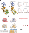
Fig. 1
Fig. 1. Structure of the Nup85•Seh1 complex
The structure of the heterodimeric Nup85•Seh1 complex is shown. Nup85 has a trunk (orange, helices α1–α3 and α12–α20) and a crown (blue, helices α4–α11) module. The β-strands at the extreme N-terminus of Nup85 form an insertion blade, which complete the Seh1 (green) β-propeller.
Fig. 2
Fig. 2. Comparison of Nup85•Seh1 and Nup145C•Sec13 and identification of the Nup84•Nup145C crown-crown binding interface
(A) The topologies of the Nup85•Seh1 (left) and Nup145C•Sec13 (right, PDB code 3BG1 (9)) complexes are shown, illustrating an overall similarity with three shared structural elements — trunk, crown, and β-propeller. Colors are assigned as in Figure 1. (B) Surface representations of the crowns of Nup85 and Nup145C are shown colored according to electrostatic surface potentials (top) and sequence conservation (bottom) in a view rotated 90° from (A). Sequence conservation is based on the phylogenetic tree of budding yeasts (39) and is colored from white (not conserved) to orange (conserved). A partial sequence alignment of helix α8 (indicated by arrows in (A)) is also shown with surface exposed residues indicated by green dots, residues buried in the hydrophobic core by blue dots, and residues not modeled in the structure by dashes. Mutations made in this helix in Nup145C are shown above the sequence alignment and the corresponding residues are outlined in the surface representations of Nup145C. (C) In the upper panels gel filtration data of Nup84 alone, Nup145C•Sec13 (wild type or -ELIEA mutant) alone, and Nup84 plus Nup145C•Sec13 (wild type or -ELIEA mutant) are shown. The shift in the Nup84 plus wild type Nup145C•Sec13 chromatogram indicates complex formation and is absent in the case of the -ELIEA mutant. In the lower panels gel filtration data of Nup145C•Sec13 alone, Nup84 alone (wild type or -DSICD mutant) alone, and Nup145C•Sec13 plus Nup84 (wild type or -DSICD mutant) are shown. The shift in the Nup145C•Sec13 plus wild type Nup84 chromatogram indicates complex formation and is absent in the case of the -DSICD mutant. (D) Isothermal titration calorimetry data illustrating high-affinity binding for wild-type Nup145C•Sec13 and Nup84 (black). Experimental values for N, KD, ΔH, and TΔS are shown. In contrast, binding is lost for both crown-surface mutants Nup84-DSICD (grey) and Nup145C-ELIEA (red).
Fig. 3
Fig. 3. Elimination of the Nup84 binding site on Nup145C results in nuclear pore assembly defects in vivo
NUP145/NUP84-GFP and NUP145-ELIEA/NUP84-GFP were grown at 24 °C and visualized by fluorescence microscopy. DIC, GFP-fluorescence, DNA (visualized with Hoechst dye), and false-colored overlay (GFP fluorescence – green, DNA – blue) images of the same field are shown in columns from left to right.
Fig. 4
Fig. 4. Architecture of ACE1
(A) ACE1 containing proteins are shown as cylinders and sheets. Crowns are shown in blue, trunks in orange, tails in green, and other domains in gray. Modules with predicted structures are shown half-transparent. (PDB codes: 2QX5, Nic96; 3BG1, Nup145C; 3CQC, Nup107 (Nup84 homolog); 2PM6, Sec31) (B) Cartoon illustrating the similarity and modular nature of the ACE1 element. The N-terminal elaborations are for Nic96 a coiled-coil domain that interacts with the Nsp1 complex, for Nup85 the Seh1-interacting insertion blade, for Nup145C the Sec13-interacting insertion blade preceded by an autocatalytic cleavage domain and Nup145N, and for Sec31 the Sec13-interacting insertion blade is preceded by its own N-terminal 7-bladed β-propeller. Sec31 has a unique proline rich insertion C-terminal to its trunk module followed by a conserved region predicted to be α-helical.
Fig. 5
Fig. 5. Lattice model for the Nup84 complex and the structural scaffold of the nuclear pore complex
The ACE1 proteins Nup85, Nup145C, Nup84, Sec31, and Nic96 are colored according to Figure 4. (A) Schematic diagram of COPII outer coat organization. The Sec31•Sec13 cuboctahedron composed of 24 edge elements (Sec31•Sec13 heterotetramers) is shown unwrapped and laid flat in 2 dimensions. The Sec31•Sec31 crown-crown interactions make edge elements while propeller-propeller interactions are vertex elements (30). (B) Schematic diagram of the predicted lattice-like organization of the structural scaffold of the NPC. The entire scaffold (8 spokes) is illustrated unwrapped and laid flat in two dimensions. The Nup84 complex comprises the nuclear and cytoplasmic rings, while the Nic96-containing complex makes up the inner ring. The relative position and interactions between the seven proteins in the Nup84 complex are shown with Sec13, Seh1, Nup133, and Nup120 colored in gray. The remainder of the complex (Nup157/170, Nup188, and Nup192) is illustrated in gray. The illustration is not meant to predict relative positions of proteins or structure of the inner ring per se, but shows the lattice-like organization of the structural scaffold similar to vesicle coating complexes.

References

    1. Tran EJ, Wente SR. Cell. 2006;125:1041. - PubMed
    1. Weis K. Cell. 2003;112:441. - PubMed
    1. D'Angelo MA, Hetzer MW. Trends Cell Biol. 2008;18:456. - PMC - PubMed
    1. Beck M, Lucic V, Forster F, Baumeister W, Medalia O. Nature. 2007;449:611. - PubMed
    1. Drummond SP, Rutherford SA, Sanderson HS, Allen TD. Can. J. Physiol. Pharmacol. 2006;84:423. - PubMed

Publication types

MeSH terms

Substances

Associated data