Review
. 2008 Oct 31;322(5902):697-701.
doi: 10.1126/science.1150451.

Structural insights into a circadian oscillator

Affiliations
Review

Structural insights into a circadian oscillator

Carl Hirschie Johnson et al. Science. .

Abstract

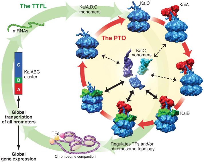
An endogenous circadian system in cyanobacteria exerts pervasive control over cellular processes, including global gene expression. Indeed, the entire chromosome undergoes daily cycles of topological changes and compaction. The biochemical machinery underlying a circadian oscillator can be reconstituted in vitro with just three cyanobacterial proteins, KaiA, KaiB, and KaiC. These proteins interact to promote conformational changes and phosphorylation events that determine the phase of the in vitro oscillation. The high-resolution structures of these proteins suggest a ratcheting mechanism by which the KaiABC oscillator ticks unidirectionally. This posttranslational oscillator may interact with transcriptional and translational feedback loops to generate the emergent circadian behavior in vivo. The conjunction of structural, biophysical, and biochemical approaches to this system reveals molecular mechanisms of biological timekeeping.


Figures

Fig. 1
Fig. 1. Rhythms in cyanobacteria from cells to molecules
(A and B) Circadian rhythms of bioluminescence in single cyanobacterial cells. Panel A shows micrographs of cyanobacterial cells at different times in constant light conditions—upper panel is the brightfield images showing growth and cell division as a function of approximate circadian time, lower panel is the luminescence emanating from these cells (the luminescence reporter was the psbAI promoter driving expression of bacterial luciferase, luxAB). Panel B shows the quantification of bioluminescence from a single cell as it divides into multiple cells as a function of time in constant light; starting at day 1.5, there are two differently colored traces as a result of cell division, the next division occurs at day 2.0, and so on (panels A and B are courtesy of I. Mihalcescu, reprinted with permission from (5). (C) Circadian rhythm of chromosomal compaction as visualized by a fluorescent DNA-binding dye (green)(red is chlorophyll autofluorescence). The chromosome is more compacted in the subjective night (hours 12-20) and less compacted in subjective day (hours 0-8, 24-28)(panel C is courtesy of S. Williams, reprinted with permission from (11). (D) Chromosomal topology shows a circadian rhythm as assayed by supercoiling of an endogenous plasmid. Topoisomers of the plasmid are more relaxed (R) in the subjective night and are more supercoiled (SC) in the subjective day (12). (E) KaiC phosphorylation in the oscillating in vitro reaction is shown by SDS PAGE in the upper panel; upper bands are hyper-phosphorylated KaiC, lower band is hypo-phosphorylated KaiC. The lower panel shows the predominant species of complexes of KaiA, KaiB, and KaiC that form during the in vitro oscillation: hypo-phosphorylated and uncomplexed KaiC hexamer (lower row) is present at all phases, but KaiC in complexes with KaiA and/or KaiB form rhythmically in concert with changes in KaiC's phosphorylation status (upper line, only the predominant complex is shown). Light blue KaiC hexamers are in the phosphorylating phase prior to monomer exchange, while dark blue KaiC hexamers are those undergoing dephosphorylation and monomer exchange.
Fig. 2
Fig. 2. KaiA-KaiC interaction
(A) The crystal structure of the KaiC hexamer shows a double doughnut shape formed by two lobes per subunit (PDB-ID 2GBL). The S-shaped loops (aa485-497)(green) dip into the central channel of the hexameric barrel. The flexible C-terminal residues (aa498-519) extend from the CII end of the hexamer. Some of the C-terminal tails are shorter than others because this region is partly disordered and only two chains have been completely traced out to the C-terminal S519 residue. ATP (gold) is bound between subunits in both the lower CI ring and the upper CII ring. (B) The six S-shaped loops (green stick representation) interact via a hydrogen bond network. Hydrogen bonds are shown in magenta and the view is perpendicular to (A) as seen from the CII side. (C) The CII end of one KaiC monomer as in the crystal structure with the S-shaped loop in green. (D) The CII end of one KaiC monomer as proposed to interact with KaiA, with the S-shaped loop pulled out (33). In parts (C) and (D) the S-shaped loop and C-terminal tail are shown as a wide ribbon. (E) Model of the KaiA-KaiC interaction based on combined structural information from x-ray crystallography, NMR, and three-dimensional electron microscopy (33). One S-shaped loop (green) is shown pulled out of the KaiC hexameric barrel. For clarity the S-shaped loop and C-terminal residues are only shown for one of the six KaiC subunits. The KaiA dimer is shown in red and purple.
Fig. 3
Fig. 3. Structural stabilization effects of phosphorylation and KaiB-KaiC interaction
(A) Ring of CII lobes from the KaiC crystal structure. Four subunits, chains a, b, e, and f (pink), are doubly phosphorylated at S431 and T432. Two subunits, chains c and d (blue), are singly phosphorylated at T432. The sidechains of S431 and T432 are shown in space filling representation and are colored by element with phosphorus atoms in cyan and oxygen atoms in red. ATP (gold) is bound between the CII lobes. (B) CII lobe of a doubly phosphorylated KaiC monomer (chain a) with the S-shaped loop in green. The view is perpendicular to (A). (C) Model of the KaiB•KaiC interaction based on combined structural information from x-ray crystallography and three-dimensional electron microscopy (34) with KaiB dimers in green and gold. (D) Model of the KaiA•KaiB•KaiC interaction with KaiA (red) and KaiB (green) dimers in orientations resembling those in the class IV KaiABC particle images from negative-stain EM (see Fig. 1D in ref. 31). (E) Hydrogen bonds (green) formed by the phosphate group of T432 in chain c (blue). (F) Hydrogen bonds formed by the phosphate groups of S431 and T432 in chain b (pink). In both (E) and (F) the neighboring chain is shown in gray. See Figs. S2 and S3 for the hydrogen bonds formed by the additional phosphate groups in chains a-f. (G) In chain f the phosphate group of S431 is leaning toward T432, rather than toward T426 as in chains a, b, and e. The position of the S431 phosphate group in chain e is shown in a faint representation. The phosphate groups of both S431 and T432 in chain f are stabilized by electrostatic interactions with R393.
Fig. 4
Fig. 4. A self-sustained post-translational oscillator (PTO) embedded within a transcription and translation feedback loop (TTFL)
The post-translational KaiABC oscillator (cycle connected by red arrows) is determined by phosphorylation of KaiC (blue hexamers) as regulated by interactions with KaiA (red dimers) and KaiB (green tetramers). Robustness is maintained by synchronization of KaiC hexameric status via exchange of KaiC monomers (23,30,31). Monomer exchange is depicted in the center of the PTO by KaiC monomers exchanging with KaiC hexamers; phase-dependent changes in the rate of monomer exchange is indicated by the thickness of the double-headed black arrows. New synthesis of KaiC feeds into the KaiABC oscillator as non-phosphorylated hexamers or as monomers that exchange into preexisting hexamers. The PTO brings KaiC to a state that regulates chromosome topology and/or transcriptional factors (“TFs”) to control global transcription of all promoters (including those driving expression of the essential clock genes kaiA, kaiB,and kaiC).

Similar articles

Cited by

References

    1. Dunlap JC, Loros JJ, DeCoursey PJ, editors. Chronobiology: Biological Timekeeping. Sinauer; Sunderland, MA: 2004.
    1. Mackey SR, Golden SS. Trends Microbiol. 2007;15:381–388. - PubMed
    1. Johnson CH. In: Methods in Enzymology. Young MW, editor. vol. 393. Academic Press; 2005. pp. 818–837. - PubMed
    1. Kondo T, et al. Proc. Natl. Acad. Sci. USA. 1993;90:5672–5676. - PMC - PubMed
    1. Mihalcescu I, Hsing W, Leibler S. Nature. 2004;430:81–85. - PubMed

Publication types

Substances