. 2008 Oct 31;322(5902):744-7.
doi: 10.1126/science.1163074.

Glia are essential for sensory organ function in C. elegans

Affiliations

Glia are essential for sensory organ function in C. elegans

Taulant Bacaj et al. Science. .

Abstract

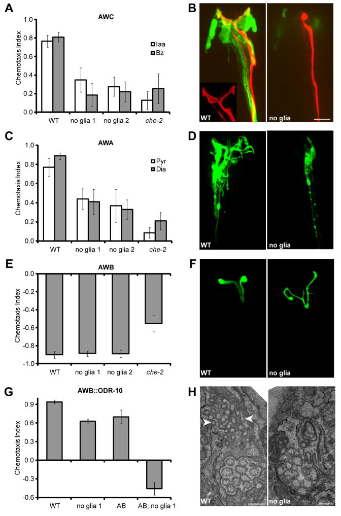
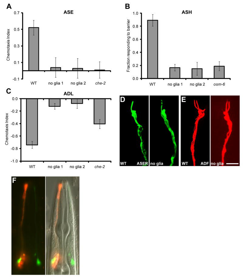
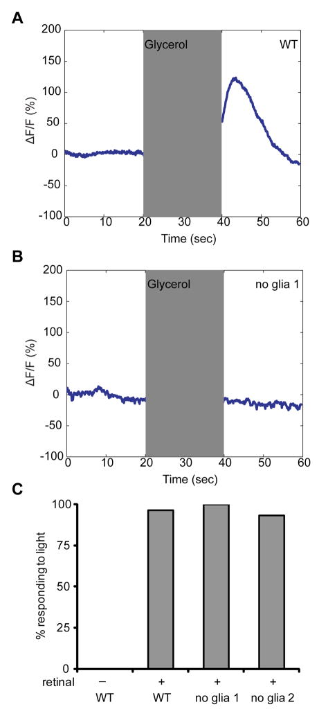
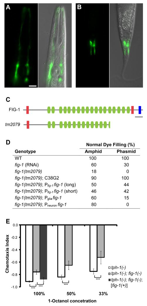
Sensory organs are composed of neurons, which convert environmental stimuli to electrical signals, and glia-like cells, whose functions are not well understood. To decipher glial roles in sensory organs, we ablated the sheath glial cell of the major sensory organ of Caenorhabditis elegans. We found that glia-ablated animals exhibit profound sensory deficits and that glia provide activities that affect neuronal morphology, behavior generation, and neuronal uptake of lipophilic dyes. To understand the molecular bases of these activities, we identified 298 genes whose messenger RNAs are glia-enriched. One gene, fig-1, encodes a labile protein with conserved thrombospondin TSP1 domains. FIG-1 protein functions extracellularly, is essential for neuronal dye uptake, and also affects behavior. Our results suggest that glia are required for multiple aspects of sensory organ function.


Figures

Fig. 1
Fig. 1
Glia are required for behavior and cilium structure. (A) Glia-ablated animals have defective AWC-mediated odortaxis towards 1% isoamyl alcohol (Iaa) and 0.5% benzahldehyde (Bz), p < 0.001 (Student’s t-test). (B) A wild-type AWC cilium (red, odr-1::RFP) ensheathed by an amphid sheath glia (green, vap-1::GFP). Glia ablation in the ontralateral amphid results in an amorphous cilium. Anterior, up. Scale bar, 5 μm. (C) Glia-ablated animals have defective AWA-mediated odortaxis towards 1% methyl pyrazine (Pyr) and 0.1% diacetyl (Dia), p < 0.001. (D) Glia removal decreases AWA cilium branching (odr-3::ODR-3p::GFP). (E) Glia are not required for AWB function, 10% 2-nonanone avoidance. (F) AWB cilium morphology appears grossly normal, although additional branching and failure of the two cilia to spread is often observed (str-1::ODR-10::GFP). (G) Animals expressing ODR-10 in AWA and AWB (AB) are attracted to diacetyl. However, dual-sensing animals lacking glia are repelled. (H) EM showing absence of AFD microvilli-like projections (arrowheads) in glia-ablated animals. Scale bar, 0.5 μm. WT, wild type; no glia, diphtheria-toxin-ablated glia; che-2, che-2(e1033) mutants; error bars, SD of 12 or more assays.
Fig. 2
Fig. 2
Glia affect channel neuron function but not morphology. (A) Glia-ablated animals fail to detect 0.2 M NaCl (p < 0.001), an ASE-mediated behavior. (B) Glia-ablated animals fail to avoid a 4M fructose ring (p < 0.001), an ASH-mediated behavior. osm-6, osm-6(p811) mutants. (C) Glia-ablated animals fail to avoid 1-octanol in a long-range assay (p < 0.001), an ADL-mediated behavior. (D and E) The morphology of amphid channel neurons is not affected by glia removal. ASER, gcy-5::GFP; ADF, T08G3.3::RFP). Scale bar, 5 μm. (F) Glia are required for neuronal uptake of DiI (red). Only the right amphid sheath glia is ablated. AWC (green, odr-1::YFP) indicates the location of the dendrite bundles. Error bars, SD of 12 or more assays.
Fig. 3
Fig. 3
Glia are required for Ca2+ responses in ASH. (A) As determined by G-CaMP fluorescence, ASH responds to application and removal of 1M glycerol (17). Shaded region, stimulus duration. (B) ASH fails to respond to glycerol in glia-ablated animals. (C) Glia are not required for neuronal function downstream of Ca2+ entry. Activation of ASH-expressed ChR2 by light causes animals to move backwards. n = 30 for each.
Fig. 4
Fig. 4
Glial fig-1 is required for neuronal dye filling and function. (A and B) fig-1 is expressed in amphid (A) and phasmid (B) sheath glia. Anterior, up. Scale bar, 5 μm. (C) FIG-1 domain structure. Red, thrombospondin type 1 domain; green, C6 repeats; blue, EGF-like type II domain; bar, 200 amino acids. The predicted protein in the fig-1(tm2079) deletion is shown. (D) fig-1 is required for DiI accumulation. One representative line shown for each condition: C38G2, cosmid containing fig-1; glial promoter, T02B11.3; neuronal promoter, sra-6; n > 40 for each. (E) fig-1 is required for 1-octanol avoidance. Assays were performed in the tph-1(mg280) background, which suppresses movement defects of fig-1(tm2079) animals. fig-1(tm2079) mutants perform worse at all three concentrations, and these defects can be rescued by fig-1(+). Asterisks, p < 0.001. Error bars, 95% confidence intervals. At least 24 assays for each condition.

Comment in

  • Neuroscience. A new glance at glia.
    Reichenbach A, Pannicke T. Reichenbach A, et al. Science. 2008 Oct 31;322(5902):693-4. doi: 10.1126/science.1166197. Science. 2008. PMID: 18974341 No abstract available.

Similar articles

Cited by

References

    1. Haydon PG. Nat Rev Neurosci. 2001;2:185. - PubMed
    1. Perea G, Araque A. Science. 2007;317:1083. - PubMed
    1. Perea G, Araque A. J Neurosci. 2005;25:2192. - PMC - PubMed
    1. Robitaille R. Neuron. 1998;21:847. - PubMed
    1. Ward S, Thomson N, White JG, Brenner S. J Comp Neurol. 1975;160:313. - PubMed

MeSH terms