Comparative Study
. 2008 Dec 5;103(12):1402-9.
doi: 10.1161/CIRCRESAHA.108.178426. Epub 2008 Oct 30.

Toll-like receptor 2 mediates apolipoprotein CIII-induced monocyte activation

Affiliations
Comparative Study

Toll-like receptor 2 mediates apolipoprotein CIII-induced monocyte activation

Akio Kawakami et al. Circ Res. .

Retraction in

  • Notice of retraction.
    [No authors listed] [No authors listed] Circ Res. 2012 Feb 17;110(4):e40. doi: 10.1161/RES.0b013e31824cec5c. Circ Res. 2012. PMID: 22343559 Free PMC article.

Abstract

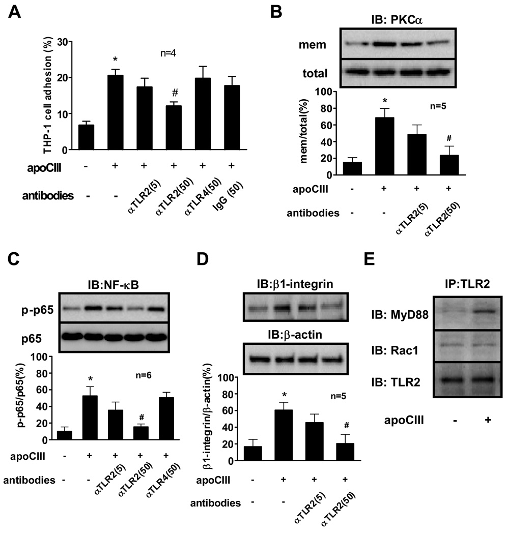
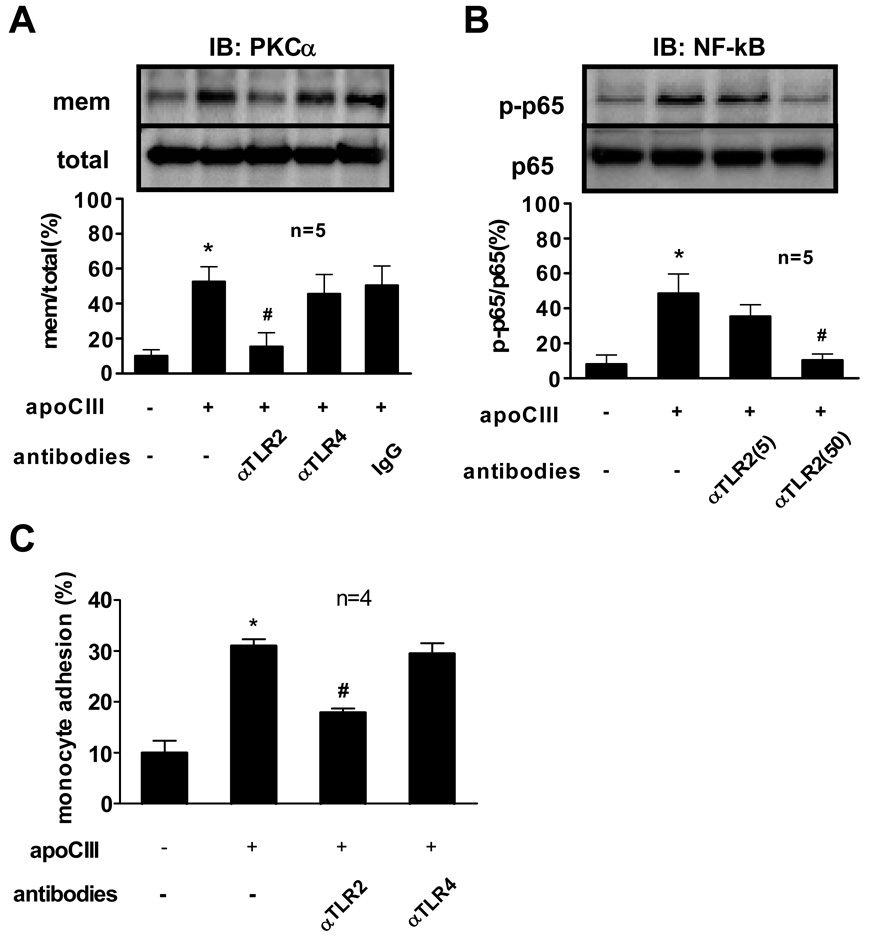
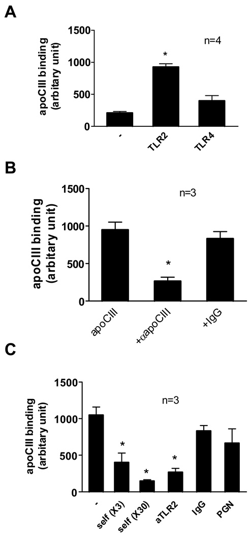
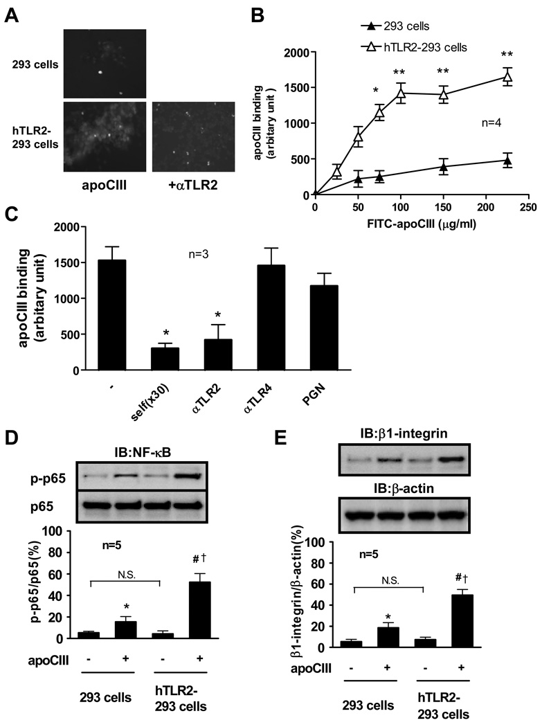
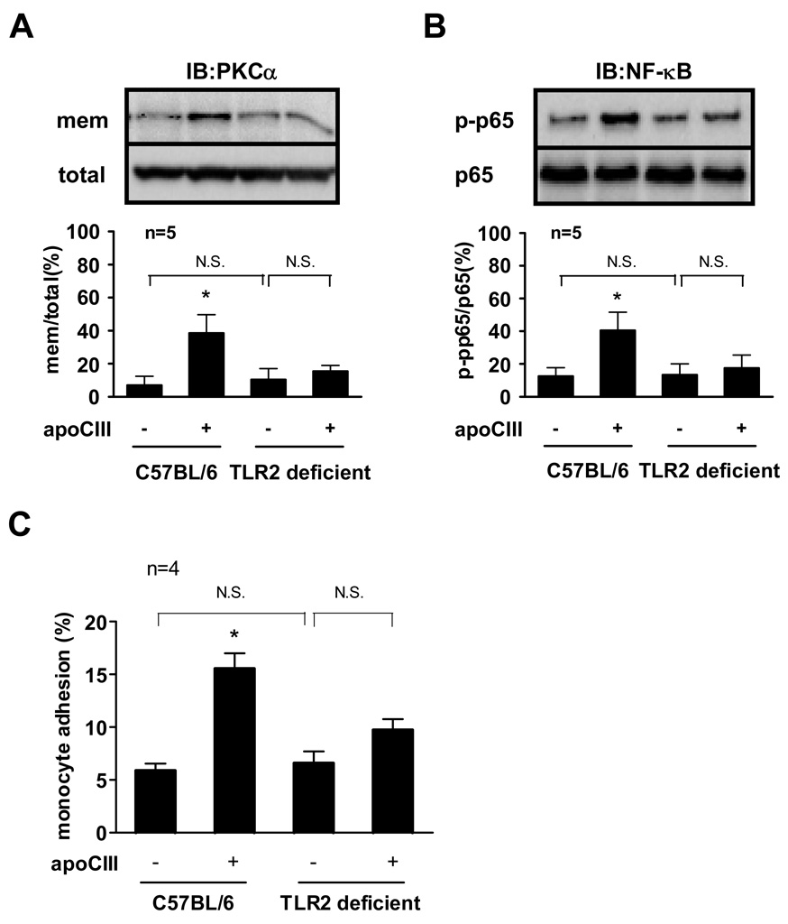
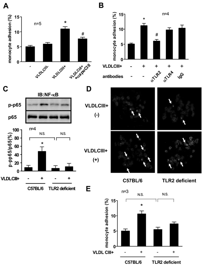
Apolipoprotein (apo)CIII predicts risk for coronary heart disease. We recently reported that apoCIII directly activates human monocytes. Recent evidence indicates that toll-like receptor (TLR)2 can contribute to atherogenesis through transduction of inflammatory signals. Here, we tested the hypothesis that apoCIII activates human monocytoid THP-1 cells through TLR2. ApoCIII induced the association of TLR2 with myeloid differentiation factor 88, activated nuclear factor (NF)-kappaB in THP-1 cells, and increased their adhesion to human umbilical vein endothelial cells (HUVECs). Anti-TLR2 blocking antibody, but not anti-TLR4 blocking antibody or isotype-matched IgG, inhibited these processes (P<0.05). ApoCIII bound with high affinity to human recombinant TLR2 protein and showed a significantly higher (P<0.05) and saturable binding to 293 cells overexpressing human TLR2 than to parental 293 cells with no endogenous TLR2. Overexpression of TLR2 in 293 cells augmented apoCIII-induced NF-kappaB activation and beta(1) integrin expression, processes inhibited by anti-apoCIII antibody as well as anti-TLR2 antibody. Exposure of peripheral blood monocytes isolated from C57BL/6 (wild-type) mice to apoCIII activated their NF-kappaB and increased their adhesiveness to HUVECs. In contrast, apoCIII did not activate monocytes from TLR2-deficient mice. Finally, intravenous administration to C57BL/6 mice of apoCIII-rich very-low-density lipoprotein (VLDL), but not of apoCIII-deficient VLDL, activated monocytes and increased their adhesiveness to HUVECs, processes attenuated by anti-TLR2 or anti-apoCIII antibody. ApoCIII-rich VLDL did not activate monocytes from TLR2-deficient mice. In conclusion, apoCIII activated monocytes at least partly through a TLR2-dependent pathway. The present study identifies a novel mechanism for proinflammatory and proatherogenic effects of apoCIII and a role for TLR2 in atherosclerosis induced by atherogenic lipoproteins.


Figures

Figure 1
Figure 1. TLR2 mediates apoCIII-induced THP-1 cell activation
(A, B, C, D) THP-1 cells were pretreated with indicated antibodies (µg/mL) for 30 minutes, and then incubated with or without apoCIII (100 µg/mL) for 8 hours (A, D) or 2 hours (B, C). “mem” indicates the membrane fraction of the cell lysates (B). (E) THP-1 cells were incubated with or without apoCIII (100 µg/mL) for 2 hours. *p<0.01 vs. apoCIII(−)/antibodies(−), #p<0.05 vs. apoCIII(+)/antibodies(−).”IB” and “IP” indicate immunoblotting and immunoprecipitation, respectively. Blots represent 4 to 6 independent experiments using apoCIII from 4 to 6 different donors that yielded similar results (B, C, D, E).
Figure 2
Figure 2. TLR2 mediates apoCIII-induced human monocyte activation
(A, B, C) Human peripheral blood monocytes were pretreated with indicated antibodies (50 µg/mL, unless otherwise indicated) for 30 minutes, and then incubated with or without apoCIII (100 µg/mL) for 2 hours (A, B) or 8 hours (C). “mem” indicates the membrane fraction of the cell lysates (A). *p<0.01 vs. apoCIII(−)/antibodies(−), #p<0.05 vs. apoCIII(+)/antibodies(−).”IB” indicates immunoblotting. Blots represent 5 independent experiments using apoCIII from 5 different donors that yielded similar results (A, B).
Figure 3
Figure 3. Binding of apoCIII with TLR2
(A) TLR2 protein or TLR4 protein were fixed on 96-well tissue-culture plates (2 µg/well), blocked, and then FITC-labeled apoCIII (100 µg/mL) was placed on it for 10 minutes. *p<0.01 vs. (−) or TLR4. (B) TLR2 protein was fixed on 96-well tissue-culture plates (2 µg/well), blocked, and then FITC-labeled apoCIII (100 µg/mL) was placed on it for 10 minutes. In experiments using antibodies, FITC-labeled apoCIII was pretreated with indicated antibodies (50 µg/mL) for 30 minutes. *p<0.05 vs. apoCIII alone. (C) TLR2 protein was fixed on 96-well tissue-culture plates (2 µg/well), blocked, and then FITC-labeled apoCIII (100 µg/mL) was placed on it for 10 minutes in the presence of excess amount of non-labeled apoCIII (self, 3- or 30-fold), indicated antibodies (50 µg/mL), or peptidoglycan (PGN) (10 µg/ml). *p<0.05 vs. (−).
Figure 4
Figure 4. Binding of apoCIII with human TLR2-transfected 293 cells
(A) FITC-labeled apoCIII (100 µg/mL) was placed on cultured 293 cells or hTLR2-293 cells for 10 minutes. Then, FITC-labeled apoCIII was observed with a fluorescent microscope. In some experiments, hTLR2-293 cells were pretreated with anti-TLR2 antibody (50 µg/mL) for 30 minutes. Photos are representative of 3 separate experiments. (B) FITC-labeled apoCIII was placed on cultured 293 cells or hTLR2-293 cells for 10 minutes at indicted concentrations. *p<0.05, **p<0.01 vs. 293 cells. (C) FITC-labeled apoCIII (100 µg/mL) was placed on cultured hTLR2-293 cells for 10 minutes in the presence of excess amount of non-labeled apoCIII (self, 30-fold), indicated antibodies (50 µg/mL), or peptidoglycan (10 µg/ml). *p<0.05 vs. (−). (D, E) Cultured 293 cells or hTLR2-293 cells were incubated with FITC-labeled apoCIII (100 µg/mL) for 2 hours (D) or 8 hours (E). *p<0.05 vs. apoCIII(−)/293 cells, #p<0.01 vs. apoCIII(−)/hTLR2-293 cells, p<0.05 vs. apoCIII(+)/293 cells. “IB” indicates immunoblotting. Blots represent 5 independent experiments using apoCIII from 5 different donors that yielded similar results.
Figure 5
Figure 5. TLR2 mediates apoCIII-induced mice monocyte activation
(A, B, C) Monocytes isolated from C57BL/6J mice or TLR2 deficient mice were incubated with or without apoCIII (100 µg/mL) for 2 hours (A, B) or 8 hours (C). “mem” indicates the membrane fraction of the cell lysates (A). *p<0.05 vs. apoCIII(−)/C57BL/6. “IB” indicates immunoblotting. Blots represent 5 independent experiments using apoCIII from 5 different donors that yielded similar results (A, B).
Figure 6
Figure 6. TLR2 mediates apoCIII-rich VLDL-induced mice monocyte activation
(A) C57BL/6J mice were injected i.v. with VLDL CIII- or VLDL CIII+ (500 µg apoB/body) from femoral veins. After 6 hours, monocytes were isolated from plasma. In some experiments, VLDL CIII+ was incubated with anti-apoCIII antibody (50 µg/mL) for 30 minutes before injection. *p<0.05 vs. VLDL CIII+(−), #p<0.05 vs. VLDL CIII+(+). (B) C57BL/6J mice were injected i.v. with indicated antibodies (200 µg/body) from femoral veins 30 minutes before VLDL CIII+ injection. Then, C57BL/6J mice were injected i.v. with VLDL CIII+ (500 µg apoB/body) from femoral veins. After 6 hours, monocytes were isolated from plasma. *p<0.05 vs. VLDL CIII+(−)/antibodies(−), #p<0.05 vs. VLDL CIII+(+)/ antibodies(−). (C, D, E) C57BL/6J mice or TLR2 deficient mice were injected i.v. with VLDL CIII+ (500 µg apoB/body) from femoral veins. After 2 hours (C, D) or 6 hours (E), monocytes were isolated from plasma. Arrows indicate the NF-κB p65 located in the nucleus (D). *p<0.05 vs. VLDL CIII+(−)/C57BL/6. “IB” indicates immunoblotting. Blots and photos represent 4 independent experiments using VLDL CIII+ from 4 different donors that yielded similar results (C, D).

Comment in

References

    1. Mullick AE, Tobias PS, Curtiss LK. Modulation of atherosclerosis in mice by Toll-like receptor 2. J Clin Invest. 2005;115:3149–3156. - PMC - PubMed
    1. Beutler B. Inferences, questions and possibilities in Toll-like receptor signalling. Nature. 2004;430:257–263. - PubMed
    1. Tobias P, Curtiss LK. Thematic review series: The immune system and atherogenesis. Paying the price for pathogen protection: toll receptors in atherogenesis. J Lipid Res. 2005;46:404–411. - PubMed
    1. Michelsen KS, Wong MH, Shah PK, Zhang W, Yano J, Doherty TM, Akira S, Rajavashisth TB, Arditi M. Lack of Toll-like receptor 4 or myeloid differentiation factor 88 reduces atherosclerosis and alters plaque phenotype in mice deficient in apolipoprotein E. Proc Natl Acad Sci U S A. 2004;101:10679–10684. - PMC - PubMed
    1. Yang QW, Mou L, Lv FL, Wang JZ, Wang L, Zhou HJ, Gao D. Role of Toll-like receptor 4/NF-kappaB pathway in monocyte-endothelial adhesion induced by low shear stress and ox-LDL. Biorheology. 2005;42:225–236. - PubMed

Publication types