. 2008;3(10):e3570.
doi: 10.1371/journal.pone.0003570. Epub 2008 Oct 30.

Halothiobacillus neapolitanus carboxysomes sequester heterologous and chimeric RubisCO species

Affiliations

Halothiobacillus neapolitanus carboxysomes sequester heterologous and chimeric RubisCO species

Balaraj B Menon et al. PLoS One. 2008.

Abstract

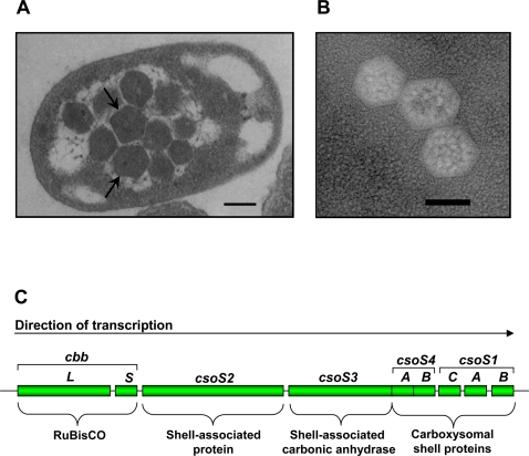
Background: The carboxysome is a bacterial microcompartment that consists of a polyhedral protein shell filled with ribulose 1,5-bisphosphate carboxylase/oxygenase (RubisCO), the enzyme that catalyzes the first step of CO2 fixation via the Calvin-Benson-Bassham cycle.

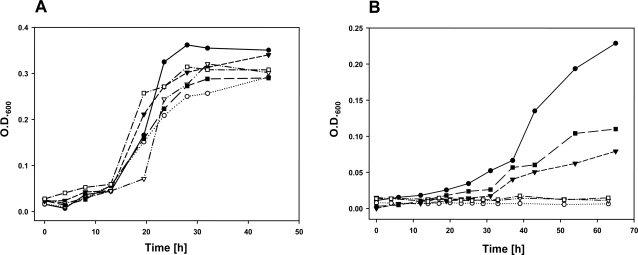
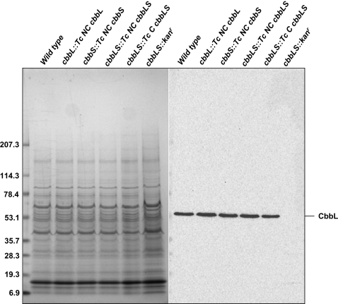
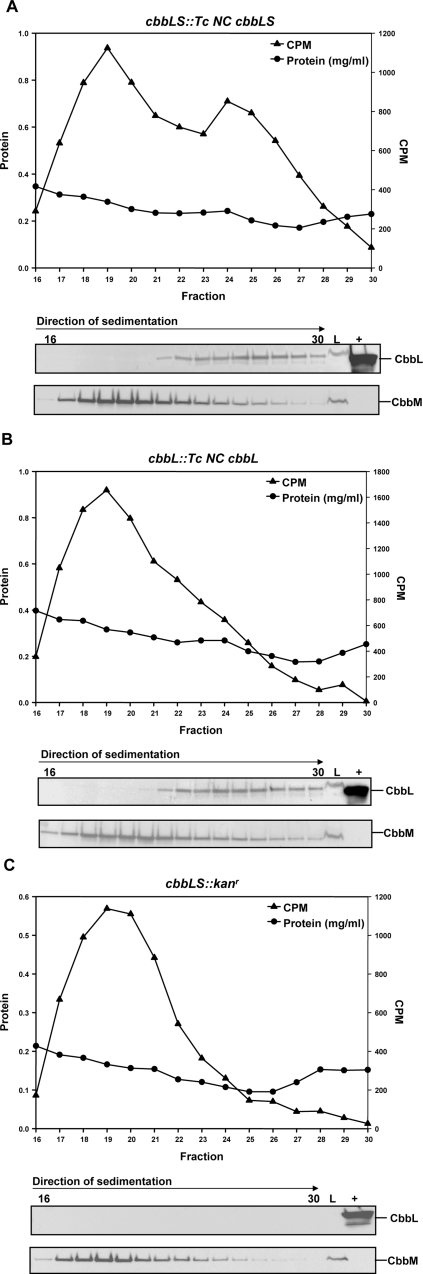
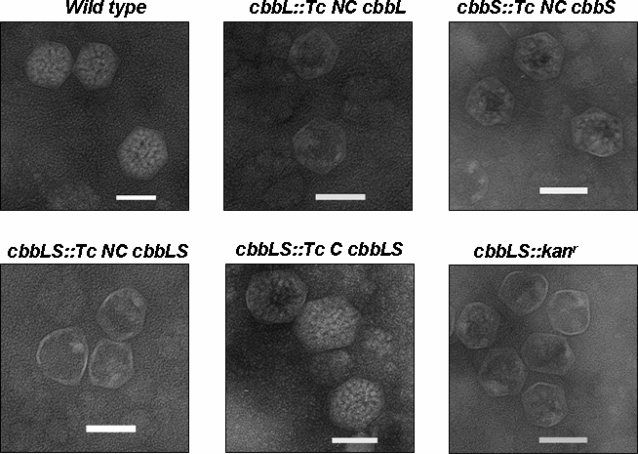
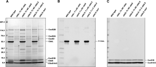
Methodology/principal findings: To analyze the role of RubisCO in carboxysome biogenesis in vivo we have created a series of Halothiobacillus neapolitanus RubisCO mutants. We identified the large subunit of the enzyme as an important determinant for its sequestration into alpha-carboxysomes and found that the carboxysomes of H. neapolitanus readily incorporate chimeric and heterologous RubisCO species. Intriguingly, a mutant lacking carboxysomal RubisCO assembles empty carboxysome shells of apparently normal shape and composition.

Conclusions/significance: These results indicate that carboxysome shell architecture is not determined by the enzyme they normally sequester. Our study provides, for the first time, clear evidence that carboxysome contents can be manipulated and suggests future nanotechnological applications that are based upon engineered protein microcompartments.


Conflict of interest statement

Competing Interests: The authors have declared that no competing interests exist.

Figures

Figure 1
Figure 1. Transmission electron micrographs of carboxysomes.
(A) Thin section of a wild type H. neapolitanus cell harboring multiple carboxysomes (arrows). (B) Negatively stained purified carboxysomes. (C) The H. neapolitanus cso operon, which contains the genes for Form I RubisCO (cbbL, cbbS) and the carboxysomal shell proteins (csoS2, csoS3, csoS4A, csoS4B, csoS1C, csoS1A, csoS1B).
Figure 2
Figure 2. H. neapolitanus RubisCO replacement and deletion mutants.
The mutants were constructed by replacing the H. neapolitanus genes for large (cbbL) and small (cbbS) subunit (green boxes) with noncarboxysomal (Tc NC; red boxes) or carboxysomal (Tc C; orange boxes) genes from T. crunogena or replacing both genes with a kanamycin resistance cassette (kanr; white boxes). All mutants carry a kanamycin cassette for selection purposes.
Figure 3
Figure 3. Growth of wild type and RubisCO mutants.
Growth of H. neapolitanus cultures in air supplemented with 5% CO2 (A) and in ambient CO2 (B); wild type (•), cbbL::Tc NC cbbL (○), cbbS::Tc NC cbbS (▾), cbbLS::Tc NC cbbLS (∇), cbbLS::Tc C cbbLS (▪), and cbbLS::kanr (□). Growth was monitored by measuring optical density of batch cultures at 600 nm.
Figure 4
Figure 4. Expression of CbbL in wild type and RubisCO mutants.
Clarified cell extracts (10 µg protein) were resolved by SDS-PAGE (left). A blot of an identical gel was probed with an anti-CbbL antibody that is specific for the large subunit of Form I RubisCO species.
Figure 5
Figure 5. RubisCO activity in cell extracts of the cbbL::Tc NC cbbL, cbbLS::Tc NC cbbLS, and cbbLS::kanr mutants.
Clarified extracts of H. neapolitanus cbbL::Tc NC cbbL (A), cbbLS::Tc NC cbbLS (B), and cbbLS::kanr (C) mutant cells were separated on 0.2–0.8 M sucrose gradients. The resulting fractions were assayed for RubisCO activity (cpm) and protein content (mg/ml). Aliquots (25 µl) of fractions 16–30 were probed for the presence of CbbL and CbbM with antibodies specific for each RubisCO type. L = 5 µg of clarified cell extract prior to gradient centrifugation; (+) = wild type carboxysome control.
Figure 6
Figure 6. Electron micrographs of wild type and RubisCO mutant carboxysomes.
Purified carboxysomes were stained with 1% ammonium molybdate and visualized by electron microscopy at 50,000 X magnification. Scale bars = 100 nm.
Figure 7
Figure 7. Polypeptide composition of purified carboxysomes.
Carboxysome proteins were separated by SDS-PAGE and stained with Gelcode Blue (A). Blots of identical gels were probed with antibodies specific for the large subunit of Form I RubisCO (B) and the CsoS1 shell proteins (C). An equal number of carboxysomes was loaded in each lane.

Similar articles

Cited by

References

    1. Shively JM, editor. Berlin/Heidelberg: Springer; 2006. Complex Intracellular Structures in Prokaryotes.
    1. Heinhorst S, Cannon GC, Shively JM. Carboxysomes and carboxysome-like inclusions. In: Shively JM, editor. Complex Intracellular Structures in Prokaryotes. Berlin/Heidelberg: Springer; 2006. pp. 141–164.
    1. Kerfeld CA, Sawaya MR, Tanaka S, Nguyen CV, Phillips M, et al. Protein structures forming the shell of primitive bacterial organelles. Science. 2005;309:936–938. - PubMed
    1. Tanaka S, Kerfeld CA, Sawaya MR, Cai F, Heinhorst S, et al. Atomic-level models of the bacterial carboxysome shell. Science. 2008;319:1083–1086. - PubMed
    1. Tsai Y, Sawaya MR, Cannon GC, Cai F, Williams EB, et al. The structure of the shell protein CsoS1A from Halothiobacillus neapolitanus and its implications for carboxysome function. PLoS Biology. 2007;5:1345–1354. - PMC - PubMed

Publication types

MeSH terms

Substances