

Interactions between HIV-1 reverse transcriptase and the downstream template strand in stable complexes with primer-template
- PMID: 18974785
- PMCID: PMC2570493
- DOI: 10.1371/journal.pone.0003561
Interactions between HIV-1 reverse transcriptase and the downstream template strand in stable complexes with primer-template
Abstract
Background: Human immunodeficiency virus type 1 reverse transcriptase (HIV-1 RT) forms stable ternary complexes in which RT is bound tightly at fixed positions on the primer-template (P/T). We have probed downstream interactions between RT and the template strand in the complex containing the incoming dNTP (+1 dNTP*RT*P/T complex) and in the complex containing the pyrophosphate analog, foscarnet (foscarnet*RT*P/T complex).
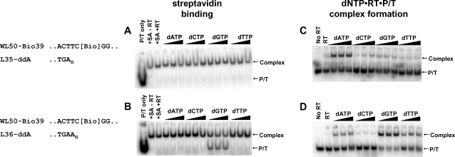
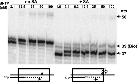
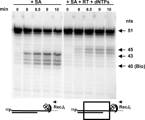
Methods and results: UV-induced cross-linking between RT and the DNA template strand was most efficient when a bromodeoxyuridine residue was placed in the +2 position (the first template position downstream from the incoming dNTP). Furthermore, formation of the +1 dNTP*RT*P/T complex on a biotin-containing template inhibited binding of streptavidin when biotin was in the +2 position on the template but not when the biotin was in the +3 position. Streptavidin pre-bound to a biotin residue in the template caused RT to stall two to three nucleotides upstream from the biotin residue. The downstream border of the complex formed by the stalled RT was mapped by digestion with exonuclease RecJ(F). UV-induced cross-linking of the complex formed by the pyrophosphate analog, foscarnet, with RT and P/T occurred preferentially with bromodeoxyuridine in the +1 position on the template in keeping with the location of RT one base upstream in the foscarnet*RT*P/T complex (i.e., in the pre-translocation position).
Conclusions: For +1 dNTP*RT*P/T and foscarnet*RT*P/T stable complexes, tight interactions were observed between RT and the first unpaired template nucleotide following the bound dNTP or the primer terminus, respectively.
Conflict of interest statement
Figures
Similar articles
-
Stable complexes formed by HIV-1 reverse transcriptase at distinct positions on the primer-template controlled by binding deoxynucleoside triphosphates or foscarnet.J Mol Biol. 2007 May 25;369(1):41-54. doi: 10.1016/j.jmb.2007.03.006. Epub 2007 Mar 12. J Mol Biol. 2007. PMID: 17400246 Free PMC article.
-
Impact of template overhang-binding region of HIV-1 RT on the binding and orientation of the duplex region of the template-primer.Mol Cell Biochem. 2010 May;338(1-2):19-33. doi: 10.1007/s11010-009-0316-x. Epub 2009 Nov 17. Mol Cell Biochem. 2010. PMID: 19921401
-
Relationship between 3'-azido-3'-deoxythymidine resistance and primer unblocking activity in foscarnet-resistant mutants of human immunodeficiency virus type 1 reverse transcriptase.J Virol. 2003 Jun;77(11):6127-37. doi: 10.1128/jvi.77.11.6127-6137.2003. J Virol. 2003. PMID: 12743270 Free PMC article.
-
K65R and K65A substitutions in HIV-1 reverse transcriptase enhance polymerase fidelity by decreasing both dNTP misinsertion and mispaired primer extension efficiencies.J Mol Biol. 2010 Aug 6;401(1):33-44. doi: 10.1016/j.jmb.2010.06.001. Epub 2010 Jun 9. J Mol Biol. 2010. PMID: 20538005 Free PMC article.
-
Protein-nucleic acid interactions and DNA conformation in a complex of human immunodeficiency virus type 1 reverse transcriptase with a double-stranded DNA template-primer.Biopolymers. 1997;44(2):125-38. doi: 10.1002/(SICI)1097-0282(1997)44:2<125::AID-BIP2>3.0.CO;2-X. Biopolymers. 1997. PMID: 9354757 Review.
Cited by
-
Potential therapeutic targets for Mpox: the evidence to date.Expert Opin Ther Targets. 2023 Jan-Jun;27(6):419-431. doi: 10.1080/14728222.2023.2230361. Epub 2023 Jul 4. Expert Opin Ther Targets. 2023. PMID: 37368464 Free PMC article.
-
Fingerprints of Modified RNA Bases from Deep Sequencing Profiles.J Am Chem Soc. 2017 Nov 29;139(47):17074-17081. doi: 10.1021/jacs.7b07914. Epub 2017 Nov 17. J Am Chem Soc. 2017. PMID: 29111692 Free PMC article.
-
The Role of Nucleotide Excision by Reverse Transcriptase in HIV Drug Resistance.Viruses. 2010 Jan 28;2(2):372-394. doi: 10.3390/v2020372. Viruses. 2010. PMID: 20523911 Free PMC article.
-
Mutations in the monkeypox virus replication complex: Potential contributing factors to the 2022 outbreak.J Autoimmun. 2022 Dec;133:102928. doi: 10.1016/j.jaut.2022.102928. Epub 2022 Oct 14. J Autoimmun. 2022. PMID: 36252459 Free PMC article.
References
-
- Wöhrl BM, Tantillo C, Arnold E, Le Grice SFJ. An expanded model of replicating human immunodeficiency virus reverse transcriptase. Biochemistry. 1995;34:5343–5350. - PubMed
-
- Tong W, Lu C-D, Sharma SK, Matsuura S, So AG, Scott WA. Nucleotide-induced stable complex formation by HIV-1 reverse transcriptase. Biochemistry. 1997;36:5749–5757. - PubMed
-
- Huang H, Chopra R, Verdine GL, Harrison SC. Structure of a covalently trapped catalytic complex of HIV-1 reverse transcriptase: implications for drug resistance. Science. 1998;282:1669–1675. - PubMed
Publication types
MeSH terms
Substances
Grants and funding
LinkOut - more resources
Full Text Sources
