. 2008;3(10):e3568.
doi: 10.1371/journal.pone.0003568. Epub 2008 Oct 30.

Silencing and nuclear repositioning of the lambda5 gene locus at the pre-B cell stage requires Aiolos and OBF-1

Affiliations

Silencing and nuclear repositioning of the lambda5 gene locus at the pre-B cell stage requires Aiolos and OBF-1

Alexander Karnowski et al. PLoS One. 2008.

Abstract

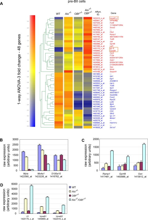
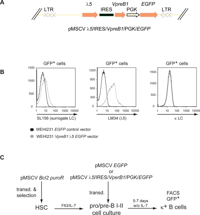
The chromatin regulator Aiolos and the transcriptional coactivator OBF-1 have been implicated in regulating aspects of B cell maturation and activation. Mice lacking either of these factors have a largely normal early B cell development. However, when both factors are eliminated simultaneously a block is uncovered at the transition between pre-B and immature B cells, indicating that these proteins exert a critical function in developing B lymphocytes. In mice deficient for Aiolos and OBF-1, the numbers of immature B cells are reduced, small pre-BII cells are increased and a significant impairment in immunoglobulin light chain DNA rearrangement is observed. We identified genes whose expression is deregulated in the pre-B cell compartment of these mice. In particular, we found that components of the pre-BCR, such as the surrogate light chain genes lambda5 and VpreB, fail to be efficiently silenced in double-mutant mice. Strikingly, developmentally regulated nuclear repositioning of the lambda5 gene is impaired in pre-B cells lacking OBF-1 and Aiolos. These studies uncover a novel role for OBF-1 and Aiolos in controlling the transcription and nuclear organization of genes involved in pre-BCR function.


Conflict of interest statement

Competing Interests: The authors have declared that no competing interests exist.

Figures

Figure 1
Figure 1. The transition from small pre-BII to immature B cells is impaired in Aio−/−/OBF-1−/− mice.
Flow cytometry analysis of bone marrow cells from 6–10 weeks old control wild type, single- and double-deficient mice. (A) Analysis of B220+ IgM+ IgD immature B cells in the bone marrow. Cells were stained with anti-B220-APC, anti-IgM-FITC, anti-IgD-Biotin followed by Streptavidin-PE. Only IgD cells are displayed and the percentages indicated are relative to the IgD cells. The percentage of IgD cells from total cells are as follows: WT, 89%; Aiolos−/−, 88%; OBF-1−/−, 92%; Aio−/−/OBF-1−/−, 95%. (B) In the left part detection of B220+ CD25+ pre-BII B cells. In the right part, pre-BII (B220+CD25+) cells were gated and their FSC analyzed. Percentages indicate the proportion of small or large cells. Data are presented from one representative experiment, out of three. (C) Pre-BII cells (B220+ CD25+ IgM) were FACS sorted from wild type and Aiolos/OBF-1 double-deficient mice by flow cytometry. Serial dilutions of the genomic DNA from the sorted cells were analyzed by PCR using primers detecting the indicated κ and λ light chain rearrangements. The relative amount of DNA used in the reaction was adjusted according to a real time PCR for the 18S gene (not shown).
Figure 2
Figure 2. Expression pattern of Aiolos and OBF-1 in the bone marrow.
The indicated B cell fractions were sorted by FACS and expression of Aiolos and OBF-1 was determined by real time RT-PCR and normalized to the expression of Ubiquitin C. Similar results were obtained when using RPII for normalization. The histograms represent the mean±SE based on the analysis of at least three independent samples per stage.
Figure 3
Figure 3. Identification of genes regulated by Aiolos and OBF-1 in pre-BII cells.
Gene expression profiles in B220+CD25+ pre-BII cells were determined by MOE430a Affymetrix GeneChip; for each genotype two RNA samples were prepared from independent pools of mice and microarray analysis was done in duplicate. (A) 48 genes showed a 3 fold expression changes in single- or double-mutant pre-BII cells compared to pre-BII cells from wild type mice (p-value cutoff: 0.05). The genes are grouped according to their expression profile in pre-BII cells from all genotypes. Low mRNA expression, blue; high mRNA expression, red. (B) Expression of genes that have been reported previously to be dependent on OBF-1 expression: Myla, Ms4a1, S100a10. (C) Expression of genes that show a strong expression increase specifically in Aio−/−/OBF-1−/− pre-B cells: Ramp1, Gpr49, Gelsolin (Gsn). (D) Expression of the surrogate light chain genes: λ5, VpreB1 and VpreB2. Figures show raw Affymetrix expression score after array normalization.
Figure 4
Figure 4. Silencing of λ5 expression at the pre-BII cell stage is impaired in absence of Aiolos and OBF-1.
(A) λ5 expression in pre-BI and pre-BII cells of the different genotypes. Pre-BI cells (B220+ckit+IgM), large or small pre-BII cells (B220+CD25+IgM, discriminated on the basis of their FSC profile) were sorted from wild type, single- and double-deficient mice and λ5 expression was analyzed by real time RT-PCR. The histograms represent the mean±SE based on the analysis of at least three independent samples per genotype/stage. (B) Downregulation of λ5 expression in Aiolos−/−/OBF-1−/− splenic mature B cells. Small pre-BII (B220+CD25+IgM) and splenic mature B (B220+IgMlowIgDhigh) cells of wild type, single- and double-deficient mice were sorted and the expression analysis was done by real time RT-PCR, as above. The histograms represent the mean±SE based on the analysis of two to three independent samples per genotype/stage. (C) Aiolos/OBF-1 double-deficient pre-BII cells fail to downregulate λ5 expression, but have a normal pre-BCR expression at the cell surface. Bone marrow cells from wild type, single- and double-mutant mice were stained for B220 together with surface pre-BCR (left panels) or intracellular surrogate light chain (λ5, right panels). Representative stainings are presented.
Figure 5
Figure 5. OBF-1 binds to the λ5 promoter region in vivo.
(A) Organization of the λ5 locus and partial sequence of the λ5 promoter region. Three putative octamer sites were identified in silico. (B) OBF-1 binds to the λ5 regulatory region. Binding of OBF-1 to three putative octamer sites (−769 to −776), (−2029 to −2036) and (−3557 to −3550) was tested by ChIP analysis. As negative controls, ChIP assays were performed with control IgGs, and control amplifications were done with a fragment of an intergenic region from chromosome 8 lacking any octamer site. Immunoprecipitated DNA was quantified by real time PCR. One representative experiment is presented.
Figure 6
Figure 6. Developmentally regulated nuclear repositioning of the λ5 locus is impaired in B lymphocytes lacking Aiolos or OBF-1.
(A) Representative pictures of the DNA FISH analysis. Confocal sections of nuclei after DNA FISH are shown, combining the λ5 probe (red staining) with a probe detecting γ-satellite DNA (blue staining). The pictures show association of zero, one or both λ5 alleles with γ-satellite DNA. (B) Localization of the λ5 locus determined by DNA FISH analysis of bone marrow pre-BI (B220+ c-kit+ IgM), small pre-BII (B220+ CD25+ IgM), and splenic resting mature B cells (CD4 CD43 Ter 119). Percentages of nuclei with one (red bar), both (yellow bar) or neither (blue bar) λ5 allele associated with γ-satellite DNA in the indicated populations of wild type, single-and double-deficient mice. (C) Percentages of alleles located at the nuclear periphery in the indicated cell populations.
Figure 7
Figure 7. Assessing the role of VpreB1 and λ5 expression for pro B cells maturation in vitro.
(A) In the upper part a schematic of the retroviral construct used to transduce pro/pre-B cells is presented. The control construct is identical, but only contains EGFP. (B) WEHI231 lymphoma B cells were transduced with either VpreB1-IRES-λ5-EGFP or an EGFP retroviral expression construct. Expression of VpreB1 and λ5 was determined by staining for surface expression of the pre-BCR (SL156) and λ5 (LM34). Possible displacement of κLC on transduced WEHI231 cells was determined by staining for surface κLC. Representative stainings are presented. (C) Schematic of the transduction experiment with bone marrow cells. Hematopoietic stem cells (HSC) were first transduced with a Bcl-2 expressing retrovirus to enhance their survival and were differentiated into pro/preB cultures in the presence of Flt3 and IL-7. Subsequently, these cultures were transduced with the indicated retroviruses and differentiated in vitro by withdrawing IL-7.

Similar articles

Cited by

References

    1. Busslinger M. Transcriptional control of early B cell development. Annu Rev Immunol. 2004;22:55–79. - PubMed
    1. Johnson K, Shapiro-Shelef M, Tunyaplin C, Calame K. Regulatory events in early and late B-cell differentiation. Molecular Immunology. 2005;42:749. - PubMed
    1. Matthias P, Rolink AG. Transcriptional networks in developing and mature B cells. Nature Reviews Immunology. 2005;5:497. - PubMed
    1. Hendriks RW, Middendorp S. The pre-BCR checkpoint as a cell-autonomous proliferation switch. Trends in Immunology. 2004;25:249. - PubMed
    1. ten Boekel E, Yamagami T, Andersson J, Rolink AG, Melchers F. The formation and selection of cells expressing preB cell receptors and B cell receptors. Curr Top Microbiol Immunol. 1999;246:3–9. discussion 9–10. - PubMed

Publication types

MeSH terms