. 2008 Oct;4(10):e1000191.
doi: 10.1371/journal.ppat.1000191. Epub 2008 Oct 31.

Repetitive N-WASP-binding elements of the enterohemorrhagic Escherichia coli effector EspF(U) synergistically activate actin assembly

Affiliations

Repetitive N-WASP-binding elements of the enterohemorrhagic Escherichia coli effector EspF(U) synergistically activate actin assembly

Kenneth G Campellone et al. PLoS Pathog. 2008 Oct.

Abstract

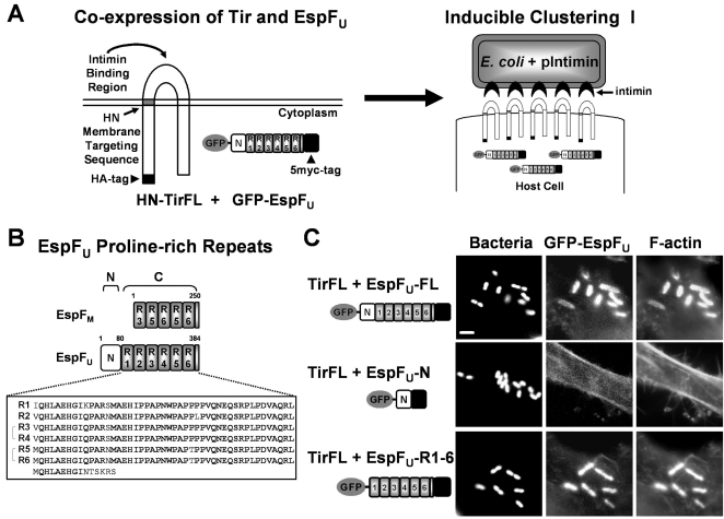
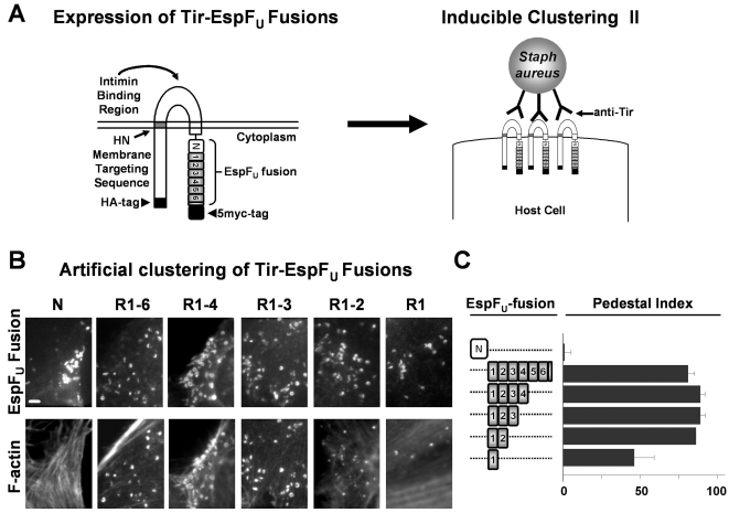
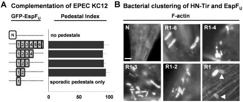
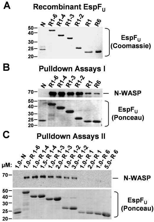
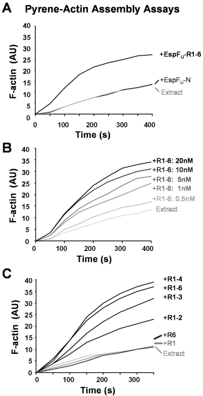
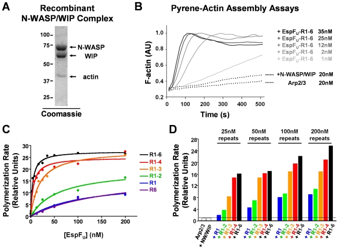
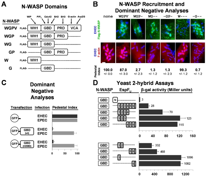
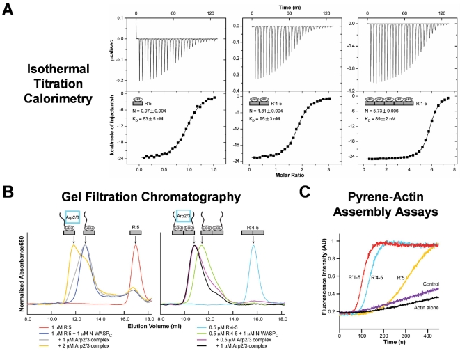
Enterohemorrhagic Escherichia coli (EHEC) generate F-actin-rich adhesion pedestals by delivering effector proteins into mammalian cells. These effectors include the translocated receptor Tir, along with EspF(U), a protein that associates indirectly with Tir and contains multiple peptide repeats that stimulate actin polymerization. In vitro, the EspF(U) repeat region is capable of binding and activating recombinant derivatives of N-WASP, a host actin nucleation-promoting factor. In spite of the identification of these important bacterial and host factors, the underlying mechanisms of how EHEC so potently exploits the native actin assembly machinery have not been clearly defined. Here we show that Tir and EspF(U) are sufficient for actin pedestal formation in cultured cells. Experimental clustering of Tir-EspF(U) fusion proteins indicates that the central role of the cytoplasmic portion of Tir is to promote clustering of the repeat region of EspF(U). Whereas clustering of a single EspF(U) repeat is sufficient to bind N-WASP and generate pedestals on cultured cells, multi-repeat EspF(U) derivatives promote actin assembly more efficiently. Moreover, the EspF(U) repeats activate a protein complex containing N-WASP and the actin-binding protein WIP in a synergistic fashion in vitro, further suggesting that the repeats cooperate to stimulate actin polymerization in vivo. One explanation for repeat synergy is that simultaneous engagement of multiple N-WASP molecules can enhance its ability to interact with the actin nucleating Arp2/3 complex. These findings define the minimal set of bacterial effectors required for pedestal formation and the elements within those effectors that contribute to actin assembly via N-WASP-Arp2/3-mediated signaling pathways.


Conflict of interest statement

The authors have declared that no competing interests exist.

Figures

Figure 1
Figure 1. Clustering of Tir and EspFU is sufficient to promote actin pedestal formation.
(A) A full-length derivative of EHEC Tir (HN-TirFL) is depicted, featuring an HA-tag, N- and C-terminal cytoplasmic regions, an extracellular intimin-binding domain, and two transmembrane segments, including one derived from the Newcastle Disease Virus HN surface protein. A derivative of EspFU tagged with GFP at its N-terminus and 5myc at its C-terminus (GFP-EspFU) is also shown. Treatment of transfected cells with a non-pathogenic strain of E. coli expressing EHEC intimin can promote clustering of membrane-localized HN-TirFL. (B) An alignment of EspFU and the EHEC pseudogene EspFM is shown, featuring an N-terminal EspFU secretion signal and six nearly identical proline-rich peptide repeats plus one partial repeat at its C-terminus. The N-terminal boundary of the EspFU repeats was assigned based upon alignment with the sequence of EspFM, which is missing the N-terminal translocation signal. (C) HeLa cells co-transfected with plasmids encoding HN-TirFL and GFP-EspFU derivatives were treated with intimin-expressing E. coli, fixed, and stained with DAPI to identify bacteria and phalloidin to detect F-actin. All scalebars are 1 µm in length.
Figure 2
Figure 2. Experimental clustering of the EspFU repeats bypasses the requirement for the Tir C-terminus during actin pedestal formation.
(A) A fusion of membrane-targeted HN-TirFL to EspFU-myc is shown. Treatment of transfected cells with Tir antibodies and S. aureus particles can promote clustering of membrane-localized Tir-EspFU fusions. (B) Murine fibroblast-like cells (FLCs) transfected with plasmids encoding Tir-EspFU fusion constructs comprising the N-terminal domain or truncations of the C-terminal repeats were treated with Tir antibodies and S. aureus, fixed, and stained with HA antibodies to identify both transfected cells and S. aureus (which binds the fluorescent antibodies) and with phalloidin to detect F-actin. (C) Pedestal formation indices were determined by calculating the percentage of transfected cells harboring five or more S. aureus particles generating actin pedestals. Data represent the mean+/−SD from three experiments. The Tir-EspFU R1 construct triggered actin assembly significantly less efficiently than the other truncations (p<0.05).
Figure 3
Figure 3. Multiple EspFU repeats are required for full activity when expressed in the presence of full-length Tir.
(A) FLCs transfected with plasmids encoding GFP-EspFU truncations containing either the N-terminal domain or the C-terminal repeats were infected with EPEC KC12, fixed, and treated with DAPI to identify bacteria and phalloidin to detect F-actin. Pedestal indices were determined by calculating the % of GFP-EspFU-expressing cells harboring five or more actin pedestals. Data represent the mean+/−SD of three experiments. (B) HeLa cells co-transfected with plasmids encoding HN-TirFL and GFP-EspFU truncations were infected with intimin-expressing E. coli, fixed, and treated with DAPI to identify bacteria and phalloidin to detect F-actin. Arrowheads indicate sites of less-intense F-actin assembly.
Figure 4
Figure 4. Multiple EspFU repeats are required for efficient binding of N-WASP in brain extract.
(A) N-terminally His10-tagged and C-terminally 5myc-tagged EspFU derivatives were expressed in E. coli, purified, resolved by SDS-PAGE, and stained with Coomassie blue. (B) Cobalt-chelated magnetic particles were coated with saturating concentrations of EspFU derivatives and subsequently incubated with porcine brain extract. The association of native N-WASP with EspFU-coated beads was assessed by SDS-PAGE followed by immunoblotting of bead eluates with antibodies to N-WASP and staining EspFU with Ponceau S. (C) His-EspFU-myc constructs were added to brain extract at the indicated concentrations and collected using cobalt-chelated magnetic particles. The association of native N-WASP and EspFU with the beads was assessed by immunoblotting of bead eluates with antibodies to N-WASP and staining EspFU with Ponceau S.
Figure 5
Figure 5. EspFU repeats cause titratable increases in actin assembly in brain extract.
(A) His-EspFU-myc constructs (20 nM each) were added to Arp2/3-enriched brain extract supplemented with 2.5 µM G-actin (10% pyrene labeled), and fluorescent actin polymerization, in arbitrary units (AU), was measured over time. EspFU did not trigger actin assembly in the absence of extract (data not shown). (B) Pyrene-actin polymerization in the presence of brain extract and various concentrations of EspFU-R1-6 was measured over time. (C) Pyrene-actin polymerization in the presence of brain extract and EspFU truncations containing different repeat segments (20 nM each) was measured over time.
Figure 6
Figure 6. EspFU repeats synergistically activate actin assembly mediated by recombinant N-WASP/WIP complex in vitro.
(A) N-terminally His6-Flag-tagged N-WASP and His6-Myc-tagged WIP were co-expressed in insect cells, purified as a stoichiometric complex, resolved by SDS-PAGE, and stained with Coomassie blue. (B) Actin (2 µM) was polymerized in the presence of Arp2/3 complex, N-WASP/WIP complex, and the indicated concentrations of EspFU. F-actin fluorescence was measured in arbitrary units (AU). (C) Actin polymerization was examined in the presence of 20 nM Arp2/3 complex, 20 nM N-WASP/WIP complex, and the indicated concentrations of EspFU derivatives. Polymerization rates at half-maximal F-actin concentrations were measured relative to the rate of polymerization in control Arp2/3+N-WASP/WIP samples lacking EspFU. Curves were fit using Prism software. (D) Actin polymerization was measured as in (C), except that EspFU concentrations have been scaled to the number of repeats in each protein.
Figure 7
Figure 7. The GTPase-binding domain (GBD) of N-WASP is a dominant negative inhibitor of EHEC pedestal formation and binds with high efficiency to a single EspFU repeat in yeast two-hybrid assays.
(A) The modular structure of N-WASP is depicted, featuring WASP homology-1 (WH1), GTPase-binding (GBD), proline-rich (PRD), and WH2/verprolin-connector-acidic (VCA) domains. Several N-WASP-binding partners are shown above their interacting domains. (B) HeLa cells transfected with plasmids encoding Flag-N-WASP constructs were infected with EHECΔdam (a mutant that binds to mammalian cells and generates pedestals with considerably higher efficiency than wild type EHEC [44]), fixed, and treated with DAPI to identify bacteria, a Flag antibody to visualize tagged N-WASP (top panels), and phalloidin to detect F-actin (bottom panels). Flag-N-WASP recruitment was only evaluated in cells expressing low levels of these tagged proteins (top panels), while effects on actin pedestal formation were only assessed in cells expressing high levels of Flag-N-WASP (bottom panels). Pedestal formation indices were determined by calculating the percentage of mock-transfected or Flag-N-WASP overexpressing cells harboring five or more actin pedestals. Data represent the mean+/−SD of three experiments. (C) HeLa cells expressing GFP alone, a GFP-tagged GBD, or a GFP-tagged GBD H208D point mutant were infected with EHECΔdam or EPEC and treated with DAPI to identify bacteria and phalloidin to detect F-actin. Pedestal formation indices were determined as in (B). (D) Plasmids encoding the N-WASP GBD fused to the LexA DNA-binding domain and EspFU fragments fused to the Gal4 transcriptional activation domain were co-transformed into a yeast two-hybrid reporter strain. Data represent the mean+/−SD of β-galactosidase activity for three co-transformants for each pairwise combination.
Figure 8
Figure 8. Increasing the number of EspFU repeats does not alter affinity for the GBD, but promotes the formation of an Arp2/3-containing complex.
(A) Isothermal titration calorimetry analyses of the interactions between WASP GBD and EspFU fragments are shown. The GBD was titrated into R′5 (left), R′4-5 (middle), or R′1-5 (right). Raw and integrated heats of injections are shown in upper and lower panels, respectively. Black lines in the lower panels show fits of data into a single-affinity, multi-site binding model. Fits of the data for R′4-5 and R′1-5 to models with two different affinities were not statistically improved over the single-affinity model. (B) Interactions between Arp2/3 complex and N-WASPC in complex with Alexa647-labeled EspFU R′5 or R′4-5 were examined by gel filtration chromatography. The A650 profile is shown. (C) Actin (4 µM) was polymerized by itself (black curve), or in the presence of 10 nM Arp2/3 complex plus either 0.5 µM WASP GBD-VCA (purple, control), 0.5 µM GBD-VCA+1 µM R′5 (yellow), or 0.5 µM GBD-VCA+0.5 µM R′4-5 (blue), or 0.5 µM GBD-VCA+0.2 µM R′1-5 (red). F-actin fluorescence was measured in arbitrary units (AU). Note that R′5, R′4-5, and R′1-5 were used at the same total repeat concentration.

Similar articles

Cited by

References

    1. Donnenberg MS, Whittam TS. Pathogenesis and evolution of virulence in enteropathogenic and enterohemorrhagic Escherichia coli. J Clin Invest. 2001;107:539–548. - PMC - PubMed
    1. Kaper JB, Nataro JP, Mobley HL. Pathogenic Escherichia coli. Nat Rev Microbiol. 2004;2:123–140. - PubMed
    1. Spears KJ, Roe AJ, Gally DL. A comparison of enteropathogenic and enterohaemorrhagic Escherichia coli pathogenesis. FEMS Microbiol Lett. 2006;255:187–202. - PubMed
    1. Deng W, Puente JL, Gruenheid S, Li Y, Vallance BA, et al. Dissecting virulence: systematic and functional analyses of a pathogenicity island. Proc Natl Acad Sci U S A. 2004;101:3597–3602. - PMC - PubMed
    1. Marches O, Nougayrede JP, Boullier S, Mainil J, Charlier G, et al. Role of tir and intimin in the virulence of rabbit enteropathogenic Escherichia coli serotype O103:H2. Infect Immun. 2000;68:2171–2182. - PMC - PubMed

Publication types

MeSH terms

LinkOut - more resources