. 2007 Jul;18(5):55102.
doi: 10.1088/0957-4484/18/5/055102.

Magnetic/luminescent core/shell particles synthesized by spray pyrolysis and their application in immunoassays with internal standard

Affiliations

Magnetic/luminescent core/shell particles synthesized by spray pyrolysis and their application in immunoassays with internal standard

Dosi Dosev et al. Nanotechnology. 2007 Jul.

Abstract

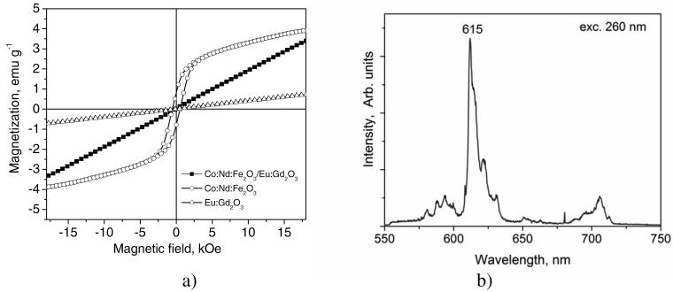
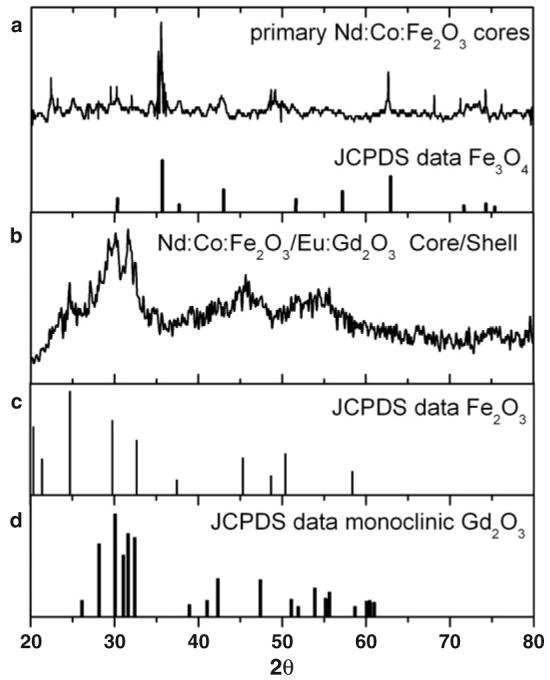
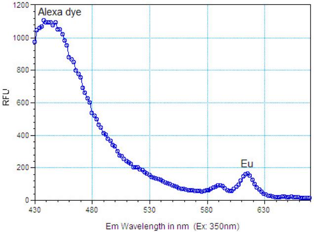
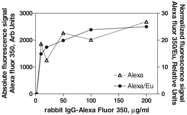
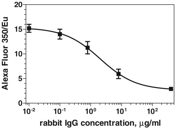
Many types of fluorescent nanoparticles have been investigated as alternatives to conventional organic dyes in biochemistry; magnetic beads also have a long history of biological applications. In this work we apply flame spray pyrolysis in order to engineer a novel type of nanoparticle that has both luminescent and magnetic properties. The particles have magnetic cores of iron oxide doped with cobalt and neodymium and luminescent shells of europium-doped gadolinium oxide (Eu:Gd(2)O(3)). Measurements by vibrating sample magnetometry showed an overall paramagnetic response of these composite particles. Luminescence spectroscopy showed spectra typical of the Eu ion in a Gd(2)O(3) host-a narrow emission peak centred near 615 nm. Our synthesis method offers a low-cost, high-rate synthesis route that enables a wide range of biological applications of magnetic/luminescent core/shell particles. Using these particles we demonstrate a novel immunoassay format with internal luminescent calibration for more precise measurements.


Figures

Figure 1
Figure 1
Magnetic characteristics of Co:Fe2O3 and Co:Nd:Fe2O3 powders synthesized by spray pyrolysis with different partial Co, Nd.
Figure 2
Figure 2
Schematic description of the synthesis of core /shell particles. Spray droplets contain solid magnetic nanoparticles and dissolved precursors of Eu and Gd passed through the hydrogen flame.
Figure 3
Figure 3
Bright field TEM image of Co:Nd:Fe2O3/Eu:Gd2O3 core/shell particles.
Figure 4
Figure 4
Properties of the magnetic/luminescent core/shell particles. (a) Comparison of the magnetic characteristics of Co:Nd:Fe2O3 powder with Co:Nd:Fe2O3/Eu:Gd2O3 core/shell particles and Eu:Gd2O3 particles; (b) emission spectrum of the Co:Nd:Fe2O3/Eu:Gd2O3 core/shell particles under excitation at 260 nm.
Figure 5
Figure 5
(a) X-ray diffraction spectrum of the primary Nd:Co:Fe2O3 particles are compared to the typical XRD peaks of Fe3O4; (b) XRD of the core/shell Nd:Co:Fe2O3/Eu:Gd2O3 particles; (c) and (d) show the typical XRD spectral peaks of Fe2O3 and monoclinic Gd2O3 respectively.
Figure 6
Figure 6
Magnetic separation of Co:Nd:Fe2O3/Eu:Gd2O3 particles from aqueous solution; (left)—before the liquid is removed, (right)—after removing the liquid.
Figure 7
Figure 7
Emission spectra of the Co:Nd:Fe2O3/Eu:Gd2O3 core/shell particles and the IgG-Alexa Fluor 350 bound to their surface (excitation at 350 nm).
Figure 8
Figure 8
Saturation of the capture antibody (anti-rabbit IgG) immobilized on the surface of the magnetic luminescent nanoparticles with rabbit IgG-Alexa Fluor 350. Absolute measured intensity of the Alexa peak (△) is compared to the intensity ratio Alexa/EuGd2O3 (●). The ratiometric approach reduces the uncertainty that arises from variations in the amount of particle separation from the sample with the magnet.
Figure 9
Figure 9
Calibration curve for the competitive magnetic immunoassay for rabbit IgG. The signal of the labelled antigen (rabbit IgG-Alexa Fluor 350) bound on the surface of the magnetic nanoparticles is normalized by the Eu luminescence of the particles.

References

    1. Alivisatos P. Nat. Biotechnol. 2004;22:47–52. - PubMed
    1. Seydack M. Biosensors Bioelectron. 2005;20:2454–69. - PubMed
    1. Parak WJ, Gerion D, Pellegrino T, Zanchet D, Micheel C, Williams SC, Boudreau R, Le Gros MA, Larabell CA, Alivisatos AP. Nanotechnology. 2003;14:R15–27.
    1. Jaiswal JK, Simon SM. Trends Cell Biol. 2004;14:497–504. - PubMed
    1. Tan MQ, Wang GL, Hai XD, Ye ZQ, Yuan JL. J. Mater. Chem. 2004;14:2896–901.