. 2008;3(10):e3579.
doi: 10.1371/journal.pone.0003579. Epub 2008 Oct 31.

The recently identified P2Y-like receptor GPR17 is a sensor of brain damage and a new target for brain repair

Affiliations

The recently identified P2Y-like receptor GPR17 is a sensor of brain damage and a new target for brain repair

Davide Lecca et al. PLoS One. 2008.

Abstract

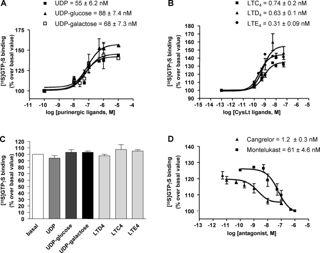
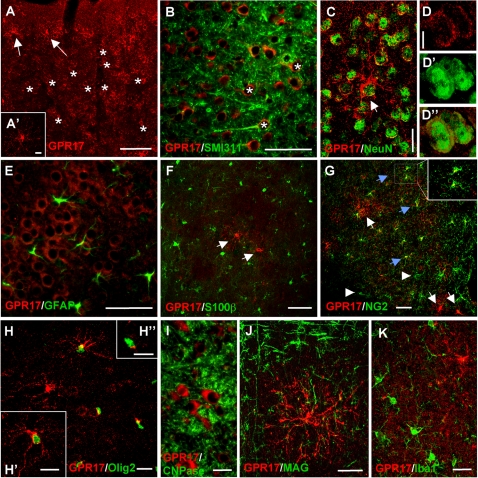
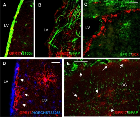
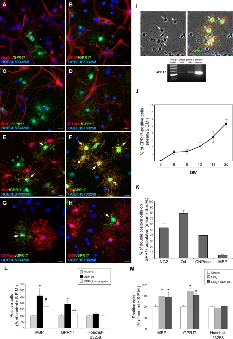
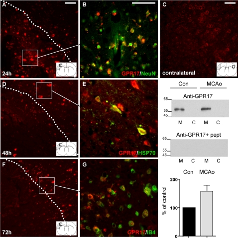
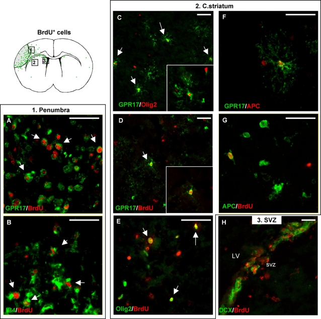
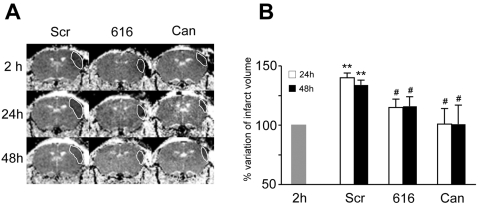
Deciphering the mechanisms regulating the generation of new neurons and new oligodendrocytes, the myelinating cells of the central nervous system, is of paramount importance to address new strategies to replace endogenous damaged cells in the adult brain and foster repair in neurodegenerative diseases. Upon brain injury, the extracellular concentrations of nucleotides and cysteinyl-leukotrienes (cysLTs), two families of endogenous signaling molecules, are markedly increased at the site of damage, suggesting that they may act as "danger signals" to alert responses to tissue damage and start repair. Here we show that, in brain telencephalon, GPR17, a recently deorphanized receptor for both uracil nucleotides and cysLTs (e.g., UDP-glucose and LTD(4)), is normally present on neurons and on a subset of parenchymal quiescent oligodendrocyte precursor cells. We also show that induction of brain injury using an established focal ischemia model in the rodent induces profound spatiotemporal-dependent changes of GPR17. In the lesioned area, we observed an early and transient up-regulation of GPR17 in neurons expressing the cellular stress marker heat shock protein 70. Magnetic Resonance Imaging in living mice showed that the in vivo pharmacological or biotechnological knock down of GPR17 markedly prevents brain infarct evolution, suggesting GPR17 as a mediator of neuronal death at this early ischemic stage. At later times after ischemia, GPR17 immuno-labeling appeared on microglia/macrophages infiltrating the lesioned area to indicate that GPR17 may also acts as a player in the remodeling of brain circuitries by microglia. At this later stage, parenchymal GPR17+ oligodendrocyte progenitors started proliferating in the peri-injured area, suggesting initiation of remyelination. To confirm a specific role for GPR17 in oligodendrocyte differentiation, the in vitro exposure of cortical pre-oligodendrocytes to the GPR17 endogenous ligands UDP-glucose and LTD(4) promoted the expression of myelin basic protein, confirming progression toward mature oligodendrocytes. Thus, GPR17 may act as a "sensor" that is activated upon brain injury on several embryonically distinct cell types, and may play a key role in both inducing neuronal death inside the ischemic core and in orchestrating the local remodeling/repair response. Specifically, we suggest GPR17 as a novel target for therapeutic manipulation to foster repair of demyelinating wounds, the types of lesions that also occur in patients with multiple sclerosis.


Conflict of interest statement

Competing Interests: MLT, PC, CV, CM and MPA are inventors in the Italian Patent Application #2004A002007 on “Modulatori del recettore GPR17 e loro impieghi terapeutici” submitted on October 21, 2004.

Figures

Figure 1
Figure 1. Effect of purinergic and leukotriene ligands on mGPR17 with [35S]GTPγS binding.
Membrane aliquots (10 µg), obtained from 1321N1 cells transfected with pcDNA3.1-Gpr17m (A,B), were incubated with the indicated concentrations of purinergic (A) or leukotriene (B) agonists. (C) Membranes from cells transfected with the pcDNA3.1 empty vector were incubated with UDP, UDP-glucose or UDP-galactose (all at 10 µM) or with LTD4, LTC4 and LTE4 (all at 50 nM), as indicated. (D) Effect of P2Y or CysLT receptor antagonist on agonist-stimulated [35S]GTPγS binding. Membranes from pcDNA3.1-Gpr17m transfected cells were pre-incubated for 10 min with graded montelukast (1 nM-1 µM) or Cangrelor (0.01 nM-100 nM) concentrations and then stimulated with 500 nM UDP or 50 nM LTD4, respectively. All data are expressed as percentage of basal [35S]GTPγS binding (set to 100%) and represent the mean±SEM of three different experiments each performed in duplicate.
Figure 2
Figure 2. Cellular localization of GPR17 in the intact mouse brain.
Double fluorescence labeling of mouse brain coronal sections with anti-GPR17 antibody (red fluorescence) and markers for different cell lineages (green fluorescence). (A) In the intact cortex, GPR17 expression is detected in large round cells present at high density (examples are illustrated by asterisks) and in sparse ramified cells (arrows and A′ inset). Virtually all GPR17+ large cell bodies display co-expression of the neuronal proteins SMI311 (B) or NeuN (C), demonstrating their neuronal identity. D panels show higher magnification images of two neurons showing immunopositivity for both GPR17 (red channel, D), and NeuN (green channel, D′); (D″) merge of the two fluorescence channels. In cortex, we found no co-localization of GPR17 with the astroglial markers GFAP (E) and S100β (F, arrows indicate GPR17+ cells); several ramified GPR17-labeled cells instead co-stained for either NG2 (blue arrows in G) or Olig2 (H, H′), which are both pre-oligodendrocyte markers. However, in the case of NG2, only a partial co-localization was found, since some cells only stained for NG2 (arrowheads in G) and some others only stained for GPR17 (white arrows in G, H). Co-localization was much higher in the case of GPR17 and Olig2 (see text for quantification). In some cases, NG2+ or Olig2+ cells show immunoreactivity for GPR17 in discrete cell body compartments (inset in G and H″, respectively; for details, see text). I and J depict no co-localization of GPR17 with myelin-related proteins (CNPase in I and MAG in J). Some “resting” microglial cells were also found in cortex, as suggested by staining with the specific marker Iba1 (K): in the intact brain, none of these cells expressed GPR17. Scale bars: A: 25 µm; A′, D, D′, D″, H′, H″: 10 µm; B, E, F, G: 50 µm; H, I, J, K: 20 µm.
Figure 3
Figure 3. GPR17 expression in the mouse germinative zones.
Micrographs depicting GPR17 staining in coronal sections of the intact anterior forebrain. (A) In the walls of the lateral ventricles (LV), GPR17 (red fluorescence) is highly expressed in ependymal cells, which are in direct contact with ventricle cavities and also express the S100β protein (green fluorescence). Cells co-expressing both GPR17 and S100β in this cell layer are indicated in yellow. (B, C) Specific GPR17 staining (red fluorescence) is also found in the subependymal layer of LV. (B): several of these cells show a morphology that is reminiscent of the GPR17+ oligodendrocyte precursors found in brain parenchyma (see also Figure 2 and panel D). These GPR17+ cells do not co-stain for the astrocytic marker GFAP (green fluorescence in B), that is also specifically expressed by multipotent adult stem cells. (C) As expected, in the subependymal layer of lateral ventricles, several double-cortin (DCX)+ neuronal precursors are evident (red fluorescence); however, these cells do not co-express GPR17 (here shown in green fluorescence). (D) An additional image of GPR17+ ramified precursors (red fluorescence) in LV subependymal layer (arrows) and in c. striatum (CST) of brain parenchyma (Hoechst 33258 staining of cell nuclei is also shown in blue). GPR17+ cells in the subependymal layer were often found to protrude their processes in the ventricular cavity. (E) Double fluorescence image showing expression of GPR17 (red fluorescence) in ramified cells in the hippocampus (DG: dentate gyrus). Also in this brain area, GPR17 immunoreactivity does not co-localize with GFAP (green fluorescence). Scale bars: A–C 50 µm; D 10 µm; E 100 µm.
Figure 4
Figure 4. Presence and activation of GPR17 in oligodendrocyte precursor cells of primary cortical cultures maintained in vitro.
Several GPR17+ ramified cells (green fluorescence) reminiscent of the precursors found in vivo were consistently found in primary neuron/glia cortical cultures. Micrographs (A–H) show their phenotype characterization. These cells (green fluorescence) did not co-express the stem cell marker nestin (A), or the astrocytic marker GFAP (B), or the neuronal markers MAP2 (C) and β-TubIII (D), but were instead positive for several pre-oligodendrocyte markers, such as NG2 (arrows in E) and O4 (arrows in F), which appear quite early during oligodendrocyte differentiation. Colocalization of GPR17 with these markers is shown in yellow-orange cells. A lower number of GPR17+ cells also expressed CNPase (arrow in G) and very few costained for MBP (arrow in H), which are markers of more mature oligodendroglia (for more details, see text). In blue, Hoechst 33258 stained nuclei. Single cell RT-PCR was used to confirm expression of GPR17 in oligodendroglial-like cells (I). For this analysis, cells were picked up from living cultures using cell morphology as a guide at the light microscope. Left micrograph: an example of a brightfield showing cells (arrows) with oligodendrocyte morphology. These cells can be distinguished from neurons based on their size (approximately 1/3 the size of neurons), phase-dark cell bodies and presence of fine ramifications. The oligodendrocyte nature of cells with this morphology was confirmed by co-immunostaining for O4 and GPR17 at a subsequent time after culture fixing (arrows in right micrograph). By following this method, one living cell or a small number of cells (up to 10) were sucked from living cultures (see MATERIALS AND METHODS) and processed for RT-PCR analysis. Significant GPR17 expression was detected in both the single-cell and the 10-cell samples, as indicated in the PCR panel. Rat cortex was utilized as a positive control (see last lane). Scale bars: 15 µm. In these primary cultures, the number of GPR17+ cells spontaneously increased as a function of time (J); DIV: days in vitro. (K) Quantification of the percentage of GPR17+ cells that also co-express the indicated oligodendrocyte markers. Total number (n) of GPR17+ cells counted from two different coverslips for each preparation: for NG2, n = 3502; for O4, n = 2685; for CNPase, n = 1738; for MBP, n = 3615. Data were obtained from five different primary culture preparations. (L) Exposure of cultures to maximal (100 µM) UDP-glucose (UDP-glc) concentrations for 72 h induced a highly significant increase in the number of mature MBP+ oligodendroglial cells. The number of GPR17+ cells was also increased. Both effects were partially counteracted in the presence of the P2Y receptor antagonist cangrelor. (M) Exposure of cultures to maximal (100 nM) LTD4 concentrations for 72 h also induced a significant increase in the number of mature MBP+ oligodendroglial cells and of GPR17+ cells. No synergistic nor additive effects were found when both agonists were combined together. There were no significant changes in the total number of cells in culture during the 72 h treatment, as shown by labeling of cells nuclei with Hoechst 33258. *p<0.05 compared to control; ** p<0.05 with respect to UDP-glc and not different from control; # p = 0.15 with respect to control and P = 0.245 with respect to UDP-glc, one-way analysis of variance (ANOVA).
Figure 5
Figure 5. Time course of GPR17 expression after MCAo in mice.
Micrographs show coronal brain slices obtained from mice 24, 48, 72 hours after MCAo. Brain drawings in insets show the areas (squares) where micrographs were taken. (A) Twenty-four h after MCAo, immunohistochemistry with an anti-GPR17 antibody (red fluorescence) show a marked up-regulation of the receptor within the ischemic lesioned area (the region delimited by the dotted line). Basal expression of GPR17 in the corresponding contralateral area is shown in C for comparison. (B) Cells showing marked GPR17 expression inside the lesioned area also express the specific neuronal marker NeuN, confirming that these cells are indeed neurons. (D) Forty-eight h after MCAo, GPR17+ neurons inside the lesioned area are markedly reduced, suggesting induction of cell death. At this time point, a marked up-regulation of GPR17 appears in neurons at the border of the lesion. (E) These cells co-localize with the neuronal damage marker HSP70 (see text for more details). Up-regulation of GPR17 in the ischemic area is also confirmed by western blot analysis in membrane (M) and cytosolic (C) fractions from intact contralateral (Con) and ipsilateral ischemic (MCAo) cortex of mice (right panel). GPR17 is identified with an anti-GPR17 antibody as an immunoreactive protein band with an approximate 50 kDa molecular weight; as expected for a membrane receptor, this band is present in membranes but not in cytosolic fractions. To confirm blot specificity, the 50 kDa protein band is abolished in the presence of the neutralizing peptide (indicated as+pept). A marked increase of the intensity of this band is found after MCAo (the quantification of this increase is shown in histograms as % of control set to 100%; results from 5 experiments). Forty-eight h (not shown) and seventy-two h after MCAo (F), there is a marked increase of GPR17+ cells at the border of the lesioned area. (G) These cells are not neurons but they are microglia/macrophages infiltrating the ischemic area, since most of them also positively stain for IB4, a microglia/macrophage marker (see also text). These GPR17+/IB4+ cells are indeed found inside the lesioned area one week after MCAo (data not shown). Scale bars: 50 µm.
Figure 6
Figure 6. Characterization of proliferating cells in adult mouse brain after induction of MCAo.
To evaluate injury-induced cell proliferation, BrdU (50 mg/Kg) was administered twice a day for 3 days to ischemic mice starting from the afternoon of MCAo. Animals were sacrified 4 h after the last BrdU injection and coronal brain slices prepared for immunohistochemical analysis (for further details, see Materials and Methods). Seventy-two h after MCAo, a marked increase of BrdU+ cells is found in the ipsilateral lesioned hemisphere with respect to the contralateral unlesioned side (compare green dots in brain's drawing). Many of these cells are also GPR17+. Two main types of BrdU+/GPR17+ cells are found. (A, B) A first, small globular cell type is mainly found in the peri-lesioned area (indicated as 1 in brain's drawing); these cells also markedly stain for IB4 and are indeed the microglia/macrophages about to infiltrate the ischemic lesion already described in Figure 5. (C–E) A second, ramified BrdU+/GPR17+ cell type (D) reminiscent of the parenchymal Olig2+ pre-oligodendrocytes (C, E; see also text) already described in the intact brain (Figure 2) is consistently found in the c. striatum (indicated as 2 in brain drawing) as well as in regions closer to the lesioned area (not shown). The number of these cells was significantly higher in the ipsilateral with respect to the unlesioned contralateral side (see text for more details). (F, G) Some of these proliferating precursor cells also positively stained for APC, a marker of mature oligodendroglia, suggesting initiation of re-myelination. (H) As expected, in the ispilateral SVZ (indicated as 3 in brain drawing), a marked increase of double-positive BrdU+/DCX+ cells is found with respect to the unlesioned hemisphere. However, none of these cells expressed GPR17 (not shown). Scale bars: 50 µm.
Figure 7
Figure 7. Effect of the in vivo knock down of GPR17, by either a biotechnological or a pharmacological strategy, on brain infarct size evolution, as determined by MRI.
(A) Representative Tr(D) images of coronal brain sections from ischemic mice treated with either an anti-sense oligonucleotide specifically designed to knock down GPR17 (616) or Cangrelor (Can), in comparison with animals treated with a “scrambled” randomly designed oligonucleotide (Scr). Images were taken from the same animals at 2, 24 and 48 h after MCAo. Dotted lines indicate the extension of the ischemic infarct. (B) Quantitative analysis of infarct size volume at 24 and 48 hours after MCAo in mice receiving Scr, 616 or Can (mean of 6 animals/group). Data are expressed as percentage variation of infarct volume at 24 and 48 h after MCAo compared to 2 h considered as 100% (see also MATERIALS AND METHODS). There were no statistically significant differences between groups in the ischemic infarct volumes at 2 h (Src = 23.4±7.47; 616 = 23.9±7.2; Can = 16.4±4.23 mm3). **P<0.001 versus 2 h; # P<0.05 versus corresponding Scr animals.

References

    1. Burnstock G, Knight GE. Cellular distribution and functions of P2 receptor subtypes in different systems. Int Rev Cytol. 2004;240:31–304. - PubMed
    1. Abbracchio MP, Boeynaems JM, Barnard EA, Boyer JL, Kennedy C, et al. Characterization of the UDP-glucose receptor (re-named here the P2Y14 receptor) adds diversity to the P2Y receptor family. Trends Pharmacol Sci. 2003;24:52–55. - PMC - PubMed
    1. Samuelsson B. The discovery of the leukotrienes. Am J Respir Crit Care Med. 2000;161:S2–6. - PubMed
    1. Drazen JM. Leukotrienes in asthma. Adv Exp Med Biol. 2003;525:1–5. - PubMed
    1. Brink C, Dahlen SE, Drazen J, Evans JF, Hay DW, et al. International Union of Pharmacology XXXVII. Nomenclature for leukotriene and lipoxin receptors. Pharmacol Rev. 2003;55:195–227. - PubMed

Publication types

MeSH terms