. 2008;3(10):e3542.
doi: 10.1371/journal.pone.0003542. Epub 2008 Oct 31.

Bridging the synaptic gap: neuroligins and neurexin I in Apis mellifera

Affiliations

Bridging the synaptic gap: neuroligins and neurexin I in Apis mellifera

Sunita Biswas et al. PLoS One. 2008.

Abstract

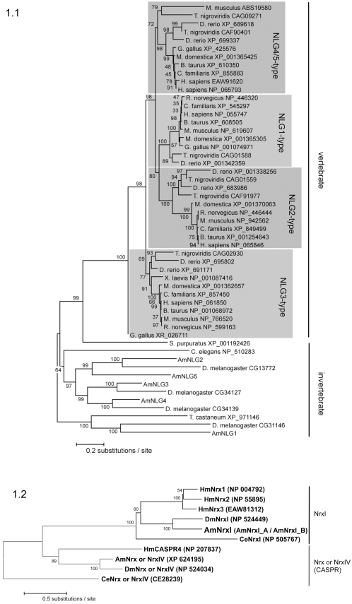
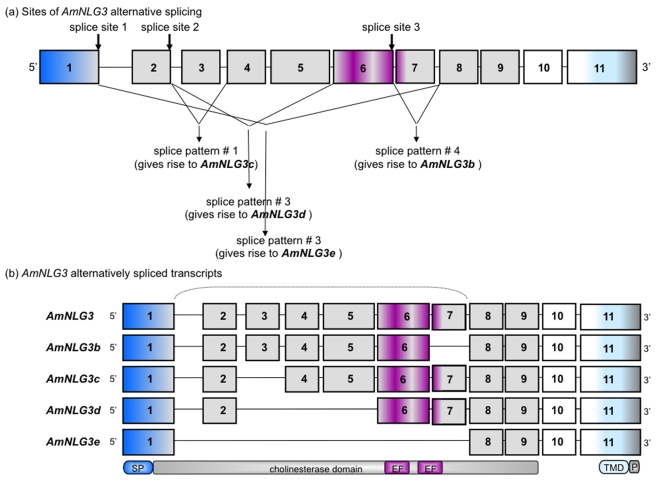
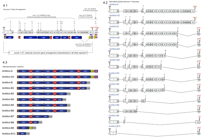
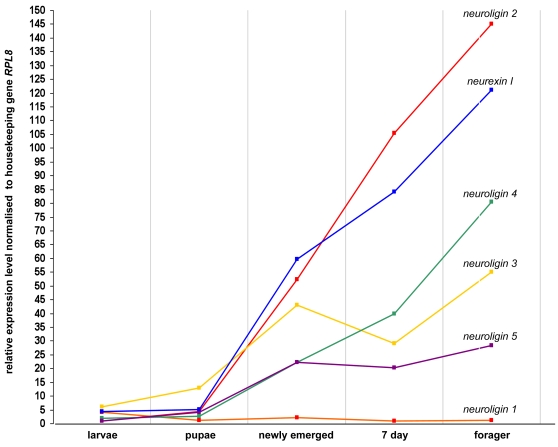
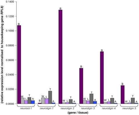
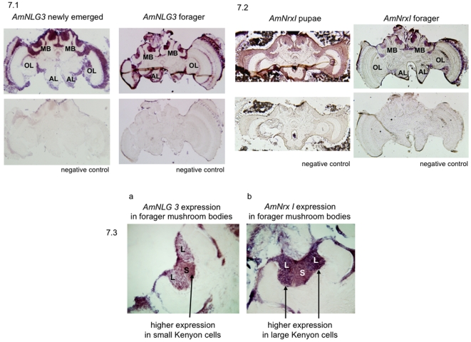
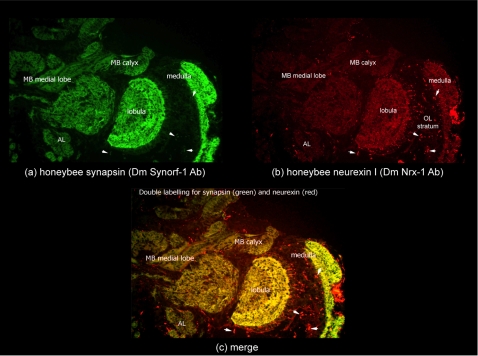
Vertebrate studies show neuroligins and neurexins are binding partners in a trans-synaptic cell adhesion complex, implicated in human autism and mental retardation disorders. Here we report a genetic analysis of homologous proteins in the honey bee. As in humans, the honeybee has five large (31-246 kb, up to 12 exons each) neuroligin genes, three of which are tightly clustered. RNA analysis of the neuroligin-3 gene reveals five alternatively spliced transcripts, generated through alternative use of exons encoding the cholinesterase-like domain. Whereas vertebrates have three neurexins the bee has just one gene named neurexin I (400 kb, 28 exons). However alternative isoforms of bee neurexin I are generated by differential use of 12 splice sites, mostly located in regions encoding LNS subdomains. Some of the splice variants of bee neurexin I resemble the vertebrate alpha- and beta-neurexins, albeit in vertebrates these forms are generated by alternative promoters. Novel splicing variations in the 3' region generate transcripts encoding alternative trans-membrane and PDZ domains. Another 3' splicing variation predicts soluble neurexin I isoforms. Neurexin I and neuroligin expression was found in brain tissue, with expression present throughout development, and in most cases significantly up-regulated in adults. Transcripts of neurexin I and one neuroligin tested were abundant in mushroom bodies, a higher order processing centre in the bee brain. We show neuroligins and neurexins comprise a highly conserved molecular system with likely similar functional roles in insects as vertebrates, and with scope in the honeybee to generate substantial functional diversity through alternative splicing. Our study provides important prerequisite data for using the bee as a model for vertebrate synaptic development.


Conflict of interest statement

Competing Interests: The authors have declared that no competing interests exist.

Figures

Figure 1
Figure 1. Neuroligin and Neurexin Phylogeny.
(1.1) shows the phylogenetic relationship of 53 neuroligin proteins from the honeybee (AmNLG1-5) and Drosophila fly together with other neuroligins described by Bolliger et al . All sequences are represented by taxon names showing species and NCBI accession numbers. The evolutionary history was inferred using the Neighbor-Joining method . An optimal tree with percentage of replicate trees in which the associated taxa clustered together in the bootstrap test (1000 replicates) is shown next to the branches . The tree is drawn to scale, with branch lengths in the same units as those of the evolutionary distances used to infer the phylogenetic tree. The evolutionary distances were computed using the JTT matrix based method and are in the units of the number of amino acid substitutions per site. All positions containing alignment gaps and missing data were eliminated only in pairwise sequence comparisons (Pairwise deletion option). There were a total of 1884 positions in the final dataset. Phylogenetic analyses were conducted in MEGA3.1 . Four radiations (shaded) represent relationship of vertebrate proteins NLG1-4 compared with invertebrate proteins. The phylogeny shows vertebrate and invertebrate neuroligin radiations arise from a single common ancestor found in the sea urchin (S. purpuratus XP_001192426), and displays a congruent topology with the Maximum-Likelihood tree reported by Bolliger et al. . Similar analysis was performed for investigating neurexin phylogeny. (1.2) shows the evolutionary relationship of two ancestrally related clades of neurexin proteins from vertebrates and invertebrates; the neurological neurexins (NrxI) and neurexin IV (also known as neurexin). Notably, the invertebrate neurexin IV proteins form an orthologous group with CASPR. There were a total of 2125 positions in the final dataset. Also shown are the NCBI and wormbase accession numbers. Abbreviations Am: Apis mellifera, honeybee.
Figure 2
Figure 2. Structural Homology Modelling.
(2.1) shows neuroligin and neurexin I homology models. The similarity between MmNLG1 and (a) AmNLG1 and (b) AmNLG3 is illustrated using colour, where sequence similarity in blue represents identical; green represents conservative; yellow represents semi-conservative and red represents dissimilar. Similarity to Mm β-Nrx1B with AmNrxI_A is illustrated using the same coloured coding. (2.2) shows the putative honeybee neuroligin dimer interfaces. The neuroligin dimer interface from (a) the crystal structure of mouse neuroligin is shown alongside the putative interfaces in (b) AmNLG1 and (c) AmNLG3. In both honeybee sequences the key residues of the ‘hydrophobic core’ are replaced by charged or polar residues. (2.3) illustrates the homology modelling of AmNLG3 aternative slice vriants. The four spliced variants (b–e) of AmNLG3 are shown. Full length AmNLG3 was modelled against mouse NLG1 . Regions missing from the alternative transcripts are highlighted in red. (2.4) shows conservation of the neuroligin-neurexin interface in the honeybee. The respective surfaces of the neuroligin-neurexin interface are shown, based on the crystal structure of the complex from mouse. Sequence similarity is shown (blue, identical; green, conservative; yellow, semi-conservative; red, dissimilar) illustrating, (a) the strong conservation in AmNrxI-A, (b) the moderate conservation in AmNLG1, and (c) the lack of conservation in AmNLG3. (d) Illustrates a potential interaction between AmNrx1-A and AmNLG1, showing the conserved salt bridges (R232-D387), hydrogen bonds (N103-D402), and the potentially complementary hydrophobic and hydrophilic regions at the centre of the interface. Amino acids are coloured by type (blue, basic; red, acidic; yellow, polar; grey, non-polar).
Figure 3
Figure 3. Honeybee Neuroligin 3 Gene Arrangement and Intron/Exon Conservation.
AmNLG3 alternatively spliced transcripts were identified by RT-PCR from honeybee brain cDNA. Arrangement and intron/exon splice sites of AmNLG3 splice variants were deciphered by both NCBI and Beebase BLAST tools against genomic DNA. Donor/acceptor splice sites were confirmed using NCBI SPIDEY. Sizes of exons, as well as intron gaps, are not drawn to scale. Exons are numbered from 5′ to 3′. (a) highlights the three sites of alternative splicing and resulting splice patterns. (b) illustrates the five alternatively spliced AmNLG3 transcripts which correspond to the splicing patterns show in (a). The bracket highlights that splicing occurs within the cholinesterase domain. Signal peptide, cholinesterase, transmembrane domains and EF hand metal and PDZ binding motif are drawn below the encoding exons. Abbreviations SP: signal peptide; EF: EF hand metal binding motif; TMD: trans-membrane domain, P: PDZ binding domain.
Figure 4
Figure 4. Gene Arrangement and Alternative Splicing of Honeybee Neurexin I.
The gene arrangement and intron/exon splice sites of honeybee neurexin I and alternatively spliced transcripts were deciphered by both NCBI and Beebase BLAST tools against genomic DNA. Donor/acceptor splice sites were confirmed using NCBI SPIDEY. Multiple protein alignment analysis by ClustalW demonstrated the conservation of intron/exon splice sites (refer to Figures S4 and S5). Structural features were deciphered through PROSITE and SMART, and from Rissone et al. ; Jeleń et al. and Sudhof et al. . Exons are numbered from 5′ to 3. The size of exons, intron gaps and protein domains (and compared to one another) are not drawn to scale. Numbered arrows indicate sites of alternative splicing. (3.1) illustrates the full length neurexin I gene arrangement. The common start codon is highlighted by ATG. The two alternate stop codons are highlighted by TAG(1) and TAG(2). Sites of alternative splicing which occur within an exon are marked by a blue dot. Exons common to both the AmNrxI-A and AmNrxI-B transcripts are indicated above the gene schematic. Exons unique to AmNrxI–A transcript with a transmembrane domain are indicated above the gene schematic. The exon unique to the AmNrxI-B is labelled as ‘28’ and indicated as unique above. Horizontal primers below the gene schematic highlight the location of primers used for RT-PCR amplification. AmNrxI-A was amplified with the forward primer at exon 1 (ATG) and the reverse primer in blue at exon 27 (TAG1). AmNrxI-B was amplified with the same forward primer at exon 1 (ATG) and the reverse primer in red at exon 28 (TAG2). The protein domains which are encoded by particular exons are shown below the specific exons. The transmembrane domain and putative PDZ domain of AmNrxI-B are shown below exon 28. (3.2) illustrates the alternatively spliced AmNrxI-A and AmNrxI-B transcripts. (3.3) illustrates the alternate isoforms of AmNrxI-A and AmNrxI-B which arise from alternative splicing. White arrowheads indicate where splicing occurs. Brackets above highlight neurexin repeats (two complete LNS domains and an EGF motif). The putative PDZ domain of AmNrxI-B is hatched. Abbreviations- SP: signal peptide; E: EGF domain; T: transmembrane domain; P: PDZ binding motif. LNS domain: Laminin, Neurexin, Sex hormone-binding globulin.
Figure 5
Figure 5. Developmental Expression Profiles of the Neuroligins and Neurexin I in Honeybee Brain.
Honeybee neuroligin and neurexin I expression was assessed by quantitative real time PCR amplification. The ribosomal gene RPL8 was used as the housekeeping gene. Methodology for data analysis and the presentation of results was taken from Collins et al ; where by expression levels were normalised by subtraction against the threshold cycle of the RPL8. Collins et al found RPL8 to be the best correlate with RNA concentration across varying developmental life stages and varying tissues of the honeybee. Expression levels were examined from whole larvae (5 day old); and brain tissue from pupae (stage P8 as outlined by Ganeshina et al [101]) 24 hour adult, 7 day adult and forager honeybees. Standards errors were negligible and less than +/−1.18 for all experimental results. The coloured lines illustrate the developmental expression profile of a single gene through development. Data points in columns illustrate the relative levels of neurexin I and neuroligin expression to one another at a particular stage of development. The developmental stage/gene with lowest expression relative to the control gene (neuroligin 1 at 7 days of age) was given an arbitrary expression level of 1. The data values are shown in Supplementary Data Table 3.
Figure 6
Figure 6. Spatial Expression of Neuroligins and Neurexin I in the Adult Honeybee.
The methodologies behind attaining these results are as described in the Figure 5 legend. Expression levels are shown relative to the house keeping gene RPL8, given an arbitrary value of 1. Expression levels were examined from the tissue of ten adults at twenty-one days of age. Standards errors were negligible and less than +/−1.22 for all experimental results. Level of gene expression in the brain expression shown in the dark purple columns marked B, wings in the light grey columns marked W, legs in the light purple columns marked L, thorax in the dark grey columns marked T and abdomen in the blue columns marked A. Raw data from the qRT-PCR experiments in Supplementary Data Table 4.
Figure 7
Figure 7. Neuroligin and Neurexin I Brain Expression.
To identify RNA transcript distribution, in situ hybridisation experiments were performed on 20 µm honeybee brain sections using gene specific digoxygenein labelled probes: (7.1) illustrates expression of AmNLG3 in the adult brain of newly emerged (left) and forager (right) bees. Top images illustrate results of anti-sense probe staining. Bottom images illustrate the results of the negative control experiments using a sense probe. (7.2) Expression of AmNrxI/AmNrxI_28 in both a P8 stage (outlined by Ganeshina et al. [101]) pupae (left) and adult forager (right) honeybee brain. Top images illustrate the results of anti-sense probe staining. Bottom images illustrate results of the negative control experiments using a sense probe. (7.3) shows contrasting AmNLG3 and AmNrxI expression in the adult mushroom body. (a) AmNLG3 expression higher in the small Kenyon cells. (b) AmNrxI expression higher in the large Kenyon cells. Abbreviations- MB: mushroom body; OL: optic lobe; AL: antennal lobe; L: large Kenyon cells; S: small Kenyon cells.
Figure 8
Figure 8. Honeybee Neurexin I Protein Expression in the Brain.
Immuno-staining of forager brain sections (a) for synapsin using SynOrf-1 antibody and incubation with Alexa-488-conjugated anti-mouse antibody allowing green colouration to highlight protein expression; (b) for honeybee neurexin-I using DmNrx-1 antibody and incubation with Alexa-546-conjugated anti-mouse antibody allowing red colouration to highlight protein expression; (c) merge shows neurexin-I and synapsin co-localise in mushroom body neuropil (MB medial lobe and MB calyx), optic lobe neuropil (medulla and lobulla) and antennal lobes. Small puncta of neurexin I expression, distinct to synapsin, are highlighted by small arrow heads within the optic lobe (OL) stratum.

Similar articles

Cited by

References

    1. Berninghausen O, Rahman MA, Silva JP, Davletov B, Hopkins C, et al. Neurexin Ibeta and neuroligin are localized on opposite membranes in mature central synapses. J Neurochem. 2007;103:1855–1863. - PMC - PubMed
    1. Taniguchi H, Gollan L, Scholl FG, Mahadomrongkul V, Dobler E, et al. Silencing of neuroligin function by postsynaptic neurexins. J Neurosci. 2007;27:2815–2824. - PMC - PubMed
    1. Ichtchenko K, Hata Y, Nguyen T, Ullrich B, Missler M, et al. Neuroligin 1: a splice site-specific ligand for beta-neurexins. Cell. 1995;81:435–443. - PubMed
    1. Bolliger MF, Frei K, Winterhalter KH, Gloor SM. Identification of a novel neuroligin in humans which binds to PSD-95 and has a widespread expression. Biochem J. 2001;356:581–588. - PMC - PubMed
    1. Gilbert M, Smith J, Roskams AJ, Auld VJ. Neuroligin 3 is a vertebrate gliotactin expressed in the olfactory ensheathing glia, a growth-promoting class of macroglia. Glia. 2001;34:151–164. - PubMed

Publication types

MeSH terms