. 2008;3(10):e3601.
doi: 10.1371/journal.pone.0003601. Epub 2008 Oct 31.

HMGB1-dependent triggering of HIV-1 replication and persistence in dendritic cells as a consequence of NK-DC cross-talk

Affiliations

HMGB1-dependent triggering of HIV-1 replication and persistence in dendritic cells as a consequence of NK-DC cross-talk

Héla Saïdi et al. PLoS One. 2008.

Abstract

Background: HIV-1 has evolved ways to exploit DCs, thereby facilitating viral dissemination and allowing evasion of antiviral immunity. Recently, the fate of DCs has been found to be extremely dependent on the interaction with autologous NK cells, but the mechanisms by which NK-DC interaction controls viral infections remain unclear. Here, we investigate the impact of NK-DC cross-talk on maturation and functions of HIV-infected immature DCs.

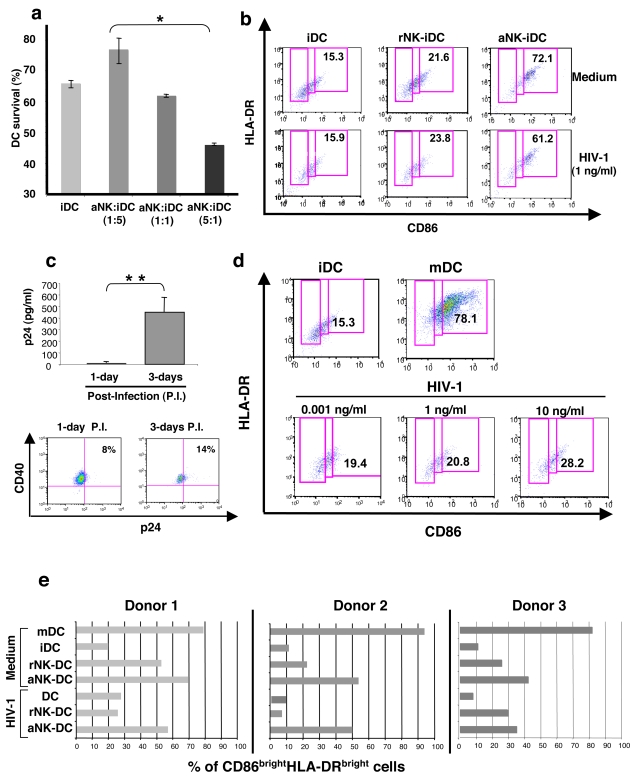
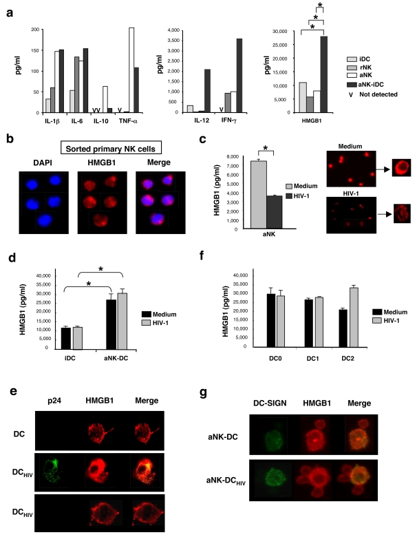
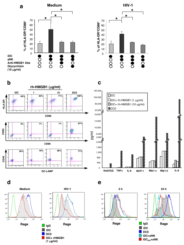
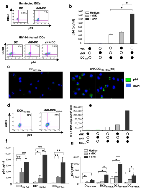
Methodology/principal findings: Immature DCs were derived from primary monocytes, cultured in the presence of IL-4 and GM-CSF. In some experiments, DCs were infected with R5-HIV-1(BaL) or X4-HIV-1(NDK), and viral replication, proviral HIV-DNA and the frequency of infected DCs were measured. Autologous NK cells were sorted and either kept unstimulated in the presence of suboptimal concentration of IL-2, or activated by a combination of PHA and IL-2. The impact of 24 h NK-DC cross-talk on the fate of HIV-1-infected DCs was analyzed. We report that activated NK cells were required for the induction of maturation of DCs, whether uninfected or HIV-1-infected, and this process involved HMGB1. However, the cross-talk between HIV-1-infected DCs and activated NK cells was functionally defective, as demonstrated by the strong impairment of DCs to induce Th1 polarization of naïve CD4 T cells. This was associated with the defective production of IL-12 and IL-18 by infected DCs. Moreover, the crosstalk between activated NK cells and HIV-infected DCs resulted in a dramatic increase in viral replication and proviral DNA expression in DCs. HMGB1, produced both by NK cells and DCs, was found to play a pivotal role in this process, and inhibition of HMGB1 activity by glycyrrhizin, known to bind specifically to HMGB1, or blocking anti-HMGB1 antibodies, abrogated NK-dependent HIV-1 replication in DCs.

Conclusion: These observations provide evidence for the crucial role of NK-DC cross-talk in promoting viral dissemination, and challenge the question of the in vivo involvement of HMGB1 in the triggering of HIV-1 replication and replenishment of viral reservoirs in AIDS.


Conflict of interest statement

Competing Interests: The authors have declared that no competing interests exist.

Figures

Figure 1
Figure 1. aNK cells induce the maturation of primary immature HIV-1-infected DCs.
(a) iDCs, generated from purified CD14+ monocytes in the presence of IL-4 and GM-CSF, were cocultured during 24 h with aNK cells at different ratios. DC survival was determined by flow cytometry with the 7-AAD assay. Surviving DCs were identified as 7AAD CD56 cells. Data represent three independent experiments and values are means±sd. (b) aNK cells induce the maturation of iDCs. Flow cytometry analysis of iDCs, which were either infected with HIV-1BaL (1 ng/ml of p24) for 3 h or uninfected, were incubated with rNK cells or aNK cells at a ratio of 1∶5. Co-staining with HLA-DR and CD86 specific antibodies allowed the identification of mature DCs (CD86brightHLA-DRbright). Data from a representative experiment out of three independent experiments are shown. (c) The conditions of infection used in this study were those of a productive infection of iDCs, as shown at day 3 by a significant p24 detection in culture supernatant of infected iDCs and intracellular detection by flow cytometry of p24 in DC targeted by CD40 expression. Experiments were performed on DCs from three independent donors, and values are means±sd. (d) HIV-1 infection does not induce by itself the maturation of iDC, as shown by CD86/HLA-DR dual staining of iDCs infected with 0.001 to 10 ng/ml p24 HIV-1BaL. The proportion of mDCs induced by LPS (DC0) (78.1% CD86brightHLA-DRbright) is shown as a positive control. (e) The proportion of mature CD86brightHLA-DRbright DCs induced in the indicated cocultures of infected or uninfected iDCs with either rNK or aNK cells are shown. These experiments have been performed on primary cells from a number of donors, and representative data from three of them are shown. When indicated, statistical analyses were made with the non-parametric Mann-Whitney test. * p<0.05, ** p = 0.02.
Figure 2
Figure 2. aNK-DC cross-talk triggers HMGB1 expression in both aNK cells and DCs.
(a) 24 h cell-free culture supernatants of iDCs, rNK cells, aNK cells (106/ml), or cocultures of aNK cells and iDCs (ratio 1∶5) were tested for cytokine content. MAP technology was used to quantify IL-1β, IL-6, IL-10, TNF-α, IL-12 and IFN-γ, whereas HMGB1 was quantified by ELISA. * p<0.05 (non-parametric Mann-Whitney test). (b) HMGB1 expression was detected by immunofluorescence (in red) in freshly sorted blood NK cells. Counterstaining with DAPI (in blue) showed the nuclear localisation of HMGB1. (c) Incubation of aNK cells with HIV-1 inhibits HMGB1 secretion. Left panel: aNK cells (106 cells/ml) were incubated in medium or with HIV-1BaL (1 ng/ml of p24) for 3 h and tested for HMGB1 production 21 h later. Data represent three independent experiments and values are means±sd. Right panel: immunofluorescence analysis of HMGB1 expression in the same preparations of aNK cells. (d) HMGB1 production during aNK-iDC cross-talk is not inhibited by HIV-1 infection of iDCs. iDCs were incubated for 3 h in medium or with HIV-1BaL (1 ng/ml of p24) and further cocultured for 21 h with aNK cells (aNK∶iDC ratio 1∶5). HMGB1 concentration was then measured in culture supernatants. Data represent the mean±sd of three independent experiments. (e) Immunofluorescence confocal analysis of HMGB1expression in uninfected or HIV-1-infected iDCs. Upper panel: non infected iDCs; middle panel: HIV-1-infected and replicating iDCs, as shown by intracellular p24 staining; lower panel: iDCs incubated with HIV-1 but negative for intracellular p24 expression. (f) Mature DCs were generated by 48 h stimulation of iDCs with LPS (DC0), soluble CD40L (DC1) or LPS+PGE2 (DC2). DC0, DC1 and DC2 were incubated for 3 h in medium or infected with HIV-1BaL (1 ng/ml of p24) and further incubated in medium for 21 h. HMGB1 quantification in culture supernatants was performed. The mean±sd of three independent experiments is shown. (g) Immunofluorescence analysis of HMGB1 expression in conjugates of aNK cells and uninfected (upper panel) or HIV-1-infected DCs (lower panel) in a 24 h coculture. DCs are DC-SIGN+ and both aNK cells and DCs express HMGB1 in these conjugates. Pictures from one representative experiment out of three conducted with different primary cell preparations are shown.
Figure 3
Figure 3. aNK-dependent maturation of HIV-1-infected iDCs is mediated by HMGB1 and involoves RAGE.
(a) Left panel: iDCs were cultured for 24 h either alone or with aNK cells, in the presence of blocking anti-HMGB1 antibodies (10 µg/ml) or glycyrrhizin (10 µg/ml). The maturation status of DCs was determined by flow cytometry with CD86 and HLA-DR –specific antibodies. Right panel: same experiment, but performed with HIV-1 infected iDCs. Data represent mean±sd of at least three independent experiments, and statistical comparisons were made with the non-parametric Mann-Whitney test. * p<0.05. (b) iDC (106 cells/ml) were cultured for 48 h with increasing concentrations (1–10 µg/ml) of rh-HMGB1. Cells were then stained with anti-CD86, -HLA-DR, -CD80, -CD83, DC-LAMP and -CD40 antibodies and analysed by flow cytometry. (c) Influence of rh-HMGB1 on cytokine and chemokine production (determined by MAP) by DCs. iDCs (106 cells/ml) were incubated for 48 h in medium or in presence of rh-HMGB1 (1 or 10 µg/ml). As a positive control, iDCs were stimulated with LPS (DC0). (d) Flow cytometry detection of surface expression of RAGE by iDCs, DC0, or iDCs incubated with rh-HMGB1 (1 µg/ml). iDCs were either non infected or infected with HIV-1BaL (1 ng/ml p24 for 3 h). (e) iDC, DC0, uninfected or HIV-1-infected iDC cocultured for 24 h with aNK cells, were incubated with rh-HMGB1 (1 µg/ml) and subsequently stained with anti-RAGE antibodies and analyzed by flow cytometry. NK cells were excluded from the analysis through the co-staining with CD3- and CD56-specific antibodies (CD3CD56+).
Figure 4
Figure 4. Impairment of NK-triggered Th1 polarization by DCs following HIV-1 infection is associated to altered IL-12 and IL-18 production.
(a) Th1 polarization by DCs triggered by NK cells was tested by incubating iDC (106/ml) for 30 mn in the presence of rNK or aNK cells (2×10⁵/ml). Naïve CD4 T cells (106/ml) were added to the cocultures and the frequency of T cells producing IFN-γ or IL-4 was determined by flow cytometry 8 days later. The experiment was performed with either uninfected iDCs (b) iDCs infected with HIV-1BaL (c), or iDC infected with HIV-1BaL in the presence of AZT (1 mM) (d). Culture supernatants of indicated cultures were tested for IL-12 (e), IL-18 (f), and IFN-g (g) content. Data represent the mean±sd of five independent experiments. Statistical comparisons were made with the non-parametric Mann-Whitney test. * p<0.05, **p = 0.03.
Figure 5
Figure 5. HMGB1-dependent triggering of HIV replication in DC as a consequence of NK-DC cross talk.
(a) Flow cytometry analysis of p24 intracellular expression in iDCs (CD40+), either uninfected (upper panel) or infected with HIV-1BaL (lower panel) following 3 day-incubation at 106/ml, either alone, or in the presence of rNK or aNK cells (2×10⁵/ml). (b) p24 concentration in culture supernatants of same cultures. Mean±sd of three independent experiments. *p<0.05, non-parametric Mann-Whitney test. (c) Immunofluorescence analysis of intracellular p24 expression in HIV-1-infected iDCs cultured for 3 days either alone or in the presence of aNK cells. Nuclei are stained with DAPI. (d) Flow cytometry intracellular p24 expression in HIV-1-infected DC0 (106/ml) cultured either alone or in the presence of aNK cells for 6 days. (e) HIV-1 proviral DNA levels, determined by light cycler analysis on cells from indicated cultures. One representative experiment out of three conducted with different primary cells preparations is shown. (f) p24 concentration in culture supernatants of mature DCs infected with HIV-1BaL and cultured for 6 days either alone or in the presence of rNK or aNK cells Mean±sd of three independent experiments. Statistical comparisons were made with the non-parametric Mann-Whitney test. * p<0.05, **p = 0.03. (g) p24 concentration in culture supernatants of either iDCs or mature DCs infected with HIV-1NDK and cultured under the same conditions as in (f). Mean±sd of three independent experiments. Statistical comparisons were made with the non-parametric Mann-Whitney test. * p<0.05.
Figure 6
Figure 6. Exogenous rh-HMGB1 triggers HIV-1 replication in iDC.
(a) iDC -infected with HIV-1BaL were cultured either alone or in the presence of aNK cells for 3 days. rh-HMGB1 (1 µg/ml) was added in some cultures. HIV replication was measured by p24 quantification in culture supernatant. (b) HIV-1-infected iDC were cultured either alone or in the presence of aNK cells for 3 days. Blocking anti-HMGB1 antibodies (10 µg/ml) or glycyrrhizin (10 µg/ml) were added at culture initiation. HIV replication was measured by p24 quantification in culture supernatant. The mean±sd of three independent experiments is shown. Statistical comparisons were made with the non-parametric Mann-Whitney test. * p<0.05.

Similar articles

Cited by

References

    1. Meng G, Wei X, Wu X, Sellers MT, Decker JM, et al. Primary intestinal epithelial cells selectively transfer R5 HIV-1 to CCR5+ cells. Nat Med. 2002;8:150–156. - PubMed
    1. Pope M, Haase AT. Transmission, acute HIV-1 infection and the quest for strategies to prevent infection. Nat Med. 2003;9:847–852. - PubMed
    1. Geijtenbeek TB, Kwon DS, Torensma R, van Vliet SJ, van Duijnhoven GC, et al. DC-SIGN, a dendritic cell-specific HIV-1-binding protein that enhances trans-infection of T cells. Cell. 2000;100:587–597. - PubMed
    1. Lee B, Leslie G, Soilleux E, O'Doherty U, Baik S, et al. cis Expression of DC-SIGN allows for more efficient entry of human and simian immunodeficiency viruses via CD4 and a coreceptor. J Virol. 2001;75:12028–12038. - PMC - PubMed
    1. Gurney KB, Elliott J, Nassanian H, Song C, Soilleux E, et al. Binding and transfer of human immunodeficiency virus by DC-SIGN+ cells in human rectal mucosa. J Virol. 2005;79:5762–5773. - PMC - PubMed

Publication types

MeSH terms