. 2008 Nov;70(4):1037-50.
doi: 10.1111/j.1365-2958.2008.06473.x. Epub 2008 Oct 2.

Intermediate filament-like proteins in bacteria and a cytoskeletal function in Streptomyces

Affiliations

Intermediate filament-like proteins in bacteria and a cytoskeletal function in Streptomyces

Sonchita Bagchi et al. Mol Microbiol. 2008 Nov.

Abstract

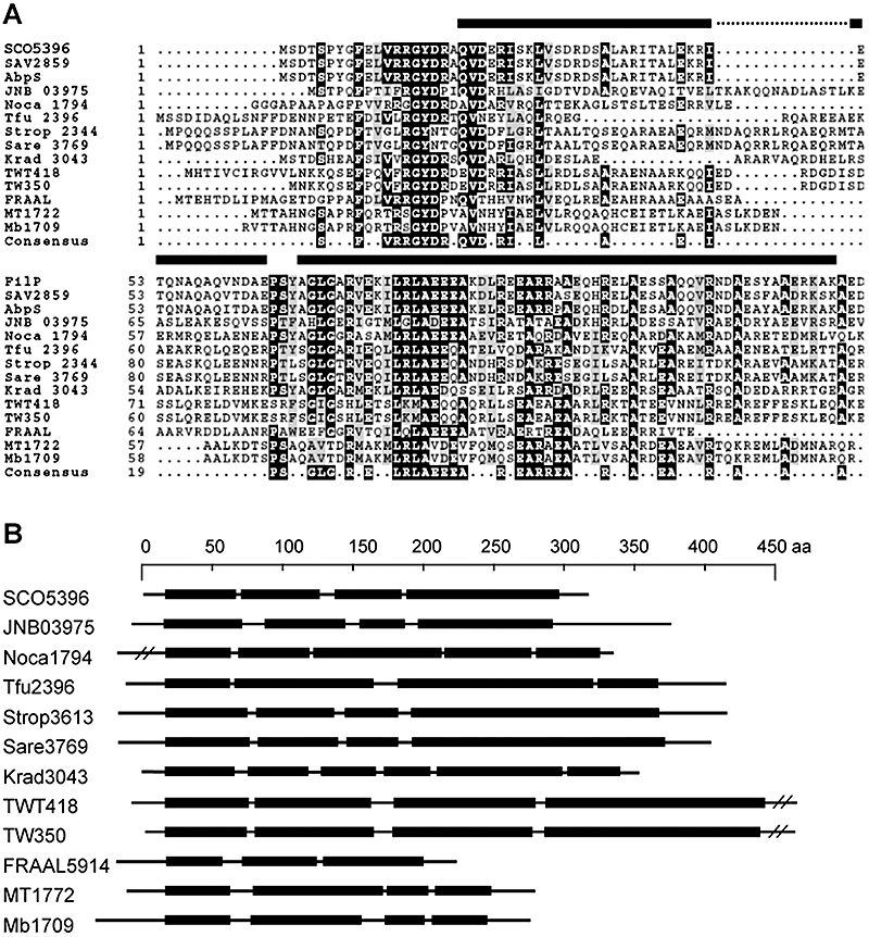
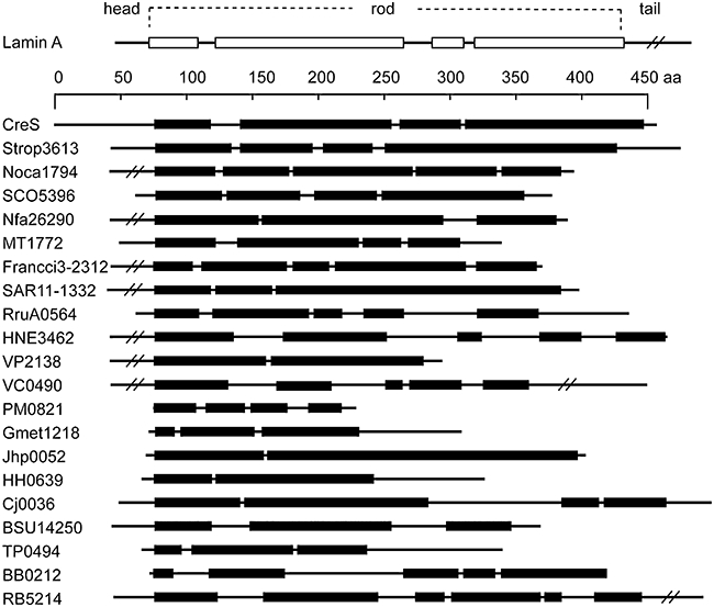
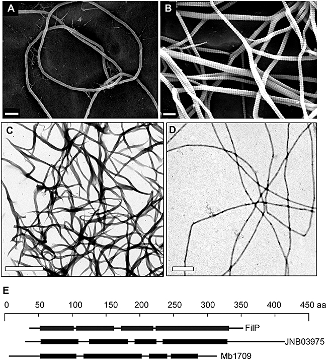
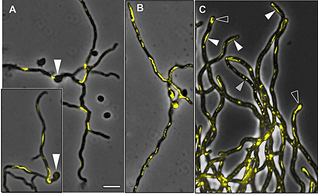
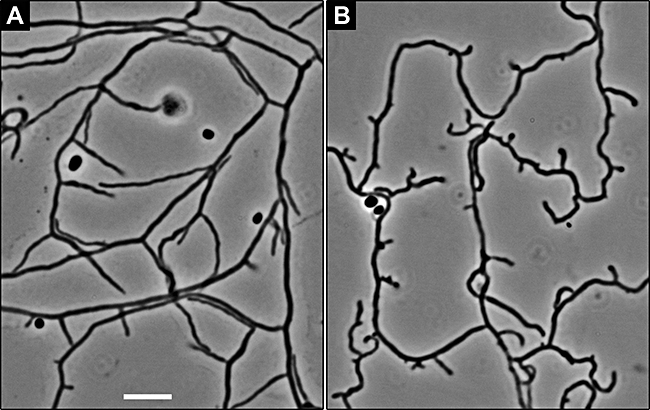
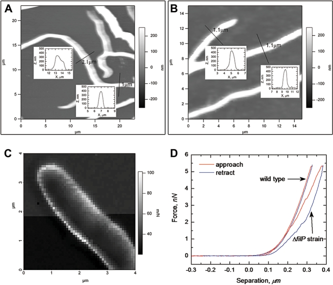
Actin and tubulin cytoskeletons are conserved and widespread in bacteria. A strikingly intermediate filament (IF)-like cytoskeleton, composed of crescentin, is also present in Caulobacter crescentus and determines its specific cell shape. However, the broader significance of this finding remained obscure, because crescentin appeared to be unique to Caulobacter. Here we demonstrate that IF-like function is probably a more widespread phenomenon in bacteria. First, we show that 21 genomes of 26 phylogenetically diverse species encoded uncharacterized proteins with a central segmented coiled coil rod domain, which we regarded as a key structural feature of IF proteins and crescentin. Experimental studies of three in silico predicted candidates from Mycobacterium and other actinomycetes revealed a common IF-like property to spontaneously assemble into filaments in vitro. Furthermore, the IF-like protein FilP formed cytoskeletal structures in the model actinomycete Streptomyces coelicolor and was needed for normal growth and morphogenesis. Atomic force microscopy of living cells revealed that the FilP cytoskeleton contributed to mechanical fitness of the hyphae, thus closely resembling the function of metazoan IF. Together, the bioinformatic and experimental data suggest that an IF-like protein architecture is a versatile design that is generally present in bacteria and utilized to perform diverse cytoskeletal tasks.


Figures

Fig. 2
Fig. 2
Conserved sequence motifs define a protein family in actinomycetes. A. MULTALIGN alignment (Corpet, 1988) of the N-termini of the FilP-family proteins. AbpS designates the Avicel-binding proteins from S. reticuli. Other proteins are identified by their locus tags shown to the left of the alignment. SCO –S. coelicolor, SAV –S. avermitilis, Krad –Kineococcus radiotolerans, Noca –Nocardioides sp., Tfu –Thermobifida fusca, FRAAL –Frankia alni, JNB –Janibacter sp., TWT – Tropheryma whipplei strain Twist, TW –Tropheryma whipplei strain TW, MT –M. tuberculosis, Mb –M. bovis, Strop –S. tropica, Sare –Salinispora arenicola. Black bars represent the positions of the two first coiled coil domains of SCO5396 (FilP). Multiple alignment was performed with default settings and the output order reflects the relatedness of the input sequences. B. Schematic representation of the various coiled coil architectures of the proteins belonging to the conserved family in (A). Black bars represent coiled coil domains, lines represent non-coiled coil sequences. Long head and tail domains are truncated in some cases.
Fig. 1
Fig. 1
Architectures of bacterial rod-domain proteins. The tripartite building plan of a human IF protein nuclear lamin A is depicted at the top of the figure. The scale bar refers to amino acid residues of crescentin (CreS), the IF-like protein from C. crescentus. Other proteins are drawn in scale, and aligned in respect to their first coiled coil domains. Boxes represent domains in coiled coil conformation and lines non-coiled coil sequences. Long head or tail domains are in some occasions truncated. Designations refer to locus tags of respective proteins from the following species (in order of occurrence): S. tropica, Nocardioides sp. JS614, S. coelicolor, N. farcinica, M. tuberculosis, Frankia sp. Ccl3, P. ubique, R. rubrum, H. neptunium, V. parahaemolyticus, V. cholerae, P. multocida, G. metallireducens, H. pylori, H. hepaticus, C. jejuni, B. subtilis, T. pallidum, B. burgdorferi, R. baltica.
Fig. 3
Fig. 3
In vitro filaments formed by rod-domain proteins of actinomycetes. A and B. Scanning electron micrographs of filaments formed by S. coelicolor protein SCO5396 (FilP) in 50 mM TrisHCl at pH 7.0. A smooth non-branching filament is shown in A and the striated branching filaments are shown in B. C and D. Transmission electron micrographs of negatively stained filaments formed by Janibacter sp. protein JNB03975 and by M. bovis protein Mb 1709 respectively. E. Schematic representation of the various rod-domain structures of the proteins in A-C. Size bars represent 200 nm for A and B, and 1 μm for C and D.
Fig. 4
Fig. 4
FilP-EGFP forms filamentous structures in growing S. coelicolor hyphae. A–C. Overlays of fluorescence and phase contrast micrographs, FilP-EGFP fluorescence is false coloured yellow. Young vegetative hyphae (12 h) of the filP-egfp strain grown in solid MS agar medium show pronounced filamentous structures of FilP-EGFP (A). White arrowheads mark germinated spores. Young vegetative hyphae (12 h) of the merodiploid filP+\filP-egfp strain are shown in B. The hybrid FilP\FilP-EGFP filaments are longer and display weaker fluorescence than those of FilP-EGFP. Hyphae of the merodiploid filP+\filP-egfp strain grown in liquid YEME medium for 14 h are slightly larger than those grown on solid MS medium and contain several types of fluorescent structures (C). White arrowheads indicate FilP\FilP-EGFP filaments, open arrowheads indicate diffuse apical spots, and grey arrowheads indicate condensed foci. Size bar corresponds to 4 μm in all panels.
Fig. 5
Fig. 5
Deletion of filP causes a morphological defect. A and B. Live hyphae of the wild-type strain M145 (A) and the †filP mutant strain (B) grown for 20 h in the angle between an inserted coverslip and the MS agar surface and visualized by phase contrast microscopy.
Fig. 6
Fig. 6
Deletion of filP causes changes in the visco-elastic properties of S. coelicolor cells. A and B. Topological images of live hyphae of the wild-type M145 (B) and the †filP strain (A) grown as in Fig. 4. Insets show height profiles of the hyphae at positions marked with perpendicular lines, indicating that mutant hyphae undergo deformation in the imaging process, whereas wild-type hyphae maintain their form. C. An image of a tip of a wild-type hypha reconstructed using data from 80 × 80 force curves. Each pixel corresponds to a compliance value calculated from an individual force curve and depicted in greyscale, as shown by a scale bar to the right. Thus, areas of the sample characterized by dark pixels are relatively stiff (low compliance), and areas with lighter pixels are softer (high compliance). D. Single representative force curves of wild-type and †filP hyphae showing that wild-type hyphae are less compliant than those of the mutant. The hysteresis effect (manifested as a loop between the load and unload curves) was consistently present in all force curves of the mutant hyphae, and absent in those of the wild-type.

Similar articles

Cited by

References

    1. Aaron M, Charbon G, Lam H, Schwarz H, Vollmer W, Jacobs-Wagner C. The tubulin homologue FtsZ contributes to cell elongation by guiding cell wall precursor synthesis in Caulobacter crescentus. Mol Microbiol. 2007;64:938–952. - PubMed
    1. Ausmees N, Kuhn JR, Jacobs-Wagner C. The bacterial cytoskeleton: an intermediate filament-like function in cell shape. Cell. 2003;115:705–713. - PubMed
    1. Bi EF, Lutkenhaus J. FtsZ ring structure associated with division in Escherichia coli. Nature. 1991;354:161–164. - PubMed
    1. Cabeen MT, Jacobs-Wagner C. Skin and bones: the bacterial cytoskeleton, cell wall, and cell morphogenesis. J Cell Biol. 2007;179:381–387. - PMC - PubMed
    1. Carballido-Lopez R. The bacterial actin-like cytoskeleton. Microbiol Mol Biol Rev. 2006;70:888–909. - PMC - PubMed

Publication types

MeSH terms

LinkOut - more resources