. 2009 Jan 16;385(2):423-31.
doi: 10.1016/j.jmb.2008.10.038. Epub 2008 Oct 19.

Head-head interaction characterizes the relaxed state of Limulus muscle myosin filaments

Affiliations

Head-head interaction characterizes the relaxed state of Limulus muscle myosin filaments

Fa-Qing Zhao et al. J Mol Biol. .

Abstract

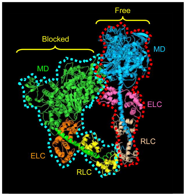
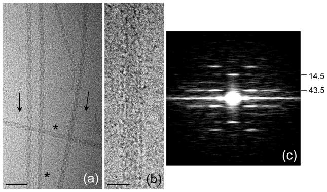
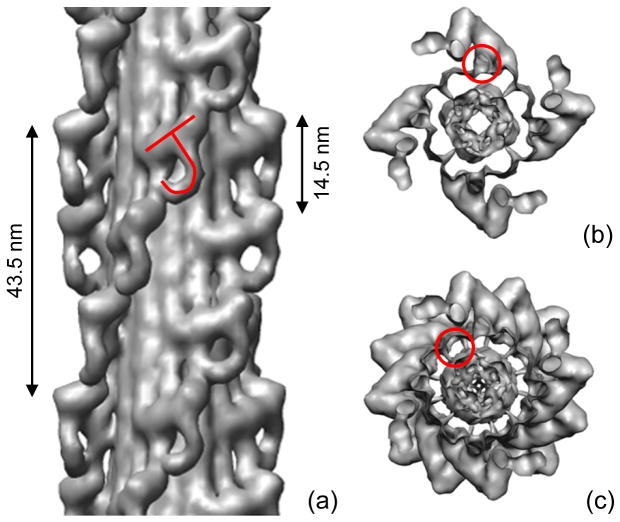
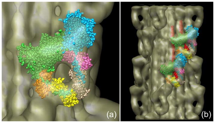
Regulation of muscle contraction via the myosin filaments occurs in vertebrate smooth and many invertebrate striated muscles. Studies of unphosphorylated vertebrate smooth muscle myosin suggest that activity is switched off through an intramolecular interaction between the actin-binding region of one head and the converter and essential light chains of the other, inhibiting ATPase activity and actin interaction. The same interaction (and additional interaction with the tail) is seen in three-dimensional reconstructions of relaxed, native myosin filaments from tarantula striated muscle, suggesting that such interactions are likely to underlie the off-state of myosin across a wide spectrum of the animal kingdom. We have tested this hypothesis by carrying out cryo-electron microscopy and three-dimensional image reconstruction of myosin filaments from horseshoe crab (Limulus) muscle. The same head-head and head-tail interactions seen in tarantula are also seen in Limulus, supporting the hypothesis. Other data suggest that this motif may underlie the relaxed state of myosin II in all species (including myosin II in nonmuscle cells), with the possible exception of insect flight muscle. The molecular organization of the myosin tails in the backbone of muscle thick filaments is unknown and may differ between species. X-ray diffraction data support a general model for crustaceans in which tails associate together to form 4-nm-diameter subfilaments, with these subfilaments assembling together to form the backbone. This model is supported by direct observation of 4-nm-diameter elongated strands in the tarantula reconstruction, suggesting that it might be a general structure across the arthropods. We observe a similar backbone organization in the Limulus reconstruction, supporting the general existence of such subfilaments.


Figures

Figure 1
Figure 1. Atomic model of head-head interaction
This ribbon model represents the best fit of the atomic model of vertebrate smooth muscle HMM (PDB1i84) into the tarantula 3D reconstruction. The actin-binding region of the blocked head, outlined in blue, binds to the converter region and essential light chain of the free head, outlined in red. Color scheme: motor domains (MD), blocked head, green; free head, blue; essential light chains (ELC), blocked head, orange; free head, pink; regulatory light chains (RLC), blocked head, yellow; free head, beige. Adapted from Ref. .
Figure 2
Figure 2. Cryo-electron microscopy of purified Limulus myosin filaments
(a) Field of filaments in relaxing conditions. Arrowheads (direction shown by arrows) result from superposition of myosin helices on front and rear of filaments as seen in projection. Bare zones are indicated by *. (b) Filament in which heads are disordered, revealing parallel strands in backbone. (c) Average Fourier transform (intensities) of the 100 filament images used in the reconstruction, showing layer lines at orders of the 43.5 nm helical repeat, confirming order visualized in A. Scale bars: (a) 100 nm; (b) 20 nm.
Figure 3
Figure 3. Three-dimensional reconstruction of Limulus thick filament
(a) Pairs of myosin heads, represented by tilted “J” motif, run in four parallel helices, with a repeat of 43.5 nm and an axial spacing between 4-fold rotationally symmetric ‘crowns’ of heads of 14.5 nm. (b) Transverse view of one 14.5 nm level of myosin heads, showing 4-fold rotational symmetric arrangement, viewed from bare zone. S2 is outlined. (c) Transverse view of one complete repeat (43.5 nm) of heads. In (b) and (c), four groups of three subfilaments appear as hollow tubes due to surface rendering (one has been outlined in (c)). The subfilaments in fact represent the highest density region of the map (Fig. 5a). In contrast, the center of the filament (interior to the tubes) appears solid but is in fact of low density (Fig. 5a).
Figure 4
Figure 4. Docking of myosin head atomic model into J-motif of reconstruction
(a) Fitting of myosin heads (PDB 1i84), each as two rigid bodies—the motor domain and the light chain domain—into the two heads of one J-motif, showing interaction of blocked head motor domain (green) with free head motor domain (blue) and essential light chain (pink). (b) Fitting of atomic model into heads at two adjacent axial levels, showing additional interactions between heads at different levels (cf.).
Figure 5
Figure 5. Backbone structure in Limulus thick filament
(a) Density projection of one complete repeat of reconstruction (43.5 nm) along filament axis (protein white). The highest protein density is in a ring of twelve rods, about 4 nm in diameter (three have been circled), running parallel to the filament axis. Each represents one subfilament. Lower density at higher radius represents myosin heads. (b) Schematic diagram showing number of tails in cross-section of a subfilament, assuming a tail length of 158 nm (see text) and that molecules within a subfilament are staggered by 43.5 nm. Top: entire tail is in subfilament; subfilament thus has 4 tails in cross-section for ~ 2/3 of each repeat. Bottom: 30 nm of tail is outside subfilament; subfilament thus has 3 tails in cross-section along almost entire repeat. (c), (d) Hypothetical backbone structure showing how paramyosin molecules ~ 130 nm long might fit into the filament core, one associated with each subfilament every 3×43.5 nm. Numbers indicate relative axial levels of subfilaments and paramyosin (in increments of 14.5 nm). Molecules in each subfilament, and in subfilaments at the same level, have the same color. In this speculative model, paramyosins (assumed diameter 2 nm) are not quite close enough to interact. Such interaction, expected in any plausible model of the filament backbone, could occur, for example, if the coiled-coil diameter were slightly larger, or the filament dimensions slightly smaller than assumed. PM = paramyosin.

References

    1. Ebashi S, Endo M, Ohtsuki I. Control of muscle contraction. Quart Rev Biophys. 1969;2:351–384. - PubMed
    1. Kendrick-Jones J, Lehman W, Szent-Gyorgyi AG. Regulation in molluscan muscles. J Mol Biol. 1970;54:313–326. - PubMed
    1. Lehman W, Szent-Gyorgyi AG. Regulation of muscular contraction. Distribution of actin control and myosin control in the animal kingdom. J Gen Physiol. 1975;66:1–30. - PMC - PubMed
    1. Szent-Gyorgyi AG, Kalabokis VN, Perreault-Micale CL. Regulation by molluscan myosins. Mol Cell Biochem. 1999;190:55–62. - PubMed
    1. Gordon AM, Homsher E, Regnier M. Regulation of contraction in striated muscle. Physiol Rev. 2000;80:853–924. - PubMed

Publication types

LinkOut - more resources