. 2008 Nov;83(5):594-603.
doi: 10.1016/j.ajhg.2008.10.014. Epub 2008 Oct 30.

Identification of a 2 Mb human ortholog of Drosophila eyes shut/spacemaker that is mutated in patients with retinitis pigmentosa

Affiliations

Identification of a 2 Mb human ortholog of Drosophila eyes shut/spacemaker that is mutated in patients with retinitis pigmentosa

Rob W J Collin et al. Am J Hum Genet. 2008 Nov.

Abstract

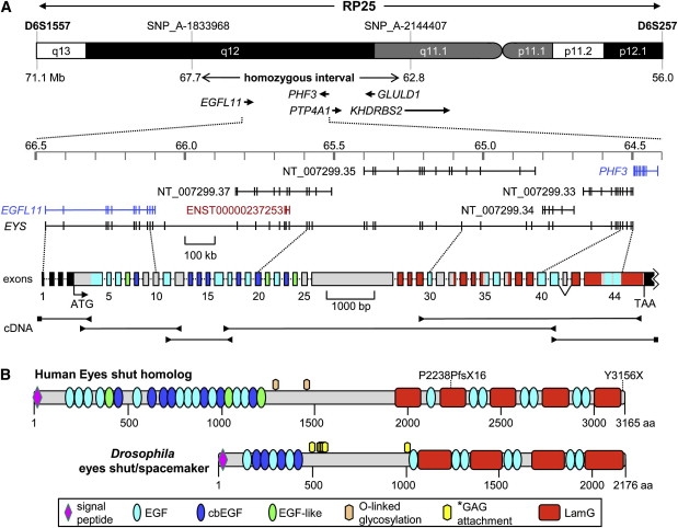
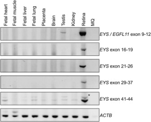
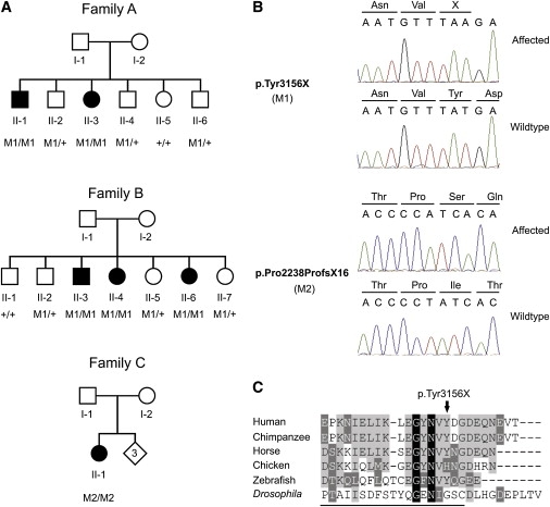
In patients with autosomal-recessive retinitis pigmentosa (arRP), homozygosity mapping was performed for detection of regions harboring genes that might be causative for RP. In one affected sib pair, a shared homozygous region of 5.0 Mb was identified on chromosome 6, within the RP25 locus. One of the genes residing in this interval was the retina-expressed gene EGFL11. Several genes resembling EGFL11 were predicted just centromeric of EGFL11. Extensive long-range RT-PCR, combined with 5'- and 3'- RACE analysis, resulted in the identification of a 10-kb transcript, starting with the annotated exons of EGFL11 and spanning 44 exons and 2 Mb of genomic DNA. The transcript is predicted to encode a 3165-aa extracellular protein containing 28 EGF-like and five laminin A G-like domains. Interestingly, the second part of the protein was found to be the human ortholog of Drosophila eyes shut (eys), also known as spacemaker, a protein essential for photoreceptor morphology. Mutation analysis in the sib pair homozygous at RP25 revealed a nonsense mutation (p.Tyr3156X) segregating with RP. The same mutation was identified homozygously in three arRP siblings of an unrelated family. A frame-shift mutation (pPro2238ProfsX16) was found in an isolated RP patient. In conclusion, we identified a gene, coined eyes shut homolog (EYS), consisting of EGFL11 and the human ortholog of Drosophila eys, which is mutated in patients with arRP. With a size of 2 Mb, it is one of the largest human genes, and it is by far the largest retinal dystrophy gene. The discovery of EYS might shed light on a critical component of photoreceptor morphogenesis.


Figures

Figure 1
Figure 1
Genomic Structure, cDNA Fragments, and Protein Domains of EYS (A) Upper panel: the RP25 chromosomal region at 6p12.1-q133, the 5.0 Mb homozygous region identified in family A, and the five known genes within the homozygous region. Exons 1 and 2 of KHDRBS2 reside in the critical region. In the middle, the exon predictions are depicted on the basis of RefSeq (in blue), Genescan (in black), and Ensembl (in red), with the use of the March 2006 UCSC genome build (hg18). Below the genomic-exon annotation is the exon structure of human EYS (exons drawn to scale; intron sizes can be found in the top panel). The complete nucleotide sequence of human EYS cDNA is presented in Figure S2. For details of the exon-intron structure, see Table S5. The 5′- and 3′- UTRs are indicated in black boxes; the colors of the protein-coding exons correspond with those of the protein domains in (B). Lower panel: reverse-transcription PCR fragments of human EYS with retina RNA and EYS-specific primers (arrowheads) or 5′- and 3′- RACE adaptor primers (squares). The 5′- UTR, the open reading frame, and the 3′-UTR altogether measure 10,475 nts (see Table S5). Exon 42 (63 bp) is alternatively spliced in retina RNA (see Figure 2). For details of RT-PCR studies, see Figure S1. (B) Protein-domain structure of EYS and its Drosophila ortholog (GenBank ID ABH07112.1). Note the conspicuous conservation of the order of EGF-like and laminin A G-like domains between human and Drosophila. The p.Pro2238ProfsX16 frame-shift mutation truncates several EGF-like and laminin A G-like domains, whereas the carboxy-terminal p.Tyr3156X mutation truncates the last ten amino acids of human EYS. Abbreviations are as follows: EGF, epidermal growth factor domain; cbEGF, calcium-binding EGF-like domain; EGF-like, EGF-like domain; LamG, laminin A G-like domain. The asterisk denotes glycosaminoglycan (GAG) attachment sites predicted by Husain and coworkers. Two putative O-glycosylation sites are predicted in the human protein (Thr1268 and Thr1424). Detailed characteristics of the human EYS protein domains are presented in Figure S3.
Figure 2
Figure 2
Tissue Distribution of EYS RT-PCR analysis was performed on total RNA from the various tissues. The expression of EYS was determined with the use of several primer pairs distributed along the transcript (see Table S4). The weak PCR product detected with primers from exon 41 to exon 44 is indicated by an asterisk and represents a transcript resulting from alternative splicing. ACTB (lower panel) was used as a control.
Figure 3
Figure 3
Mutation Analysis of EYS in RP Patients (A) Pedigrees of three families with individuals affected with RP. Below the individuals, genotypes are presented for either the p.Tyr3156X change (M1, families A and B) or the p.Pro2238ProfsX16 change (M2, family C) detected to segregate with the RP. M1/M1 and M2/M2 represent homozygous mutants; M1/+ indicates heterozygous carriers, whereas +/+ indicates individuals carrying two wild-type alleles. (B) Upper panel: partial sequence of the EYS gene showing the nonsense c.9486T → A change, in an affected individual (family A, II-1) and an unaffected sibling (family A, II-5). The mutation replaces a tyrosine residue by a termination codon (p.Tyr3156X). Preceding amino acids are indicated above the sequence trace. Lower panel: partial sequence of the EYS gene showing the c.6714 delT change, in an affected individual (family C, II-1) and a control individual. The mutation results in a frame shift and, finally, in premature termination of the protein (p.Pro2238ProfsX16). Amino acids are indicated above the sequence trace. (C) Sequence comparison of the 25 most C-terminal amino acids of the human EYS protein and several vertebrate and invertebrate orthologs. Residues identical in all sequences are white on a black background, whereas similar amino acids are white on a gray background. Residues that are present in at least three of the six proteins are indicated in black on a light gray background. Residues constituting the most C-terminal laminin A G-like domain in the Drosophila protein are underlined. Accession numbers of the protein sequences used for sequence comparison are as follows: chimpanzee, XM_527426.2 (RefSeq); horse, XM_001918159.1 (RefSeq); chicken, XM_426198.2 (RefSeq); zebrafish, BX005106.5 (EMBL); Drosophila, ABH07112.1 (GenBank).
Figure 4
Figure 4
Clinical Characteristics of RP Patients with a Homozygous p.Tyr3156X Mutation in EYS (A) Fundus photograph of the right eye of patient II-1 of family A, showing mild pallor of the optic disc, a peripapillary crescent, attenuated retinal vessels, and bone-spicule pigmentations. An area of sharply demarcated chorioretinal atrophy is located nasal to the fovea, with similar atrophic lesions along the vascular arcades, conflating to diffuse atrophy in the midperiphery. (B) Fundus photograph of the posterior pole and nasal peripheral retina of the right eye of patient II-6 of family B, showing mild pallor of the optic disc, severely attenuated vessels, pronounced atrophic changes in the (mid) periphery that spare the posterior pole, and extensive bone spicules in the peripheral retina. (C) Scotopic and photopic ERG of the right eye of patient II-3 of family A and a normal subject. Scotopic mixed response (ISCEV measurement; 2500 mcds/m2) had a b-wave amplitude of 274 μV (normal > 195 μV, mean 424 μV). The b-wave amplitude of the photopic response (ISCEV measurement; 2500 mcds/m2) was 58.8 μV (normal > 69 μV, mean 79 μV).

References

    1. Hartong D.T., Berson E.L., Dryja T.P. Retinitis pigmentosa. Lancet. 2006;368:1795–1809. - PubMed
    1. Daiger S.P., Shankar S.P., Schindler A.B., Sullivan L.S., Bowne S.J., King T.M., Daw E.W., Stone E.M., Heckenlively J.R. Genetic factors modifying clinical expression of autosomal dominant RP. Adv. Exp. Med. Biol. 2006;572:3–8. - PMC - PubMed
    1. Barragan I., Abd El-Aziz M.M., Borrego S., El-Ashry M.F., O'Driscoll C., Bhattacharya S.S., Antinolo G. Linkage validation of RP25 using the 10K genechip array and further refinement of the locus by new linked families. Ann. Hum. Genet. 2008;72:454–462. - PubMed
    1. Ruiz A., Borrego S., Marcos I., Antinolo G. A major locus for autosomal recessive retinitis pigmentosa on 6q, determined by homozygosity mapping of chromosomal regions that contain gamma-aminobutyric acid-receptor clusters. Am. J. Hum. Genet. 1998;62:1452–1459. - PMC - PubMed
    1. Abd El-Aziz M.M., El-Ashry M.F., Barragan I., Marcos I., Borrego S., Antinolo G., Bhattacharya S.S. Molecular genetic analysis of two functional candidate genes in the autosomal recessive retinitis pigmentosa, RP25, locus. Curr. Eye Res. 2005;30:1081–1087. - PubMed

Publication types

MeSH terms