. 2008 Nov;83(5):572-81.
doi: 10.1016/j.ajhg.2008.10.003. Epub 2008 Oct 30.

A homozygous mutation in human PRICKLE1 causes an autosomal-recessive progressive myoclonus epilepsy-ataxia syndrome

Affiliations

A homozygous mutation in human PRICKLE1 causes an autosomal-recessive progressive myoclonus epilepsy-ataxia syndrome

Alexander G Bassuk et al. Am J Hum Genet. 2008 Nov.

Abstract

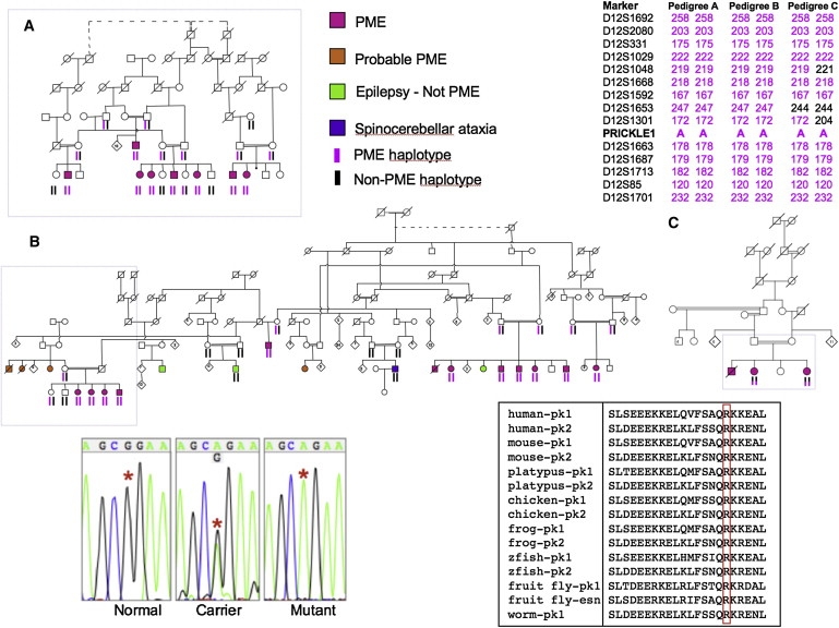
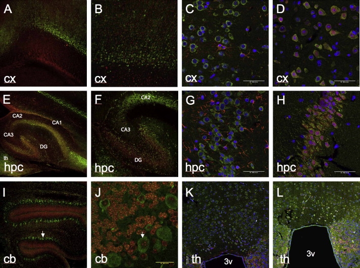
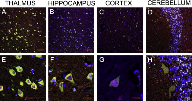
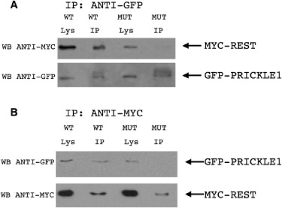
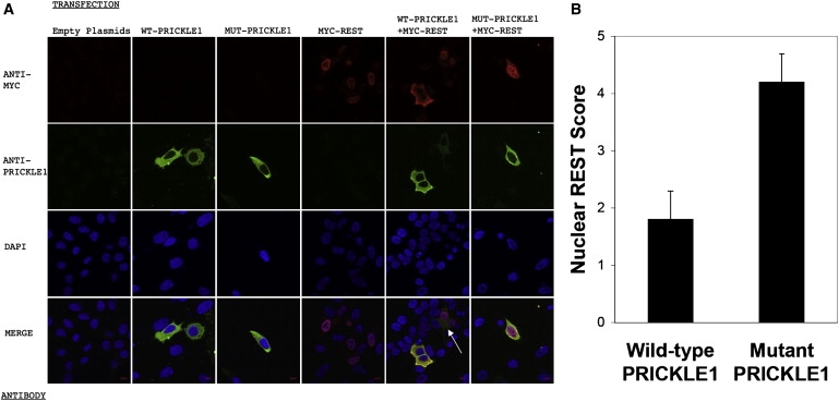
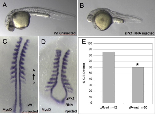
Progressive myoclonus epilepsy (PME) is a syndrome characterized by myoclonic seizures (lightning-like jerks), generalized convulsive seizures, and varying degrees of neurological decline, especially ataxia and dementia. Previously, we characterized three pedigrees of individuals with PME and ataxia, where either clinical features or linkage mapping excluded known PME loci. This report identifies a mutation in PRICKLE1 (also known as RILP for REST/NRSF interacting LIM domain protein) in all three of these pedigrees. The identified PRICKLE1 mutation blocks the PRICKLE1 and REST interaction in vitro and disrupts the normal function of PRICKLE1 in an in vivo zebrafish overexpression system. PRICKLE1 is expressed in brain regions implicated in epilepsy and ataxia in mice and humans, and, to our knowledge, is the first molecule in the noncanonical WNT signaling pathway to be directly implicated in human epilepsy.


Figures

Figure 1
Figure 1
Pedigrees of the Affected Families, Representative Sequences, and Evolutionary Comparison of the Altered PRICKLE1 Amino Acid Nine nuclear families from three pedigrees including 23 subjects with progressive myoclonus epilepsy and ataxia (pink symbol). Boxes on pedigrees indicate individuals previously reported by Berkovic et al. (A), El-Shanti et al. (B), and Straussberg et al. (C). Dotted lines indicate individuals believed to be related, but the exact relationship was unknown. Subjects who probably had the familial syndrome but were not personally examined are shown in orange. Shared chromosome 12 haplotypes of affected subjects are shown on the top right. Haplotypes were remarkably stable within nuclear families and extended pedigrees. Individuals with epilepsy or ataxia, clinically distinct from the familial syndrome (green and purple symbols), did not share the haplotypes or have the PRICKLE1 mutation. Representative DNA sequence chromograms from normal, carrier, and affected (mutant) individuals are in the bottom left panel with the red asterisks denoting the position of the abnormal nucleotide. Amino acid sequence alignment surrounding the altered amino acid for PRICKLE proteins in multiple species. pk1, Prickle1 protein; pk2, Prickle2 protein; esn, espinas protein; zfish, zebrafish. Accession numbers for the protein sequences are: human-pk1, NP_694571; human-pk2, NP_942559; mouse-pk1, NP_001028389; mouse-pk2, NP_001074615; platypus-pk1, XP_001505284; platypus-pk2, XP_001508261; chicken-pk1, XP_416036; chicken-pk2, XP_001234704; frog-pk1, NP_001016939; frog-pk-2, NP_001103517; zfish-pk1, NP_899185; zfish-pk2, NP_899186; fruit fly-pk1, NP_724534; fruit fly-esn, CAB64381; worm-pk1, NP_741435. The amino acid altered in the families and the corresponding amino acid in Prickle proteins from other species are boxed in red.
Figure 2
Figure 2
Expression of Prickle1 in Postnatal Mouse Brain Prickle1 expression in cortex (A–D), hippocampus (E–H), cerebellum (I, J), and thalamus (K, L). For (A), (B), (E), (F), (I), and (J), Prickle1 antibody, raised against epitope corresponding to amino acids 339–514 of Prickle1, is labeled with green secondary, nuclear staining is in red. For (C), (D), (G), (H), (K), and (L), Prickle1 antibody, raised against epitope corresponding to amino acids 808–822 of Prickle1, is labeled in green. In (C), (D), (G), (H), (K), and (L), nuclear staining is in blue. In (C), (G), and (K), GFAP staining is in red. In (D), (H), and (L), NeuN, staining is in red. Arrows point to representative cerebellar Purkinje cells. 3v, third ventricle from coronal section; CA1, CA2, CA3, hippocampal Cornu Ammonis subregions; cb, cerebellum; cx, cerebral cortex; DG, dentate gyrus; hpc, hippocampus; th, thalamus. Magnification is 10× in (A), (E), and (I); 20× in (B), (F), (J), (K), and (L); 60× in (C), (D), (G), and (H). (A), (B), (E), (F), (I), and (J) are from P7 brain, (C), (D), (G), (H), (K), and (L) are from adult brain. (A)–(J) are from sagittal sections, (K) and (L) are coronal sections. A representative sample of sagittal sections from a P19 brain at multiple magnifications can be found in Figure S1.
Figure 3
Figure 3
Expression of PRICKLE1 in Adult Human Brain Low-power (A–D) and high-power (E–H) confocal images from immunostaining of adult human thalamus (A, E), hippocampus (B, F), cerebral cortex (C, G), and cerebellum (D, H). PRICKLE1 staining is in green, NeuN is in red, and nuclear staining is in blue. Scale markers are represented on the bottom right corner of each image.
Figure 4
Figure 4
R104Q Mutant PRICKLE1 Has Impaired NRSF/REST Binding Coimmunoprecipitation of REST with wild-type (WT) or R104Q mutant (MUT) encoding PRICKLE1 demonstrates decreased REST binding for MUT PRICKLE1. MYC-REST and either GFP-tagged WT or R104Q MUT PRICKLE1 plasmids were transfected into HeLa cells. Cell lysates were prepared in RIPA buffer and immunoprecipitated with agarose-conjugated anti-GFP antibody (A) or anti-MYC antibody (B). Immunoprecipitates (IP) were subjected to SDS-PAGE followed by western blotting (WB) with MYC or GFP antibodies. WB antibody noted to left of gels. The input (1/5) of immunoprecipitation is shown in the “Lys” lanes. Arrows to the right of the gels note the position of MYC-REST and GFP-PRICKLE1.
Figure 5
Figure 5
Subcellular Localization of Recombinant MYC-REST and WT or MUT PRICKLE1 (A) HeLa cells were transfected with MYC-REST and WT PRICKLE1 or MUT PRICKLE1, as noted on the top of the images in (A). Antibody staining with ANTI-MYC antibody (red), ANTI-PRICKLE1 antibody (green), DAPI nuclear staining (blue), and MERGED confocal images are noted on the left. Scale bar represents 9 microns. All confocal images were captured with identical exposure settings. The white arrow in the merged WT-PRICKLE1+MYC-REST marks a cell with relatively less WT PRICKLE1 expressed versus the two other WT PRICKLE1-expressing cells in the same panel. Note that increased REST in the nucleus is inversely proportional to WT PRICKLE1 expression, whereas the nuclear REST signal is strong even in the presence of a strong MUT PRICKLE1 signal (bottom right). (B) Quantification of nuclear REST in PRICKLE1 versus mutant PRICKLE1 cotransfections. Results are the mean (±SD) of 100 cells for each group.
Figure 6
Figure 6
Mutant prickle1 Shows Decreased Activity In Vivo in Zebrafish (A–D) Equivalent amounts of RNA encoding wild-type zebrafish prickle1 (pk-wt) or zebrafish prickle1 encoding the human R104Q homologous amino acid (pk-mut) were injected into zebrafish embryos and assayed for convergence extension defects by morphology (A) wild-type compared to (B) pk-overexpression, anterior to the left and with molecular markers to axial tissues in (C) wild-type and (D) pk-overexpressing embryos, anterior to the top. (E) Decreased gastrulation defects are observed in mutant prickle1 expressing embryos compared to wild-type prickle expressing embryos. The mutant Prickle1 showed significantly decreased overexpression phenotype, with a p value of 0.01 by the Fisher's exact test.

Comment in

References

    1. Berkovic S.F., Mazarib A., Walid S., Neufeld M.Y., Manelis J., Nevo Y., Korczyn A.D., Yin J., Xiong L., Pandolfo M. A new clinical and molecular form of Unverricht-Lundborg disease localized by homozygosity mapping. Brain. 2005;128:652–658. - PubMed
    1. El-Shanti H., Daoud A., Sadoon A.A., Leal S.M., Chen S., Lee K., Spiegel R. A distinct autosomal recessive ataxia maps to chromosome 12 in an inbred family from Jordan. Brain Dev. 2006;28:353–357. - PMC - PubMed
    1. Straussberg R., Basel-Vanagaite L., Kivity S., Dabby R., Cirak S., Nurnberg P., Voit T., Mahajnah M., Inbar D., Saifi G.M. An autosomal recessive cerebellar ataxia syndrome with upward gaze palsy, neuropathy, and seizures. Neurology. 2005;64:142–144. - PubMed
    1. Ciani L., Salinas P.C. WNTs in the vertebrate nervous system: from patterning to neuronal connectivity. Nat. Rev. Neurosci. 2005;6:351–362. - PubMed
    1. Veeman M.T., Axelrod J.D., Moon R.T. A second canon. Functions and mechanisms of beta-catenin-independent Wnt signaling. Dev. Cell. 2003;5:367–377. - PubMed

Publication types