Review
. 2009 Jan 1;481(1):1-15.
doi: 10.1016/j.abb.2008.10.013. Epub 2008 Oct 17.

Bacterial heme-transport proteins and their heme-coordination modes

Affiliations
Review

Bacterial heme-transport proteins and their heme-coordination modes

Yong Tong et al. Arch Biochem Biophys. .

Abstract

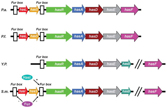
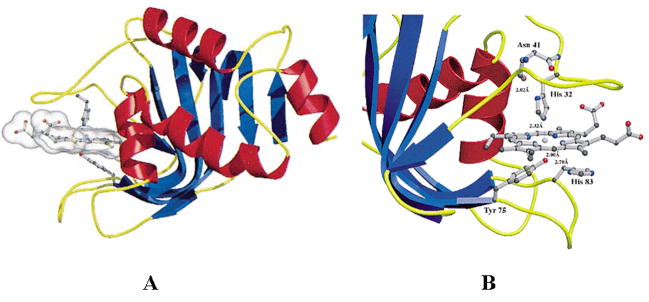
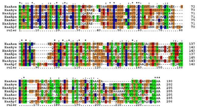
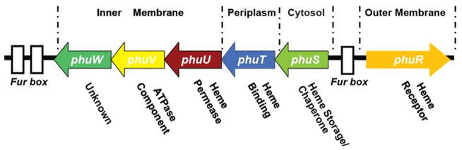
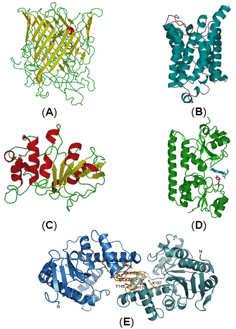
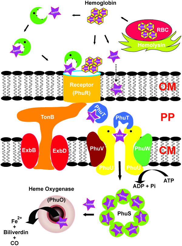
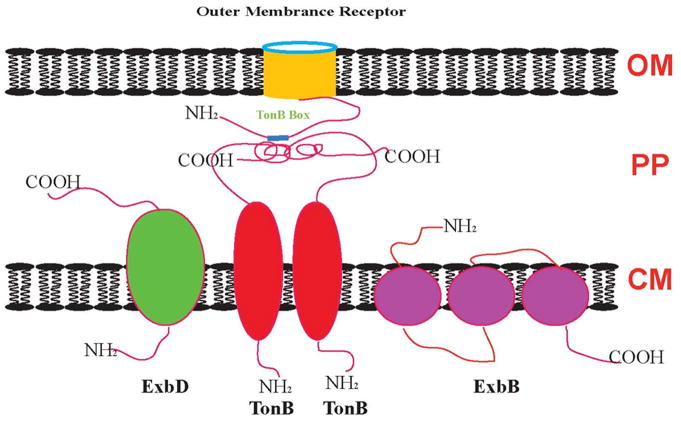
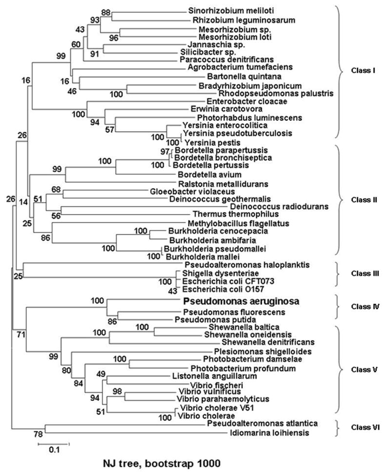
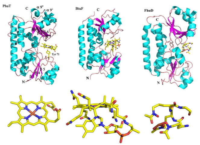
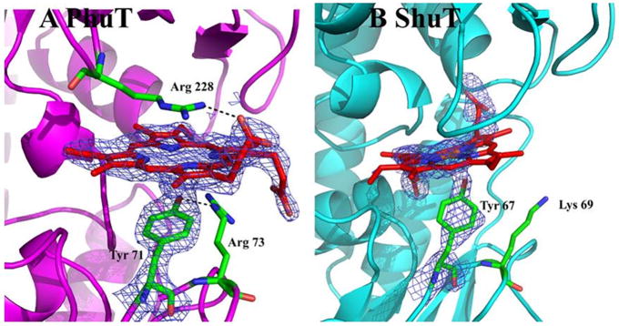
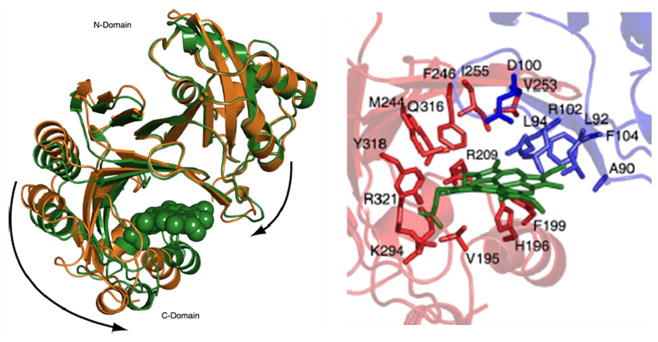
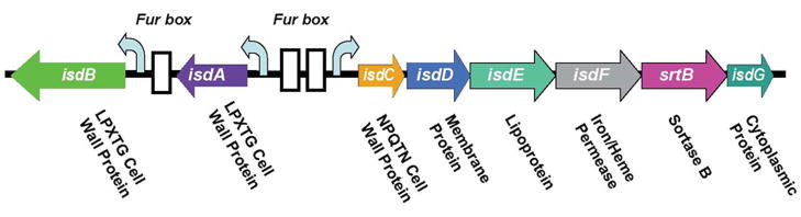
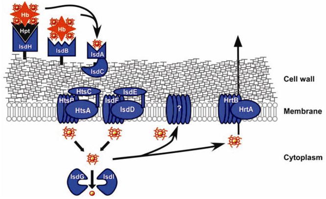
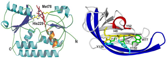
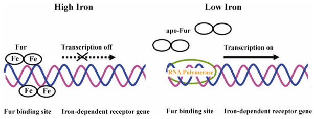
Efficient iron acquisition is critical for an invading microbe's survival and virulence. Most of the iron in mammals is incorporated into heme, which can be plundered by certain bacterial pathogens as a nutritional iron source. Utilization of exogenous heme by bacteria involves the binding of heme or hemoproteins to the cell surface receptors, followed by the transport of heme into cells. Once taken into the cytosol, heme is presented to heme oxygenases where the tetrapyrrole ring is cleaved in order to release the iron. Some Gram-negative bacteria also secrete extracellular heme-binding proteins called hemophores, which function to sequester heme from the environment. The heme-transport genes are often genetically linked as gene clusters under Fur (ferric uptake regulator) regulation. This review discusses the gene clusters and proteins involved in bacterial heme acquisition, transport and processing processes, with special focus on the heme-coordination, protein structures and mechanisms underlying heme-transport.


Figures

Figure 1
Figure 1
Genetic organization of the has systems in different bacteria. P.a., Pseudomonas aeruginosa; P.f., Pseudomonas fluorescens; Y.p., Yersinia pestis; S.m., Serratia marcescens. HasI and hasS encode sigma and antisigma factors, respectively. The white boxes indicate consensus Fur boxes. The has operon is directly controlled by Fur (Ferric uptake regulator) [26] and positively regulated by sigma and antisigma factors [158]. HasA is secreted by an ABC transporter consisting of three envelope proteins: HasD, HasE and HasF. HasD, an inner membrane ATPase (the ABC protein) provides the energy for the substrate export; HasE is another inner membrane component and also a membrane fusion protein; HasF, an outer membrane component, is homologous to TolC, which creates a channel in ~ 30Å diameter through the periplasmic space and the outer membrane through which the secreted protein is most likely translocated [159]. Like most proteins secreted by an ABC exporter, hemophores have an α-helical C-terminal secretion signal which can be accessed by SecB, a cytoplasmic chaperone required for hemophore secretion [160]. The secretion signal is recognized by the ABC protein HasD, through modulating its ATPase activity, a HasA-HasDEF multi-protein complex will be formed [161]. Once translocated into the extracellular medium, HasA will bind heme and return it to the HasR receptor. Apo-HasA will be released into the extracellular medium through interaction with TonB-dependent outer membrane receptor.
Figure 2
Figure 2
Crystal structures of holo-HasASM (PDB ID: 1b2v). A: Ribbon diagram with helices colored in red and strands in blue. Heme and the ligands of heme are shown in ball-and-stick representation. B: View of the residues in the heme binding site. Adapted from [58].
Figure 3
Figure 3
Multiple sequence alignment of HasAs from Serratia marcescens, Yersinia enterocolitica strain 8081, Yersinia pseudotuberculosis, Pseudomonas aeruginosa PAO1, Erwinia carotovora subsp. Atroseptica, Yersinia pestis and Pseudomonas fluorescens.
Figure 4
Figure 4
Map of the P. aeruginosa heme uptake locus containing the phuR gene and the phuSTUVW operon. Three Fur binding elements are shown as white boxes (adapted from [49]).
Figure 5
Figure 5
Homology models and structures of heme transport proteins of P. aeruginosa in the phu locus: PhuR (A); PhuU (B); PhuV (C); Crystal structure of PhuT (D) [48] and Crystal structure of a PhuW analogue, ChaN (adapted from [123]) (E).
Figure 6
Figure 6
Carton showing the organization and the key elements of heme (purple star) acquisition systems in Gram negative bacteria P. aeruginosa (adpted from [49]).
Figure 7
Figure 7
Schematic illustration of the protein complex involved in energy transduction from the cytoplasmic membrane to the outer membrane in Gram-negative bacteria. OM: outer membrane; PP: periplasmic space; CM: cytoplasmic membrane (modified from [97]).
Figure 8
Figure 8
Unrooted NJ tree of HTP sequences of 52 taxa was conducted using MEGA version 3.1. Eight clusters are indicated and are grouped into 6 classes. Pseudomonas aeruginosa (P.ae), AAC13287; Pseudomonas fluorescens Pf-5, YP_262338; Pseudomonas putida KT2440, NP_746798; Bordetella avium, AAM28270; Burkholderia cenocepacia AU 1054, ZP_00457560; Burkholderia pseudomallei 668 (B.ps), ZP_00488203; Burkholderia ambifaria AMMD, ZP_00689266; Escherichia coli CFT073, NP_756175; Burkholderia mallei SAVP1, ZP_00449888; Shigella dysenteriae (S.dy), AAC27815; Escherichia coli O157:H7 EDL933, NP_290082; Bordetella pertussis Tohama I, NP_879218; Vibrio vulnificus CMCP6, NP_763480; Bordetella parapertussis 12822, NP_886318; Bordetella bronchiseptica RB50, NP_891189; Ralstonia metallidurans CH34 (R.me), ZP_00596240; Gloeobacter violaceus PCC 7421, BAC88521; Vibrio parahaemolyticus RIMD 2210633, NP_799933; Shewanella baltica OS155, ZP_00581516; Vibrio cholerae V51, ZP_00751095; Shewanella oneidensis MR-1, NP_719214; Paracoccus denitrificans PD1222 (P.de), ZP_00628787; Photobacterium damselae subsp. Piscicida, CAE46553; Listonella anguillarum serovar O1, CAF25487; Photobacterium profundum SS9, YP_130306; Erwinia carotovora subsp. Atroseptica SCRI1043, CAG74748; Pseudoalteromonas haloplanktis TAC125, YP_341564; Chloroflexus aurantiacus J-10-fl, ZP_00768862; Sinorhizobium meliloti, CAC47008; Vibrio fischeri ES114 (V. fi), YP_204605; Shewanella denitrificans OS217, EAN70978; Shewanella putrefaciens CN-32, EAO95810; Vibrio cholerae, AAB94547; Jannaschia sp. CCS1, ZP_00557202; Deinococcus geothermalis DSM 11300, ZP_00395505; Rhizobium leguminosarum, CAC34393; Plesiomonas shigelloides, AAK38770; Methylobacillus flagellatus KT, ZP_00566306; Deinococcus radiodurans R1, NP_051557; Yersinia enterocolitica, CAA54866; Yersinia pseudotuberculosis IP 32953, YP_068884; Bradyrhizobium japonicum, CAC38745; Enterobacter cloacae (E. cl), CAD61864; Mesorhizobium sp. BNC1, EAN05942; Silicibacter sp. TM1040, ZP_00621030; Yersinia pestis biovar Medievalis str. 91001, AAS60707; Agrobacterium tumefaciens str. C58, NP_533132; Pseudoalteromonas atlantica T6c, ZP_00775180; Thermus thermophilus HB8, YP_145459; Photorhabdus luminescens subsp. Laumondii TTO1, NP_929869; Rhodopseudomonas palustris CGA009, NP_947465; Mesorhizobium loti MAFF303099, NP_102804; Idiomarina loihiensis L2TR (I.lo), YP_154505; Bartonella quintana str. Toulouse, CAF25913. The values at each node are bootstrap value on 1000 replications.
Figure 9
Figure 9
Structure of PhuT compared with BtuF and FhuD. The major elements of the secondary structure according to the BtuF structure are shown on the PhuT structure. The ligands for each protein are also shown. The PDB accession code for BtuF and FhuD are, 1N2Z and 1K7S, respectively (modified from [48]). Figures were made with PyMOL [162].
Figure 10
Figure 10
Heme binding pockets in PhuT and ShuT. The maps shown are 2FoFc maps contoured at 1.0 σ. (A) In PhuT, Arg228 H-bonds with one heme propionate while Arg73, in the proximal pocket, is H-bonding with the Tyr71 ligand. (B) The partially bound heme in the ShuT structure has Lys69 nearby. In order to form a H-bond with the Tyr ligand, an unfavorable bending of the side chain would be required (adapted from [48]).
Figure 11
Figure 11
(A) Crystal structure reveals HemS conformation switches between apo, open state and the heme-bond closed state. The holo-form is shown in green, and the apo-structure is shown in gold. (B) Heme-binding pocket in the heme-HemS complex: His196 coordinates to the heme iron; Arg-102 extends over the porphyrin plane to interact with the heme propionates by electrostatic interactions (adapted from [127]).
Figure 12
Figure 12
Organization of the isd locus of S. aureus encodes surface proteins, lipoprotein, membrane transporter, and cytoplasmic proteins (modified from [146]).
Figure 13
Figure 13
Gram-positive bacteria S. aureus heme-acquisition system (adapted from [163]).
Figure 14
Figure 14
The overall structure of the IsdE-heme (A) and heme-IsdC complex (B). (A) Schematic representation of bi-lobed IsdE. Secondary structural elements of the protein are represented by strands, loops, and helices colored in blue, green, and cyan, respectively. Propionate stabilizing of helix-1 is represented in orange, and protein termini are labeled with N or C. Heme is shown as sticks within the binding pocket. Heme carbon and oxygen are shown as red, and nitrogen and iron are shown as blue and orange, respectively (adapted from [149]). (B) Schematic representation of IsdC highlights the lip region, shown in red, with the distal Ile48 and Tyr52 over the heme. The prominent β-hairpin structure is in dark blue, with Tyr136 hydrogen-bonded to the proximal Tyr132, both in cyan. Lastly, Trp77, Ile78, and Ile117, in green, pack along the equatorial plane of the porphyrin, effectively interlocking the heme into its binding pocket (adapted form [148]).
Figure 15
Figure 15
Schematic representation of Fur regulated gene expression. At high iron concentration, both iron-loaded Fur dimers bind Fur box on opposite faces of DNA. Transcription of the iron-dependent receptor gene will be turned off. When iron concentration decrease, iron is released from Fur and apo-Fur is displaced from Fur box. RNA polymerase binds at the promoter region initiating the transcription.

Similar articles

Cited by

References

    1. Heinemann IU, Jahn M, Jahn D. Arch Biochem Biophys. 2008;474:238–251. - PubMed
    1. Rouault TA. Science. 2004;305:1577–1578. - PubMed
    1. Raymond KN, Dertz EA, Kim SS. Proc Natl Acad Sci U S A. 2003;100:3584–3588. - PMC - PubMed
    1. Braun V. Int J Med Microbiol. 2001;291:67–79. - PubMed
    1. Wandersman C, Delepelaire P. Annu Rev Microbiol. 2004;58:611–647. - PubMed

Publication types

LinkOut - more resources