

BDNF-induced synaptic delivery of AMPAR subunits is differentially dependent on NMDA receptors and requires ERK
- PMID: 18977306
- PMCID: PMC2649981
- DOI: 10.1016/j.nlm.2008.10.002
BDNF-induced synaptic delivery of AMPAR subunits is differentially dependent on NMDA receptors and requires ERK
Abstract
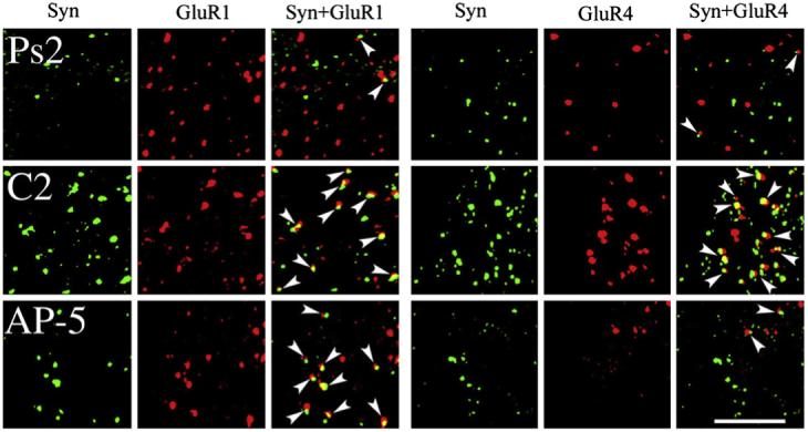
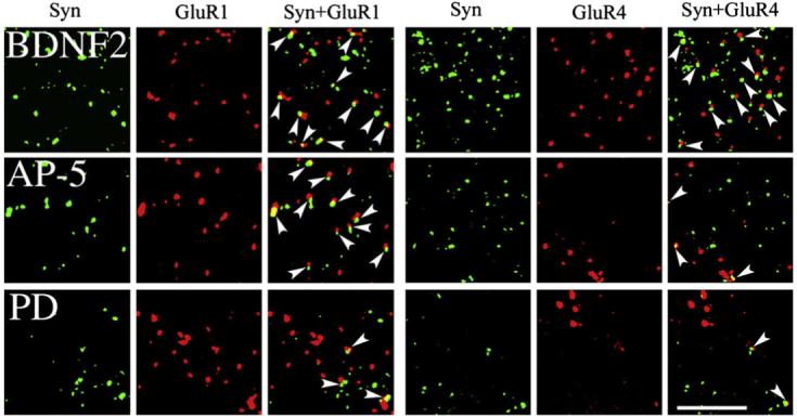
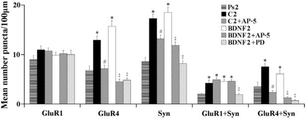
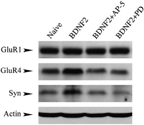
Previous studies using an in vitro model of eyeblink classical conditioning in turtles suggest that increased numbers of synaptic AMPARs supports the acquisition and expression of conditioned responses (CRs). Brain-derived neurotrophic factor (BDNF) and its associated receptor tyrosine kinase, TrkB, is also required for acquisition of CRs. Bath application of BDNF alone induces synaptic delivery of GluR1- and GluR4-containing AMPARs that is blocked by coapplication of the receptor tyrosine kinase inhibitor K252a. The molecular mechanisms involved in BDNF-induced AMPAR trafficking remain largely unknown. The aim of this study was to determine whether BDNF-induced synaptic AMPAR incorporation utilizes similar cellular mechanisms as AMPAR trafficking that occurs during in vitro classical conditioning. Using pharmacological blockade and confocal imaging, the results show that synaptic delivery of GluR1 subunits during conditioning or BDNF application does not require activity of NMDARs but is mediated by extracellular signal-regulated kinase (ERK). In contrast, synaptic delivery of GluR4-containing AMPARs during both conditioning and BDNF application is NMDAR- as well as ERK-dependent. These findings indicate that BDNF application mimics AMPAR trafficking observed during conditioning by activation of some of the same intracellular signaling pathways and suggest that BDNF is a key signal transduction element in postsynaptic events that mediate conditioning.
Figures
Similar articles
-
PKA has a critical role in synaptic delivery of GluR1- and GluR4-containing AMPARs during initial stages of acquisition of in vitro classical conditioning.J Neurophysiol. 2009 May;101(5):2539-49. doi: 10.1152/jn.91282.2008. Epub 2009 Mar 4. J Neurophysiol. 2009. PMID: 19261706 Free PMC article.
-
Coordinate action of pre- and postsynaptic brain-derived neurotrophic factor is required for AMPAR trafficking and acquisition of in vitro classical conditioning.Neuroscience. 2008 Aug 26;155(3):686-97. doi: 10.1016/j.neuroscience.2008.06.043. Epub 2008 Jun 25. Neuroscience. 2008. PMID: 18639615 Free PMC article.
-
Protein kinase C-dependent and independent signaling pathways regulate synaptic GluR1 and GluR4 AMPAR subunits during in vitro classical conditioning.Neuroscience. 2008 Oct 28;156(4):872-84. doi: 10.1016/j.neuroscience.2008.08.042. Epub 2008 Aug 27. Neuroscience. 2008. PMID: 18809472 Free PMC article.
-
Regulation of neuronal PKA signaling through AKAP targeting dynamics.Eur J Cell Biol. 2006 Jul;85(7):627-33. doi: 10.1016/j.ejcb.2006.01.010. Epub 2006 Feb 28. Eur J Cell Biol. 2006. PMID: 16504338 Review.
-
Synaptic Mechanisms of Delay Eyeblink Classical Conditioning: AMPAR Trafficking and Gene Regulation in an In Vitro Model.Mol Neurobiol. 2023 Dec;60(12):7088-7103. doi: 10.1007/s12035-023-03528-z. Epub 2023 Aug 2. Mol Neurobiol. 2023. PMID: 37531025 Review.
Cited by
-
Different roles of BDNF in nucleus accumbens core versus shell during the incubation of cue-induced cocaine craving and its long-term maintenance.J Neurosci. 2013 Jan 16;33(3):1130-42. doi: 10.1523/JNEUROSCI.3082-12.2013. J Neurosci. 2013. PMID: 23325250 Free PMC article.
-
Regulation of AMPA receptor trafficking by secreted protein factors.Front Cell Neurosci. 2023 Nov 27;17:1271169. doi: 10.3389/fncel.2023.1271169. eCollection 2023. Front Cell Neurosci. 2023. PMID: 38089145 Free PMC article. Review.
-
Identification of a functionally distinct truncated BDNF mRNA splice variant and protein in Trachemys scripta elegans.PLoS One. 2013 Jun 25;8(6):e67141. doi: 10.1371/journal.pone.0067141. Print 2013. PLoS One. 2013. PMID: 23825634 Free PMC article.
-
Coincidence detection in a neural correlate of classical conditioning is initiated by bidirectional 3-phosphoinositide-dependent kinase-1 signalling and modulated by adenosine receptors.J Physiol. 2015 Apr 1;593(7):1581-95. doi: 10.1113/jphysiol.2014.282947. Epub 2015 Feb 11. J Physiol. 2015. PMID: 25639253 Free PMC article.
-
Age-related changes in synaptic markers and monocyte subsets link the cognitive decline of APP(Swe)/PS1 mice.Front Cell Neurosci. 2012 Nov 1;6:51. doi: 10.3389/fncel.2012.00051. eCollection 2012. Front Cell Neurosci. 2012. PMID: 23125823 Free PMC article.
References
-
- Anderson CW, Keifer J. Properties of conditioned abducens nerve responses in a highly reduced in vitro brain stem preparation from the turtle. Journal of Neurophysiology. 1999;81:1242–1250. - PubMed
-
- Boehm J, Kang MG, Johnson RC, Esteban J, Huganir RL, Malinow R. Synaptic incorporation of AMPA receptors during LTP is controlled by a PKC phosphorylation site on GluR1. Neuron. 2006;51:213–225. - PubMed
-
- Caldeira MV, Melo CV, Pereira DB, Carvalho R, Correia SS, Backos DS, et al. Brain-derived neurotrophic factor regulates the expression and synaptic delivery of alpha-amino-3-hydroxy-5-methyl-4-isoxazole propionic acid receptor subunits in hippocampal neurons. Journal of Biological Chemistry. 2007;282:12619–12628. - PubMed
-
- Carlezon WA, Jr., Duman RS, Nestler EJ. The many faces of CREB. Trends in Neuroscience. 2005;28:436–445. - PubMed
Publication types
MeSH terms
Substances
Grants and funding
LinkOut - more resources
Full Text Sources
Miscellaneous
