Review
. 2009 Feb;20(1):90-104.
doi: 10.1016/j.semcdb.2008.10.001. Epub 2008 Oct 14.

Stripes and belly-spots -- a review of pigment cell morphogenesis in vertebrates

Affiliations
Review

Stripes and belly-spots -- a review of pigment cell morphogenesis in vertebrates

Robert N Kelsh et al. Semin Cell Dev Biol. 2009 Feb.

Abstract

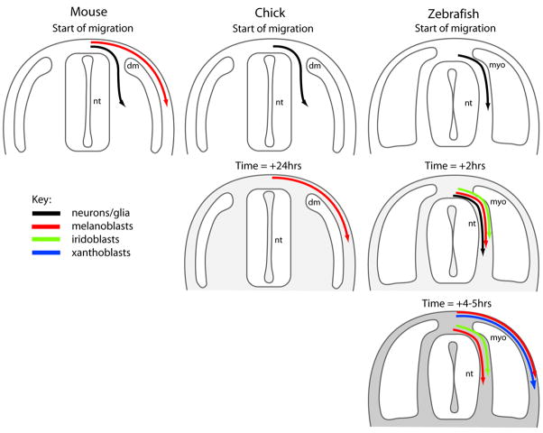
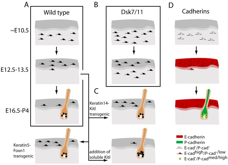
Pigment patterns in the integument have long-attracted attention from both scientists and non-scientists alike since their natural attractiveness combines with their excellence as models for the general problem of pattern formation. Pigment cells are formed from the neural crest and must migrate to reach their final locations. In this review, we focus on our current understanding of mechanisms underlying the control of pigment cell migration and patterning in diverse vertebrates. The model systems discussed here - chick, mouse, and zebrafish - each provide unique insights into the major morphogenetic events driving pigment pattern formation. In birds and mammals, melanoblasts must be specified before they can migrate on the dorsolateral pathway. Transmembrane receptors involved in guiding them onto this route include EphB2 and Ednrb2 in chick, and Kit in mouse. Terminal migration depends, in part, upon extracellular matrix reorganization by ADAMTS20. Invasion of the ectoderm, especially into the feather germ and hair follicles, requires specific signals that are beginning to be characterized. We summarize our current understanding of the mechanisms regulating melanoblast number and organization in the epidermis. We note the apparent differences in pigment pattern formation in poikilothermic vertebrates when compared with birds and mammals. With more pigment cell types, migration pathways are more complex and largely unexplored; nevertheless, a role for Kit signaling in melanophore migration is clear and indicates that at least some patterning mechanisms may be highly conserved. We summarize the multiple factors thought to contribute to zebrafish embryonic pigment pattern formation, highlighting a recent study identifying Sdf1a as one factor crucial for regulation of melanophore positioning. Finally, we discuss the mechanisms generating a second, metamorphic pigment pattern in adult fish, emphasizing recent studies strengthening the evidence that undifferentiated progenitor cells play a major role in generating adult pigment cells.


Figures

Figure 1
Figure 1. Schematic of neural crest cell migration in transverse sections
Significant differences are observed in the timing and pathways of neural crest cell migration when comparing mouse, chick and zebrafish during early embryogenesis (see text for details).
Figure 2
Figure 2. Chromatoblast migration in zebrafish
Transverse sections of trunk of prim-5 (A,B) or prim-15 (C,D) stage wild-type embryos showing mRNA expression for melanoblast (dct, A, B), iridoblast (ltk, C) and xanthoblast (gch, D) markers. Note how iridoblast migration is restricted to the medial pathway, whereas xanthoblasts occupy the lateral pathway and melanoblasts migrate on both pathways (cf mouse and chick). m, muscle; nt, neural tube. Red arrow, medial pathway; green arrow, lateral pathway. Scale bar: 20 μm (A, B); 35 μm (C, D). A, B) reproduced with permission from [134].
Figure 3
Figure 3. Wild-type zebrafish pigment patterns and pigment pattern morphogenesis
Lateral views of wild-type early larval (A) pigment pattern. B–E) Pigment pattern metamorphosis. B) The larval pattern consists of 4 melanophore stripes, but only one, the lateral stripe (LS) is shown in this close-up of the lateral flank (horizontal black bars). C) From 14 dpf, early metamorphic melanophores (small, grey stars) differentiate in the vicinity of the horizontal myoseptum over a period of around 7 days; some larval melanophores (large black stars) emerge onto the flank from the early larval stripes (e.g. LS) and may persist. D) Melanophores aggregate dorsal and ventral to the myoseptum to form the first two stripes, while xanthophores (yellow stars) form adjacent to the horizontal myoseptum. Melanophores in the interstripe region, including those from the larval pattern’s lateral stripe in the myoseptum itself, either migrate into the nascent stripes, or die in situ (faint grey). E) Late metamorphic phase of melanophore and xanthophore production increases the pigment cell density within the stripes and interstripes; a second interstripe of xanthophores begins to form ventral to the primary melanophore stripes. Anterior to the left, dorsal up. Stages not drawn to scale. Scale bar in A): 0.5 mm. C–F) adapted from [21, 114].
Figure 4
Figure 4. Regulation of melanoblast patterning in mouse skin
Schematic of embryonic skin in wild type (A), transgenic or mutant (B, C), and cadherin-immunolabeled (C) embryos. A) Melanoblasts from the dermis (light gray) sequentially invade the epidermis (dark gray) and then the hair follicle (brown). B) In Dsk or Gnaq/11 mutants melanoblasts are produced in elevated numbers during early melanogenesis which results in ectopic pigmentation of the dermis. These results provide a basis for the regulation of cell number in maintaining normal patterning. C) Transgenic mice expressing Kitl or Foxn1 under keratin promotors results in the retention of melanocytes in the epidermis. This suggests that both play a role in the maintenance or localization of melanocytes to this tissue. Exogenous Kitl applied to skin explants causes a chemokinetic increase of melanocytes to the hair follicle. D) Cadherin expression by melanoblasts correlates with cadherin expression patterns seen in the skin and suggests an extrinsic regulation of melanoblast localization that is coincident with the development of the skin.
Figure 5
Figure 5. Pigment pattern mutants in zebrafish
A) choker mutants at 5 dpf show ectopic melanophores in the anterior trunk region (left panels), and lack the lateral stripe melanophores of the body (right). B) Adult pigment pattern phenotypes of wild-type and various homozygous mutant strains. See text for further details. (Adapted, with permission, from Kelsh and Parichy 2007).

References

    1. Le Douarin NM, Kalcheim C. The Neural Crest. 2. Cambridge: Cambridge University Press; 1999.
    1. Fox MH. Analysis of some phases of melanoblast migration in barred plymouth rock embryos. Physiol Zool. 1949;22:1–22. - PubMed
    1. Ris H. An experimental study of the origin of melanophores in birds. Physiol Zool. 1941;14:48–66.
    1. Watterson RL. The morphogenesis of down feathers with special reference to the developmental history of melanophores. Physiol Zool. 1942;15:234–259.
    1. Willier BH, Rawles ME. The control of feather color pattern by melanophores grafted from one embryo to another of a different breed of fowl. Physiol Zool. 1940;13:177–202.

Publication types

LinkOut - more resources