. 2008 Nov 4;14(5):408-19.
doi: 10.1016/j.ccr.2008.10.011.

Overexpression of interleukin-1beta induces gastric inflammation and cancer and mobilizes myeloid-derived suppressor cells in mice

Affiliations

Overexpression of interleukin-1beta induces gastric inflammation and cancer and mobilizes myeloid-derived suppressor cells in mice

Shuiping Tu et al. Cancer Cell. .

Erratum in

  • Cancer Cell. 2008 Dec 9;14(6):494
  • Cancer Cell. 2011 Jan 18;19(1):154

Abstract

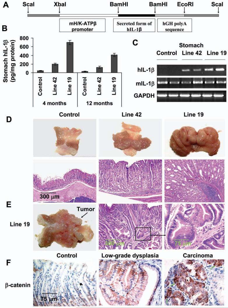
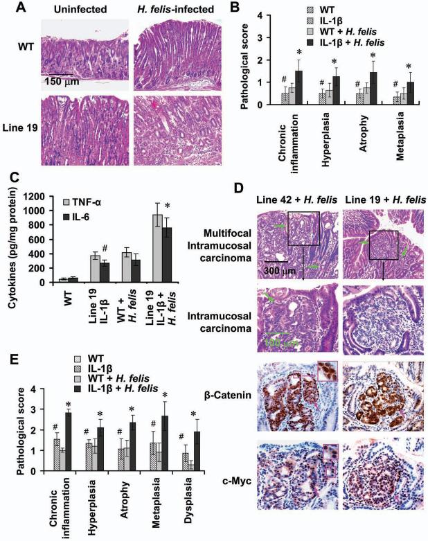
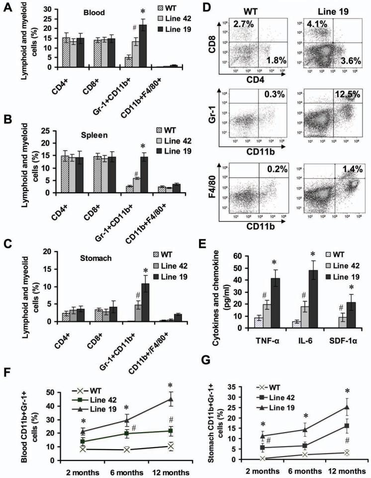
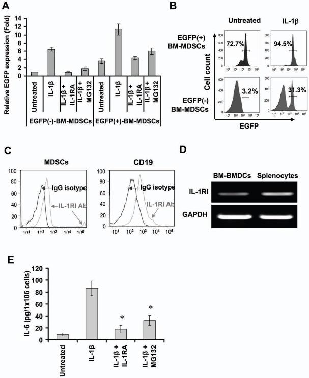
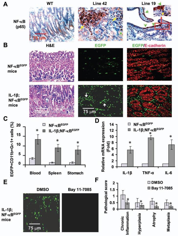
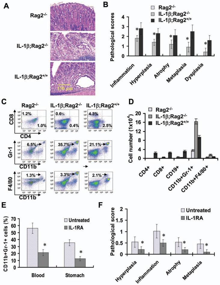
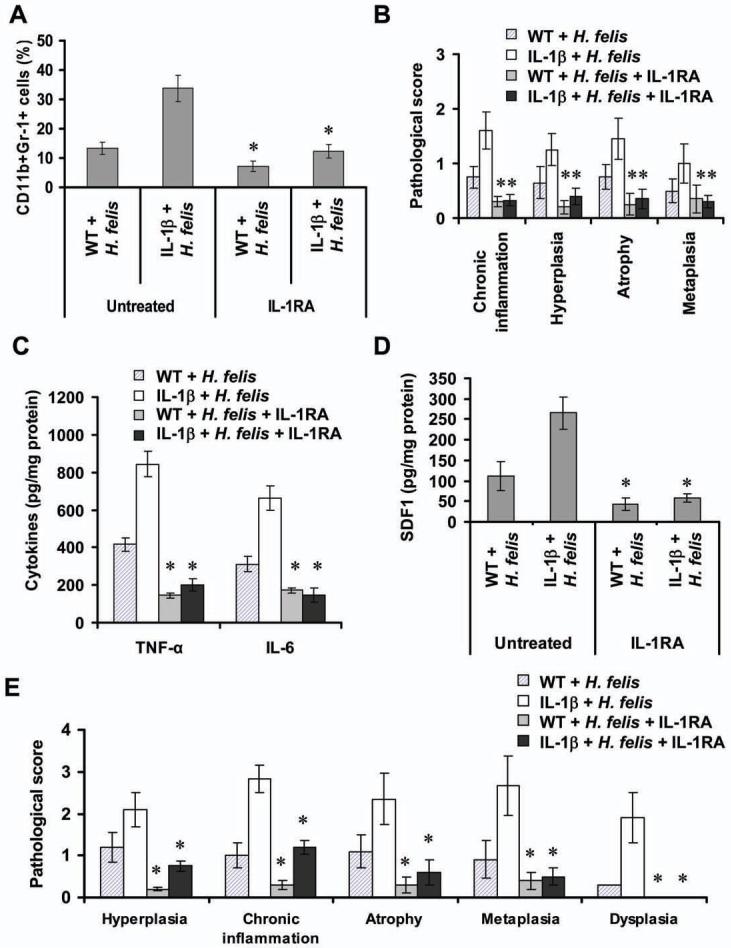
Polymorphisms of interleukin-1beta (IL-1beta) are associated with an increased risk of solid malignancies. Here, we show that stomach-specific expression of human IL-1beta in transgenic mice leads to spontaneous gastric inflammation and cancer that correlate with early recruitment of myeloid-derived suppressor cells (MDSCs) to the stomach. IL-1beta activates MDSCs in vitro and in vivo through an IL-1RI/NF-kappaB pathway. IL-1beta transgenic mice deficient in T and B lymphocytes develop gastric dysplasia accompanied by a marked increase in MDSCs in the stomach. Antagonism of IL-1 receptor signaling inhibits the development of gastric preneoplasia and suppresses MDSC mobilization. These results demonstrate that pathologic elevation of a single proinflammatory cytokine may be sufficient to induce neoplasia and provide a direct link between IL-1beta, MDSCs, and carcinogenesis.


Figures

Figure 1
Figure 1. IL-1β transgenic mice develop gastric inflammation and dysplasia/carcinoma
(A) The construct pBS/HKATPase/ß globin/IL-1β, which contains the mouse H+/K+-ATPase ß subunit gene and secreted form of hIL-1β cDNA, was microinjected into fertilized mouse oocytes. (B) Expression of hIL-1β in the stomachs of 4 and 12 month old IL-1β transgenic mice and control mice was determined using a human-specific IL-1β ELISA kit. Data are the mean ± SD of 10 mice. (C) Expression of human (transgenic) and mouse (endogenous) IL-1β mRNA in stomach as assessed by RT-PCR. (D) IL-1β transgenic mice develop gastric hyperplasia (upper lane) and gastritis (H&E staining, lower lane). (E) Male Line 19 IL-1β transgenic mice develop stomach cancer. (F) Activation and relocalization of β-catenin in gastric cancer of IL-1β transgenic mice. The stomach sections were stained with an anti-β-catenin antibody. Arrows show nuclear β-catenin+ cells (brown).
Figure 2
Figure 2. Overexpression of IL-1β accelerates the development of gastric inflammation and carcinoma in the setting of H. felis infection
(A) Stomach sections from Line 19 IL-1β transgenic and control mice infected with H. felis for 5 months were stained with H&E. (B) Pathological scores from the stomach of above mice were graded according to the diagnostic criterion described in Methods. The data represent the mean ± SD of 16 mice (#p < 0.05, vs WT mice; *p < 0.01, vs uninfected WT mice). (C) Expression of mouse TNF-α and IL-6 in stomach tissue from mice was determined by ELISA. The data represent the mean ± SD of 6 mice (#p < 0.05, vs WT mice; *p < 0.01, vs uninfected WT mice). (D) IL-1β transgenic mice infected with H. felis for 12 months developed dysplasia and carcinoma with activation of β-catenin. The sections from intramucosal/invasive gastric cancer were stained with anti-β-catenin and anti-c-Myc antibodies. Green arrows indicate invasive gastric cancer and pink arrows show nuclear β-catenin- or c-Myc-positive cells (brown), respectively. (E) Gastric inflammation and pathology scores were graded in Line 19 IL-1β transgenic and control mice infected with H. felis for 12 months. The data represent the mean ± SD of 10 animals (#p < 0.05, vs control mice; *p < 0.01, vs uninfected WT mice).
Figure 3
Figure 3. Overexpression of IL-1β in the stomach leads to mobilization and recruitment of MDSCs
The frequencies of lymphoid and myeloid cells in peripheral blood (A), spleen (B) and stomach (C) from 2 month old IL-1β mice and age-matched WT mice were measured by FACS. The data are the mean ± SD of 6 mice (#p < 0.05, p < 0.01, vs WT mice). (D) Representative FACS blots for detecting lymphoid and myeloid cells in the stomach from WT and Line 19 IL-1β mice. (E) Expression of mouse TNF-α, IL-6 and SDF-1α in the gastric tissue from 2 month old mice was determined by ELISA. The data represent the mean ± SD of 6 animals (*p < 0.01, vs WT mice). (F-G) The kinetics of MDSCs was determined by FACS in peripheral blood (F) and stomach (G) from IL-1β mice and WT mice at different time points. The data represent the mean ± SD of 6 animals (#p < 0.05; *p < 0.01, vs WT mice).
Figure 4
Figure 4. IL-1β activates MDSCs through an NF-κB signal pathway
(A) IL-1β upregulates EGFP mRNA expression by activates NF-κB in MDSCs. EGFP+ and EGFP- MDSCs from NF-κBEGFP mice were treated with IL-1β in absence or presence of 50 ng/ml IL-1RA or 1 μM MG132 for 3 hours. mRNA expression was determined by real time-PCR. The data are normalized to untreated EGFP- MDSCs and represent the mean ± SD of four independent experiments. (B) IL-1β upregulates expression of EGFP protein in MDSCs. EGFP+ and EGFP-MDSCs were treated with IL-1β for 24 hours. The EGFP fluorescence intensities were detected by FACS. (C) Expression of IL-1RI protein in MDSCs was determined by FACS using PE-IL-1RI antibody. (D) IL-1RI mRNA expression in MDSCs measured by RT-PCR. (E) Blocking IL-1β /NF-κB signal pathway inhibits IL-1β-stimulates secretion of IL-6 in MDSCs. EGFP+ MDSCs were treated with IL-1β in absence or presence of IL-1RA or MG132 for 36 hours. The level of IL-6 were measured by ELISA. The data represent the mean ± SD of four independent experiments (*p < 0.01, vs IL-1β treated group).
Figure 5
Figure 5. Overexpression of IL-1β activates NF-κB in MDSCs in vivo
(A) NF-κB activation in the stomachs of IL-1β mice. Sections were stained a NF-κB p65 antibody. Arrows indicate p65+ cells. (B) Enhanced EGFP expression in IL-1β;NF-κBEGFP mice. Frozen gastric sections from 6 month old indicated mice were subjected to H&E staining and double staining with anti-EGFP (green) and E-cadherin (red) antibodies. The red staining indicates epithelial cells. Localization of EGFP+ cells is mostly confined to stromal region. (C) Increased frequencies of EGFP+ MDSCs in peripheral blood, spleen and stomach tissues in IL-1β;NF-κBEGFP mice as analyzed by FACS. The data represent the mean ± SD of 6 animals (*p < 0.05, vs NF-κBEGFP mice). (D) Increased expression of cytokines in stomach MDSCs in IL-1β;NF-κBEGFP mice. Stomach MDSCs sorted from 6 month old NF-κBEGFP and IL-1β;NF-κBEGFP mice were restimulated with PMA for 4 hours. mRNA expression was determined by real-time PCR. The data are normalized to MDSCs of NF-κBEGFP mice and represent the means ± SD of three independent experiments (*p < 0.01, vs NF-κBEGFP mice). (E) Blocking NF-κB activity by injecting i.p. Bay 11-7085 prevents EGFP expression in 3 month old IL-1β;NF-κBEGFP mice. Representative photos were taken from frozen gastric sections under fluorescence microscope. (F) Blocking NF-κB activity inhibits the development of gastritis. The pathological scores were graded in Bay 11-7085 or DMSO-treated IL-1β;NF-κBEGFP mice (*p < 0.05, vs DMSO treated mice, n = 8).
Figure 6
Figure 6. MDSCs are implicated in IL-1β-induced chronic gastritis and dysplasia
(A) IL-1β;Rag2-/- mice develop spontaneous gastritis and dysplasia (H&E staining). (B) The pathological scores were graded in old Rag2-/-, IL-1β;Rag2-/- and IL-1β;Rag2+/+ mice (>12 months). The data represent the mean ± SD of 10 animals (*p <0.01, vs Rag2-/- mice). (C-D) Increased the number of myeloid cells in the stomach of 6 month old IL-1 β;Rag2-/- mice. Representative FACS blots (C) and the data shown are the mean ± SD of cell number per stomach derived from 6 animals (D). (E) IL-1RA treatment inhibits the mobilization and recruitment of MDSCs in IL-1β;Rag2-/- mice. Three month old IL-1β;Rag2-/- mice were treated with IL-1RA for 6 weeks. Single nucleated cells isolated were stained with APC-CD11b and PerCP-Gr-1 antibodies and analyzed by FACS. (F) IL-1RA treatment inhibits the development of gastritis in IL-1β;Rag2-/- mice. The pathological scores were graded in above indicated mice The data represent the mean ± SD of 6 animals (*p < 0.05, vs untreated mice).
Figure 7
Figure 7. IL-1RA treatment improves gastric pathology and blocks MDSC recruitment in H. felis infected IL-1β mice
(A) IL-1RA treatment inhibits the mobilization of MDSCs. Single nucleated cells in peripheral blood were isolated from Line 19 IL-1β transgenic and control mice infected with H. felis for 5 months with or without preventive administration of IL-1RA. Cells were stained with fluorescence-labeled CD11b and Gr-1 antibodies and analyzed by FACS (*p < 0.05, vs untreated mice, n = 6). (B) IL-1RA treatment inhibits the development of gastric inflammation and preneoplasia in H. felis-infected mice. Histopathological scores assessed in mice (*p < 0.05, vs untreated mice, n = 6). Gastric expression of mouse TNF-α and IL-6 (C) and SDF-1α (D) in indicated mice were determined by ELISA. (E) IL-1RA treatment inhibits the development of gastric dysplasia in H. felis-infected IL-1β mice. Stomach sections were from Line 19 IL-1β transgenic and WT mice infected with H. felis for 12 months with or without IL-1RA treatment for 3 months. The histopathologic scores were graded (*p < 0.01, vs untreated mice, n = 10). Error bar in (A)-(E) indicate ± SD.

References

    1. Ahn GO, Brown JM. Matrix metalloproteinase-9 is required for tumor vasculogenesis but not for angiogenesis: role of bone marrow-derived myelomonocytic cells. Cancer Cell. 2008;13:193–205. - PMC - PubMed
    1. Almand B, Clark JI, Nikitina E, van Beynen J, English NR, Knight SC, Carbone DP, Gabrilovich DI. Increased production of immature myeloid cells in cancer patients: a mechanism of immunosuppression in cancer. J Immunol. 2001;166:678–689. - PubMed
    1. Arend WP. The balance between IL-1 and IL-1Ra in disease. Cytokine Growth Factor Rev. 2002;13:323–340. - PubMed
    1. Balkwill F. TNF-alpha in promotion and progression of cancer. Cancer Metastasis Rev. 2006;25:409–416. - PubMed
    1. Barber MD, Powell JJ, Lynch SF, Fearon KC, Ross JA. A polymorphism of the interleukin-1 beta gene influences survival in pancreatic cancer. Br J Cancer. 2000;83:1443–1447. - PMC - PubMed

Publication types

MeSH terms