

Neural mechanisms for illusory filling-in of degraded speech
- PMID: 18977448
- PMCID: PMC2653101
- DOI: 10.1016/j.neuroimage.2008.09.045
Neural mechanisms for illusory filling-in of degraded speech
Abstract
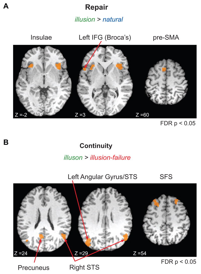
The brain uses context and prior knowledge to repair degraded sensory inputs and improve perception. For example, listeners hear speech continuing uninterrupted through brief noises, even if the speech signal is artificially removed from the noisy epochs. In a functional MRI study, we show that this temporal filling-in process is based on two dissociable neural mechanisms: the subjective experience of illusory continuity, and the sensory repair mechanisms that support it. Areas mediating illusory continuity include the left posterior angular gyrus (AG) and superior temporal sulcus (STS) and the right STS. Unconscious sensory repair occurs in Broca's area, bilateral anterior insula, and pre-supplementary motor area. The left AG/STS and all the repair regions show evidence for word-level template matching and communicate more when fewer acoustic cues are available. These results support a two-path process where the brain creates coherent perceptual objects by applying prior knowledge and filling-in corrupted sensory information.
Figures


Similar articles
-
Contributions of sensory input, auditory search and verbal comprehension to cortical activity during speech processing.Cereb Cortex. 2004 Mar;14(3):247-55. doi: 10.1093/cercor/bhg124. Cereb Cortex. 2004. PMID: 14754865 Clinical Trial.
-
The Motor Network Reduces Multisensory Illusory Perception.J Neurosci. 2018 Nov 7;38(45):9679-9688. doi: 10.1523/JNEUROSCI.3650-17.2018. Epub 2018 Sep 24. J Neurosci. 2018. PMID: 30249803 Free PMC article.
-
Neural Prediction Errors Distinguish Perception and Misperception of Speech.J Neurosci. 2018 Jul 4;38(27):6076-6089. doi: 10.1523/JNEUROSCI.3258-17.2018. Epub 2018 Jun 11. J Neurosci. 2018. PMID: 29891730 Free PMC article.
-
[Auditory perception and language: functional imaging of speech sensitive auditory cortex].Rev Neurol (Paris). 2001 Sep;157(8-9 Pt 1):837-46. Rev Neurol (Paris). 2001. PMID: 11677406 Review. French.
-
Stimulus-dependent activations and attention-related modulations in the auditory cortex: a meta-analysis of fMRI studies.Hear Res. 2014 Jan;307:29-41. doi: 10.1016/j.heares.2013.08.001. Epub 2013 Aug 11. Hear Res. 2014. PMID: 23938208 Review.
Cited by
-
The Connectivity Fingerprints of Highly-Skilled and Disordered Reading Persist Across Cognitive Domains.Front Comput Neurosci. 2021 Feb 12;15:590093. doi: 10.3389/fncom.2021.590093. eCollection 2021. Front Comput Neurosci. 2021. PMID: 33643016 Free PMC article.
-
Plasticity in auditory categorization is supported by differential engagement of the auditory-linguistic network.Neuroimage. 2019 Nov 1;201:116022. doi: 10.1016/j.neuroimage.2019.116022. Epub 2019 Jul 13. Neuroimage. 2019. PMID: 31310863 Free PMC article.
-
Left temporal alpha-band activity reflects single word intelligibility.Front Syst Neurosci. 2013 Dec 27;7:121. doi: 10.3389/fnsys.2013.00121. eCollection 2013. Front Syst Neurosci. 2013. PMID: 24416001 Free PMC article.
-
A roadmap for the study of conscious audition and its neural basis.Philos Trans R Soc Lond B Biol Sci. 2017 Feb 19;372(1714):20160103. doi: 10.1098/rstb.2016.0103. Epub 2017 Jan 2. Philos Trans R Soc Lond B Biol Sci. 2017. PMID: 28044014 Free PMC article. Review.
-
Retained capacity for perceptual learning of degraded speech in primary progressive aphasia and Alzheimer's disease.Alzheimers Res Ther. 2018 Jul 25;10(1):70. doi: 10.1186/s13195-018-0399-2. Alzheimers Res Ther. 2018. PMID: 30045755 Free PMC article.
References
-
- Bamiou DE, Musiek FE, Luxon LM. The insula (Island of Reil) and its role in auditory processing. Literature review. Brain Res Brain Res Rev. 2003;42:143–154. - PubMed
-
- Bashford JA, Jr, Meyers MD, Brubaker BS, Warren RM. Illusory continuity of interrupted speech: speech rate determines durational limits. J Acoust Soc Am. 1988;84:1635–1638. - PubMed
-
- Bashford JA, Jr, Warren RM, Brown CA. Use of speech-modulated noise adds strong “bottom-up” cues for phonemic restoration. Percept Psychophys. 1996;58:342–350. - PubMed
-
- Binder JR, Liebenthal E, Possing ET, Medler DA, Ward BD. Neural correlates of sensory and decision processes in auditory object identification. Nat Neurosci. 2004;7:295–301. - PubMed
-
- Binder JR, McKiernan KA, Parsons ME, Westbury CF, Possing ET, Kaufman JN, Buchanan L. Neural correlates of lexical access during visual word recognition. J Cogn Neurosci. 2003;15:372–393. - PubMed
Publication types
MeSH terms
Grants and funding
LinkOut - more resources
Full Text Sources
Other Literature Sources
Medical
