. 2009 Jan 2;284(1):404-413.
doi: 10.1074/jbc.M807027200. Epub 2008 Oct 31.

Transcriptional activation in yeast in response to copper deficiency involves copper-zinc superoxide dismutase

Affiliations

Transcriptional activation in yeast in response to copper deficiency involves copper-zinc superoxide dismutase

L Kent Wood et al. J Biol Chem. .

Abstract

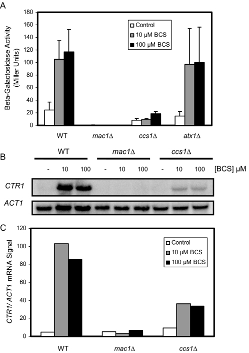
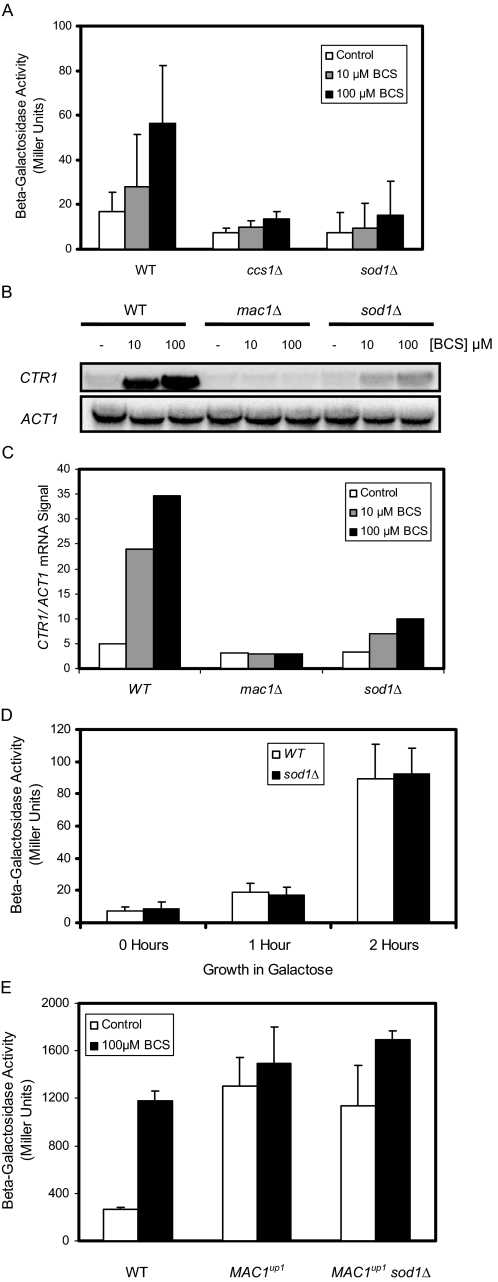
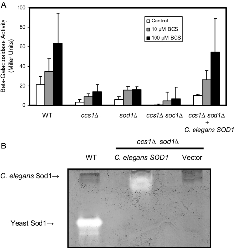
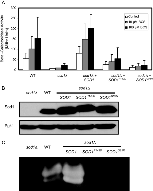
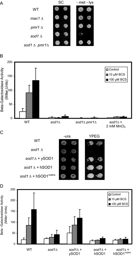
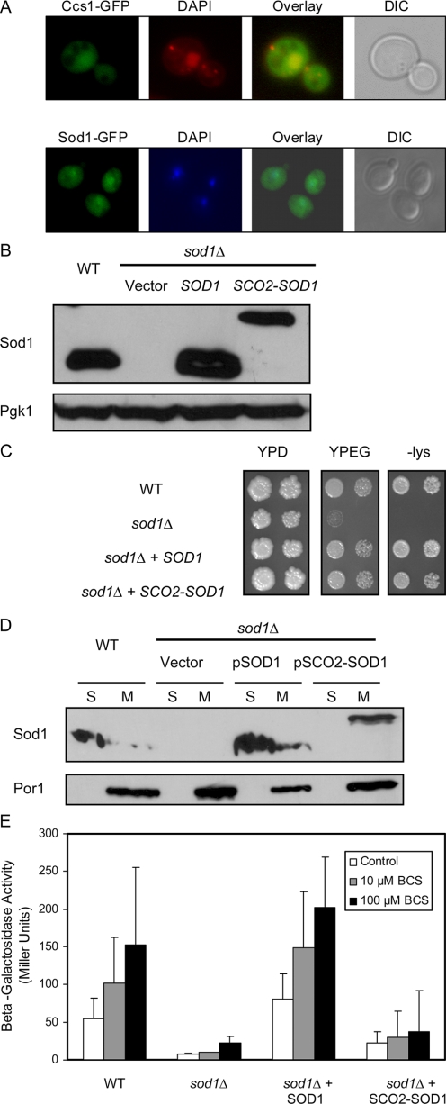
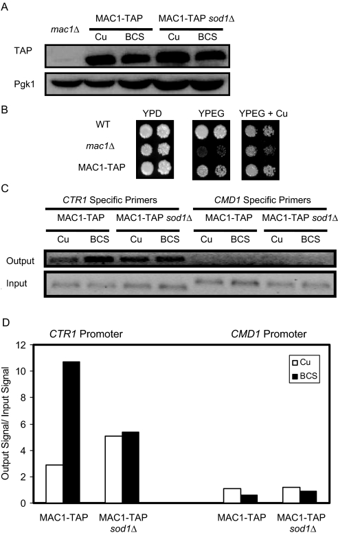
Copper is an essential trace element, yet excess copper can lead to membrane damage, protein oxidation, and DNA cleavage. To balance the need for copper with the necessity to prevent accumulation to toxic levels, cells have evolved sophisticated mechanisms to regulate copper acquisition, distribution, and storage. In Saccharomyces cerevisiae, transcriptional responses to copper deficiency are mediated by the copper-responsive transcription factor Mac1. Although Mac1 activates the transcription of genes involved in high affinity copper uptake during periods of deficiency, little is known about the mechanisms by which Mac1 senses or responds to reduced copper availability. Here we show that the copper-dependent enzyme Sod1 (Cu,Zn-superoxide dismutase) and its intracellular copper chaperone Ccs1 function in the activation of Mac1 in response to an external copper deficiency. Genetic ablation of either CCS1 or SOD1 results in a severe defect in the ability of yeast cells to activate the transcription of Mac1 target genes. The catalytic activity of Sod1 is essential for Mac1 activation and promotes a regulated increase in binding of Mac1 to copper response elements in the promoter regions of genomic Mac1 target genes. Although there is precedent for additional roles of Sod1 beyond protection of the cell from oxygen radicals, the involvement of this protein in copper-responsive transcriptional regulation has not previously been observed. Given the presence of both Sod1 and copper-responsive transcription factors in higher eukaryotes, these studies may yield important insights into how copper deficiency is sensed and appropriate cellular responses are coordinated.


Figures

FIGURE 1.
FIGURE 1.
The Ccs1 copper chaperone is required for robust activation of Mac1. A, ccs1Δ cells show defects in the induction of a Mac1 reporter plasmid upon copper depletion. WT, mac1Δ, and ccs1Δ cells transformed with the pCm64CTR3-LacZ reporter plasmid were grown to mid-log phase in synthetic complete media with no supplementation (Control) or media treated with either 10 or 100 μm copper chelator BCS. β-Galactosidase assays were performed and analyzed in triplicate. The data are representative of at least three independent experiments. B, the induction of Mac1 target mRNA upon copper depletion is decreased in ccs1Δ cells. RNA blotting analysis for the CTR1 transcript was carried out from cells grown under control conditions (-) or in the presence of the indicated concentration of the copper-specific chelator BCS. The ACT1 transcript is shown as an RNA loading control. C, quantification of mRNA blots from B.
FIGURE 2.
FIGURE 2.
Cu,Zn-superoxide dismutase functions in the activation of Mac1. A, sod1Δ cells exhibit defects in the induction of a Mac1 reporter plasmid upon copper depletion. WT, ccs1Δ, and sod1Δ cells transformed with the CTR3-LacZ reporter plasmid were grown to mid-log phase in complete media or media with 10 μm or 100 μm BCS and β-galactosidase assays were performed. ccs1Δ cells and sod1Δ display similar defects in the ability to activate the Mac1 reporter in response to limiting copper. Samples were analyzed in triplicate and data are representative of at least three independent experiments. B, the induction of Mac1 target mRNA upon copper depletion is decreased in sod1Δ cells. RNA blotting analysis for the CTR1 transcript also indicates that sod1Δ mutants show decreased activation of Mac1 in response to copper deprivation. C, quantification of mRNA blots from B. D, sod1Δ cells WT and sod1Δ cells transformed with a galactose-inducible reporter plasmid were grown for 0, 1, or 2 h in media containing galactose as the sole carbon source. β-Galactosidase activity assays demonstrate that β-galactosidase is transcribed/translated at similar levels to WT in sod1Δ mutants. E, WT, MAC1up1, and isogenic MAC1up1 sod1Δ cells transformed with the CTR3-LacZ reporter plasmid were grown to mid-log phase in synthetic complete media supplemented with 1 μm CuSO4 or 100 μm BCS. MAC1up1 and MAC1up1 sod1Δ cells show similar levels of Mac1 reporter activity which is higher than WT control cells.
FIGURE 3.
FIGURE 3.
C. elegans SOD1 rescues Mac1 activation in a Ccs1-independent manner. A, the SOD1 gene from C. elegans (C. elegans SOD1) rescues the activation of Mac1 protein in ccs1Δ sod1Δ mutants. WT, ccs1Δ, sod1Δ, and ccs1Δ sod1Δ cells co-transformed with a CTR3-LacZ reporter plasmid and an empty pRS415 vector. ccs1Δ sod1Δ double mutants were also co-transformed with a vector carrying the SOD1 gene from C. elegans (C. elegans SOD1). Growth to mid-log phase in complete media or media with 10 or 100 μm BCS and subsequentβ-galactosidase assays indicates the C. elegans SOD1 rescues the activation of Mac1 in a CCS1-independent manner. B, consistent with previous reports that copper is inserted into the C. elegans Sod1 protein in a Ccs1-independent manner, superoxide dismutase in-gel activity assays demonstrate that the C. elegans Sod1 protein has catalytic activity in the absence of Ccs1, and the ccs1Δ sod1Δ double mutant shows no detectable superoxide dismutase activity.
FIGURE 4.
FIGURE 4.
Activation of Mac1 requires catalytically-active Sod1. A, catalytic activity of Sod1 is necessary for robust activation of Mac1. WT cells were co-transformed with a CTR3-LacZ reporter and an empty vector, and sod1Δ cells were co-transformed with CTR3-LacZ reporter and an empty vector, a plasmid encoding the yeast SOD1 gene (SOD1), or a plasmid encoding the yeast SOD1 gene with either a mutation that disrupts the proton channel of the Sod1 enzyme (SOD1R143D) or a mutation corresponding to a human mutation identified in familial amytrophic lateral sclerosis (SOD1G85R). Cells were grown to mid-log phase in complete media or media with 10 or 100 μm BCS, and β-galactosidase assays were performed. Although the wild type yeast Sod1 can rescue Mac1 activation in sod1Δ cells, neither of the catalytically inactive Sod1 mutants' proteins are able to restore Mac1 reporter activity. B, immunoblot analysis using an antibody against yeast Sod1 (Sod1) demonstrates that catalytically inactive Sod1 proteins are expressed at levels equivalent to wild type Sod1. Immunoblot analysis of 3-phosphoglycerate kinase (Pgk1) shows that loading of each protein sample is equivalent. C, the Sod1R143D and the Sod1G85R proteins are catalytically inactive as determined using superoxide dismutase in-gel activity assays. Cells transformed with either an empty vector or the mutant SOD1R143D gene or the SOD1G85R gene exhibit no detectable superoxide dismutase activity, whereas WT cells and sod1Δ expressing the WT SOD1 gene show considerable superoxide dismutase activity.
FIGURE 5.
FIGURE 5.
Cytosolic reduction in oxidative stress does not rescue Mac1 activation. A, deletion of PMR1 is able to rescue the sensitivity to oxygen seen in sod1Δ cells. Serial dilutions of WT, mac1Δ, pmr1Δ, sod1Δ, and sod1Δ pmr1Δ cells were plated on synthetic complete (SC) media or on SC -methionine–lysine media. Deletion of PMR1 is able to rescue the growth defect of sod1Δ cells on SC media without methionine or lysine. B, neither the deletion of PMR1 nor the addition of exogenous MnCl2, both of which rescue the oxidative stress phenotypes associated with SOD1 deletion, is able to complement the defect in Mac1 activation seen in sod1Δ mutants. C, yeast or human Sod1 rescues the oxidative stress phenotype associated with SOD1 deletion. Serial dilutions spotted onto plates containing ethanol and glycerol (YPEG) indicated that sod1Δ cells expressing either the yeast SOD1 gene or the wild type or the mitochondrial matrix tethered human SOD1 gene are able to grow on the nonfermentable carbon sources. D, the expression of the yeast, but not the human, SOD1 gene is able to restore Mac1 activity in sod1Δ cells. Although the transformation of sod1Δ cells with a vector encoding yeast SOD1 gene restores activity of a Mac1 reporter to wild type levels, transformation with either an empty vector or wild type or the mitochondrial matrix-tethered human SOD1 does not restore the ability of sod1Δ cell to activate the reporter.
FIGURE 6.
FIGURE 6.
Ccs1 and Sod1 proteins partially localize to the yeast nucleus. A, after staining with the DNA dye 4′,6-diamidino-2-phenylindole (DAPI), CCS1-GFP and SOD1-GFP cells were examined by fluorescence microscopy to visualize the whole cell (differential interference contrast; DIC), the nucleus (DAPI), or either the Ccs1-GFP or the Sod1-GFP fusion protein. Nuclear localization of Ccs1-GFP and Sod1-GFP is evident from the overlay image of the 4′,6-diamidino-2-phenylindole and GFP images. B, immunoblot analysis using an antibody against yeast Sod1 (Sod1) demonstrates that Triton-solubilized whole cell protein extracts from WT cells and sod1Δ cells expressing either the SOD1 gene or the SCO2-SOD1 gene from a low copy centromeric plasmid contain similar amounts of Sod1 protein. Immunoblot analysis of 3-phosphoglycerate kinase (Pgk1) shows that loading of each protein sample is equivalent. C, serial dilution on plates containing ethanol and glycerol (YPEG) or plates lacking lysine (-lys) indicates that sod1Δ cells expressing either the yeast SOD1 gene (SOD1) or the mitochondrial tethered yeast SOD1 (SCO2-SOD1) gene are able to grow on the nonfermentable carbon sources or in the absence of lysine. D, yeast cells were fractionated to separate the mitochondria (M) from the soluble protein extract (S), and proteins were solubilized with 2% Triton. Immunoblot analysis using an antibody against yeast Sod1 (Sod1) demonstrates that a significant portion of Sod1 protein from either WT cells or sod1Δ cells expressing wild type Sod1 is found in the soluble fraction, whereas some Sod1 protein is also localized to the mitochondria. In yeast expressing Sco2-Sod1, the Sod1 fusion protein is localized exclusively to the mitochondria. Immunoblot analysis of the mitochondrial membrane protein Porin (Por1) demonstrates that fractionation results in distinct separation of the soluble and mitochondrial portions of the cell. E, WT cells co-transformed with a CTR3-LacZ reporter and an empty vector and sod1Δ cells co-transformed with CTR3-LacZ reporter and an empty vector, a plasmid encoding the yeast SOD1 gene (SOD1), or a plasmid encoding the tethered yeast SCO2-SOD1 fusion gene (SCO2-SOD1) were grown to mid-log phase in complete media or media with 10 μm or 100 μm BCS, and β-galactosidase assays were performed.
FIGURE 7.
FIGURE 7.
Sod1 is required for copper deficiency-induced Mac1 DNA binding. A, immunoblot analysis with an antibody against the TAP tag (TAP) indicates that either WT or sod1Δ cells with a genomic TAP-tag of the MAC1 gene (MAC1-TAP) express similar levels of the Mac1 protein. Expression of 3-phosphoglycerate kinase (Pgk1) was used as a loading control. B, serial dilutions of WT, mac1Δ, and MAC1-TAP demonstrate that WT and MAC1-TAP cells can grow on nonfermentable carbon sources (YPEG), whereas mac1Δ cells show a severe growth defect on YPEG. C, enrichment of the CTR1 promoter DNA by ChIP PCR is evident after treatment of MAC1-TAP with 500 μm BCS for 15 min. Isogenic MAC1-TAP sod1Δ cells do not demonstrate significant enrichment of the CTR1 promoter. There is no apparent binding of Mac1 at the calmodulin (CMD1) promoter. D, quantification of ChIP results from C.

Similar articles

Cited by

References

    1. Prohaska, J. R., and Gybina, A. A. (2004) J. Nutr. 134 1003-1006 - PubMed
    1. Rees, E. M., and Thiele, D. J. (2004) Curr. Opin. Microbiol. 7 175-184 - PubMed
    1. Halliwell, B., and Gutteridge, J. M. (1984) Biochem. J. 219 1-14 - PMC - PubMed
    1. Balamurugan, K., and Schaffner, W. (2006) Biochim. Biophys. Acta 1763 737-746 - PubMed
    1. Rutherford, J. C., and Bird, A. J. (2004) Eukaryot. Cell 3 1-13 - PMC - PubMed

Publication types

MeSH terms