. 2008 Dec;36(21):6934-43.
doi: 10.1093/nar/gkn824. Epub 2008 Oct 31.

Coordination of two sequential ester-transfer reactions: exogenous guanosine binding promotes the subsequent omegaG binding to a group I intron

Affiliations

Coordination of two sequential ester-transfer reactions: exogenous guanosine binding promotes the subsequent omegaG binding to a group I intron

Penghui Bao et al. Nucleic Acids Res. 2008 Dec.

Abstract

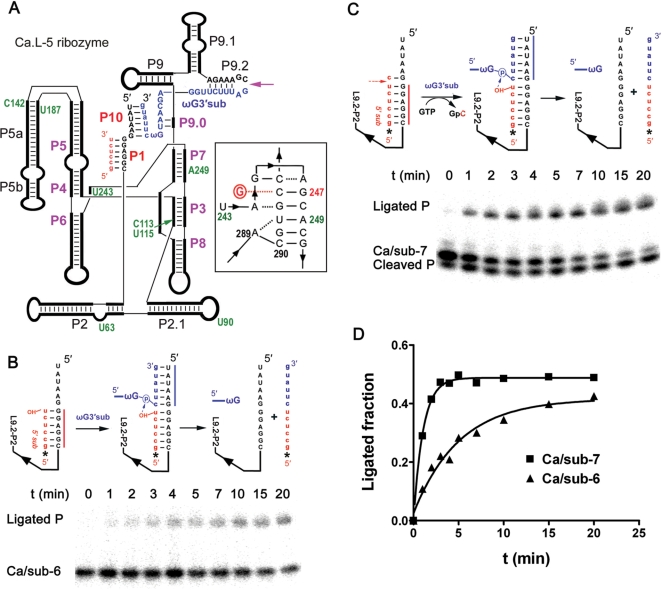
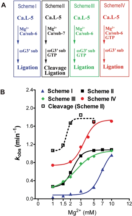
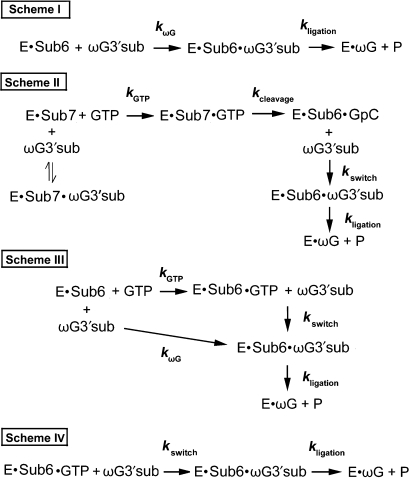
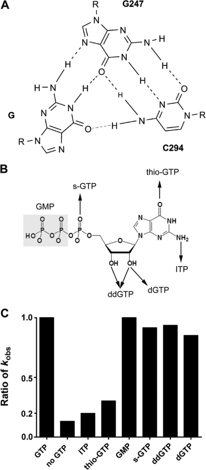
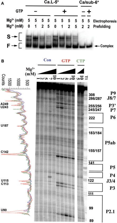
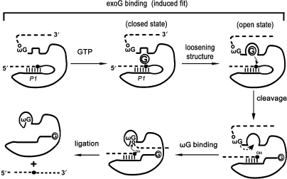
Self-splicing of group I introns is accomplished by two sequential ester-transfer reactions mediated by sequential binding of two different guanosine ligands, but it is yet unclear how the binding is coordinated at a single G-binding site. Using a three-piece trans-splicing system derived from the Candida intron, we studied the effect of the prior GTP binding on the later omegaG binding by assaying the ribozyme activity in the second reaction. We showed that adding GTP simultaneously with and prior to the esterified omegaG in a substrate strongly accelerated the second reaction, suggesting that the early binding of GTP facilitates the subsequent binding of omegaG. GTP-mediated facilitation requires C2 amino and C6 carbonyl groups on the Watson-Crick edge of the base but not the phosphate or sugar groups, suggesting that the base triple interactions between GTP and the binding site are important for the subsequent omegaG binding. Strikingly, GTP binding loosens a few local structures of the ribozyme including that adjacent to the base triple, providing structural basis for a rapid exchange of omegaG for bound GTP.


Figures

Figure 1.
Figure 1.
A three-piece trans-splicing system derived from the Candida ribozyme. (A) Simplified secondary structure of the Ca.L-5 ribozyme. The thick black lines represent the ribozyme and the paired regions are labeled. The Ca.L-5 ribozyme is truncated at U6 in L1 and C362 in L9.2 (the purple arrow). Paired core structures of Ca.L-5 are labeled by purple bold letters while peripheral structures by black regular ones. Red lowercase letters refer to the 5′ substrate, which basepairs with the internal guide sequence of Ca.L-5 to form P1. The 3′ substrate (blue), named as ωG3′sub, forms P10–P9.0–P9.2 helixes; the lowercase letters refer to the 3′ exon. Positions indicated by green numbers refer to the sites that become more opened in the presence of GTP (Figure 5B). The proposed base triples encompassing the G-binding site are drawn according to the crystal structure of the Tetrahymena ribozyme (33) (insert on the right); circled red G refers to exoG. (B) Exon ligation reaction of Ca/sub-6. Ca.L-5 ribozyme (100 nM) was denatured and folded with [5′-32P] Ca/sub-6 (25 nM) at 37°C for 10 min. Then, the ligation reaction was initiated by adding ωG3′sub (250 nM). Upper panels illustrate the reaction process. The asterisk refers to the 5′ substrate radio-labeled at the 5′-end. Folding of Ca.L-5 ribozyme (black) with Ca/sub-6 (red) forms a ribozyme–substrate complex (left in the upper panel). ωG3′sub (blue) is added and binds to the complex to form an intermediate complex (middle in the upper panel). The intermediate complex undergoes the exon ligation reaction in which the 3′-hydroxyl group of Ca/sub-6 attacks the 3′ splice site in ωG3′sub, resulting in a free ribozyme and a ligated product (right in the upper panel). Lower panel is a representative gel of the exon ligation reaction of Ca/sub-6 at 3 mM MgCl2. Ligated P (12 nt) refers to the product of the ligation reaction. (C) Trans-splicing reaction of Ca/sub-7. The reaction was similarly performed as that of Ca/sub-6, except that Ca/sub-7 was used and 0.1 mM GTP was included. Upper panel illustrates the reaction process. Ca.L-5 (black) folds with Ca/sub-7 (red) to form a ribozyme–substrate complex (left in the upper panel). The complex undergoes the cleavage reaction when GTP is added and attacks the 5′ splice site in Ca/sub-7, resulting in a product of GpC. ωG3′sub (blue) binds to the complex and forms the same intermediate complex and final products (middle and right in the upper panel) as in (B). Lower panel is a representative gel of the trans-splicing reaction of Ca/sub-7 at 3 mM MgCl2. Cleaved P (6 nt) refers to the product from the cleavage at 5′ splice site, which is the same as Ca/sub-6; Ligated P (12 nt) refers to the product of the exon ligation reaction. (D) Quantification of the exon ligation reaction shown in (B) and (C). The ligation reactions were analyzed by calculating the fraction of Ligated P among the total substrate for Ca/sub-6 (black triangle) and the total cleaved product for Ca/sub-7 (black square), and then plotted against reaction time. The kobs for the exon ligation reaction of Ca/sub-7 and Ca/sub-6 were 0.94 and 0.17 min–1, respectively.
Figure 2.
Figure 2.
Facilitation of the exon ligation at 3′ splice site by GTP. (A) Experiment procedures of the exon ligation reaction with or without GTP. Scheme I (blue) represents the direct exon ligation reaction where the ribozyme initially folded with Ca/sub-6. Scheme II (black) represents the cleavage-coupled exon ligation reaction where GTP was present and the ribozyme initially folded with Ca/sub-7, and the trans-splicing reaction occurred in this scheme. Scheme III (green) and IV (red) represent the GTP-facilitated ligation reactions. Scheme II is the same to Scheme I except for the simultaneous addition of 0.1 mM GTP and ωG3′sub in the reaction. Scheme IV differs from Scheme III in that GTP was added during the ribozyme folding. (B) The cleavage and exon ligation activities of Ca.L-5 ribozyme from four Schemes were plotted as described in Figure 1D. Same color was used to represent Schemes in (A). Dashed line refers to the cleavage of Scheme II. The fraction of Ca/sub-7 being cleaved by the ribozyme among the total substrate was calculated and plotted similarly as of the ligation product. kobs of each time-dependent reaction versus Mg2+ concentration was fitted to the Hill equation kobs = kmax × [Mg2+]n/(Mg1/2n + [Mg2+]n).
Figure 3.
Figure 3.
Illustration of kinetic steps of exon ligation in all the reactions listed in Figure 2. E refers to Ca.L-5 ribozyme. Sub6 and Sub7 refer to Ca/sub-6 and Ca/sub-7, respectively. In Scheme I, E·Sub6 represents the complex initially formed during ribozyme folding. ωG3′sub binding to this complex forms E·Sub6·ωG3′sub complex at a rate constant of kωG. The three-piece complex performs the exon ligation reaction at a rate constant of kligation. In Scheme II, E·Sub7 complex was formed during ribozyme folding. And the subsequent GTP binding to the complex forms E·Sub7·GTP (kGTP), which is followed by the cleavage reaction at the 5′ splice site (kcleavage). The resulted E·Sub6·GpC complex is bound by ωG3′sub, and then ωG switches GpC from the G-binding site (kswitch), resulting in the E·Sub6·ωG3′sub complex that undergoes the ligation reaction (kligation). ωG3′sub can also binds to E·Sub7 to form E·Sub7·ωG3′sub complex, but this complex is inactive and unable to undergo the splicing events. In Scheme III, two pathways of exon ligation are shown. E·Sub6 complex can bind to ωG3′sub and initiate the ligation pathway the same as that in Scheme I. Alternatively, GTP binds E·Sub6 and forms E·Sub6·GTP. Then, ωG3′sub binds to E·Sub6·GTP, and ωG switches GTP from the G-binding site (kswitch), resulting in the E·Sub6·ωG3′sub undergoing the ligation reaction (kligation). In Scheme IV, E, GTP and Sub6 initially form E·Sub6·GTP complex, the binding of ωG3′sub allows ωG to switch GTP from the G-binding site (kswitch), resulting in the E·Sub6·ωG3′sub complex to undergo exon ligation (kligation).
Figure 4.
Figure 4.
GTP binding facilitates ωG binding through forming a base triple at the G-binding site. (A) Base-triple interactions between guanosine and the active site of the Candida ribozyme. G247 and C294 locate at P7. R refers to ribose of the nucleotide. Hydrogen bonds between N2 of GTP and N7 of G247, N1 of GTP and O6 of G247 have been proven by Michel et al. (27). The hydrogen bond between O6 of GTP and N4 of C294 (red dashed line) is proposed based on its importance in promoting ωG binding (this study) and in supporting the self-splicing reaction (29). All three hydrogen bonds are formed between ωG and the binding site in the crystal structure of Tetrahymena ribozyme (33). (B) Simplified diagrams of guanosine analogs. (C) Exon ligation activities in the presence of GTP analogs. The reaction was performed as in Scheme IV in Figure 2 in the presence of 2 mM Mg2+. Ratio of kobs was obtained by dividing the ligation activity of GTP analog by that of GTP. No GTP refers to the exon ligation reaction in the absence of GTP.
Figure 5.
Figure 5.
GTP binding to the ribozyme induces a looser core structure. (A) Native gel electrophoresis of the folded Ca.L-5 ribozyme with (+) or without GTP (–). Both the native polyacrylamide gel and the TB buffer contained 5 mM MgCl2. The asterisk referred the radio-labeled RNA species. F and S refer to fast and slow-migrated ribozyme species, and complex represents the ribozyme bound by Ca/sub-6. (B) Hydroxyl radical footprinting analysis of the tertiary structures of Ca.L-5 in the presence and absence of 0.1 mM GTP and varying concentrations of magnesium. Hydroxyl radical footprinting was performed as described in ‘Materials and Methods’ section. The band intensity profiles of lanes with 5 mM and 10 mM Mg2+ on the footprinting gels were obtained as described previously (23) and shown in the left. Opened regions of the Ca.L-5 ribozyme in the presence of GTP are labeled. Red and brown refer to the ribozyme folding in the presence of GTP and CTP, respectively, while blue refers to that in the absence of both GTP and CTP (Con).
Figure 6.
Figure 6.
GTP binding converts the ribozyme to an open state competent for ωG binding. Dashed lines and solid lines represent exons and introns, respectively. The base-paired P1 helix is shown. Solid black dot indicates the 5′ splice site while open dot indicates the 3′ splice site. Circled G refers to exogenous GTP. The induced-fit binding of guanosine to the Tetrahymena ribozyme has been previously reported (13).

Similar articles

Cited by

References

    1. Cech TR. Self-splicing of group I introns. Annu. Rev. Biochem. 1990;59:543–568. - PubMed
    1. Doudna JA, Cech TR. The chemical repertoire of natural ribozymes. Nature. 2002;418:222–228. - PubMed
    1. Narlikar GJ, Herschlag D. Mechanistic aspects of enzymatic catalysis: lessons from comparison of RNA and protein enzymes. Annu. Rev. Biochem. 1997;66:19–59. - PubMed
    1. Thirumalai D, Lee N, Woodson SA, Klimov D. Early events in RNA folding. Annu. Rev. Phys. Chem. 2001;52:751–762. - PubMed
    1. Treiber DK, Williamson JR. Beyond kinetic traps in RNA folding. Curr. Opin. Struct. Biol. 2001;11:309–314. - PubMed

Publication types