. 2008 Oct;20(10):2696-713.
doi: 10.1105/tpc.108.059857. Epub 2008 Oct 31.

EFD Is an ERF transcription factor involved in the control of nodule number and differentiation in Medicago truncatula

Affiliations

EFD Is an ERF transcription factor involved in the control of nodule number and differentiation in Medicago truncatula

Tatiana Vernié et al. Plant Cell. 2008 Oct.

Abstract

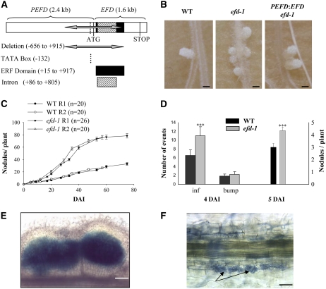
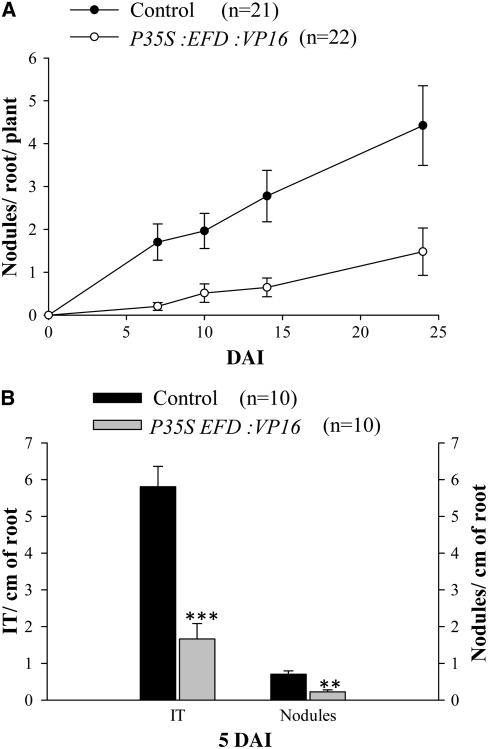
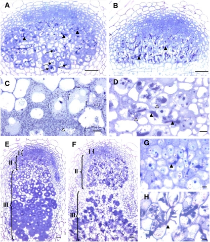
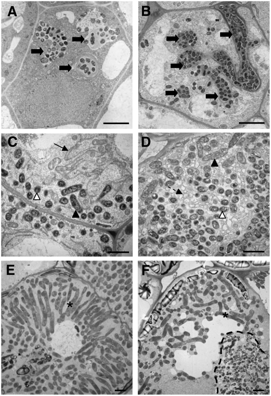
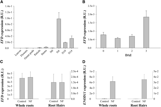
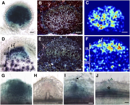
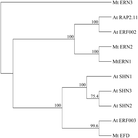
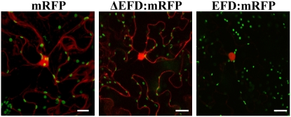
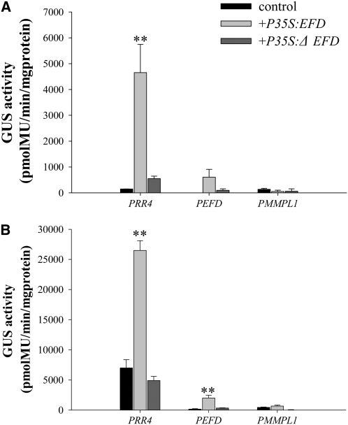
Mechanisms regulating legume root nodule development are still poorly understood, and very few regulatory genes have been cloned and characterized. Here, we describe EFD (for ethylene response factor required for nodule differentiation), a gene that is upregulated during nodulation in Medicago truncatula. The EFD transcription factor belongs to the ethylene response factor (ERF) group V, which contains ERN1, 2, and 3, three ERFs involved in Nod factor signaling. The role of EFD in the regulation of nodulation was examined through the characterization of a null deletion mutant (efd-1), RNA interference, and overexpression studies. These studies revealed that EFD is a negative regulator of root nodulation and infection by Rhizobium and that EFD is required for the formation of functional nitrogen-fixing nodules. EFD appears to be involved in the plant and bacteroid differentiation processes taking place beneath the nodule meristem. We also showed that EFD activated Mt RR4, a cytokinin primary response gene that encodes a type-A response regulator. We propose that EFD induction of Mt RR4 leads to the inhibition of cytokinin signaling, with two consequences: the suppression of new nodule initiation and the activation of differentiation as cells leave the nodule meristem. Our work thus reveals a key regulator linking early and late stages of nodulation and suggests that the regulation of the cytokinin pathway is important both for nodule initiation and development.


Figures

Figure 1.
Figure 1.
Symbiotic Phenotype of the efd-1 Mutant. (A) The efd-1 deletion encompasses 1571 bp within the promoter (PEFD) and the coding region (EFD), including the ERF domain. (B) Appearance of 27-d-old nodules. From left to right: the wild type (Fix+ and elongated), efd-1 (Fix and more spherical), and efd-1 complemented by a PEFD:EFD construct (Fix+ and elongated). All three samples were collected from the same experiment. Bars = 1 mm. (C) Time course of S. meliloti–induced nodule production in wild-type M. truncatula versus the efd-1 mutant. Two biological repetitions are represented (R1 and R2). Error bars represent se. (D) Number of infections, bumps, and nodules counted at 4 DAI (left panel; n = 10) or 5 DAI (right panel; n = 29) in wild-type M. truncatula versus efd-1. Statistically significant differences are indicated by asterisks (Mann and Whitney test, P < 0.001). Error bars represent se. (E) Example of a multilobe nodule (5 DAI) found in the efd-1 mutant: nonsectioned nodule induced by S. meliloti hemA-lacZ, stained in blue following a β-galactosidase assay. Bar = 160 μm. (F) The efd-1 mutant exhibits a normal mycorrhization phenotype. Interaction between M. truncatula roots and G. intradices at 22 DAI. Arrows indicate arbuscules. Bar = 20 μm.
Figure 2.
Figure 2.
The Number of Infections and Nodules Is Strongly Decreased in Roots Overexpressing EFD Fused to the VP16 Activator Domain. (A) One-month-old transgenic roots expressing P35S:EFD:VP16 or P35S:VP16 (Control) constructs were inoculated by wild-type S. meliloti. Nodules were counted until 23 DAI. Graphs represent the average of two biological repetitions, and error bars represent se. (B) Histograms of infection threads (IT) and nodules number per root at 5 DAI, determined on a third biological repetition. Error bars represent se. Asterisks indicate statistically significant differences (Mann and Whitney test, P < 0.001 [***] and P < 0.01[**]).
Figure 3.
Figure 3.
Microscopy Characterization of efd-1 Nodules Reveals Defects in Symbiosome Formation and Tissue Differentiation. (A) and (B) Four-micrometer sections of 5-d-old nodules, following inclusion in Technovit, induced by S. meliloti in wild-type M. truncatula (A) or the efd-1 mutant (B). Arrows in (A) point to bacterial release in the proximal infection region. (C) and (D) Four-micrometer sections showing infected cells from 7-d-old nodules (zone II) induced by S. meliloti in the wild type (C) or efd-1 mutant (D) (epon inclusions). (E) to (H) Sections of 10-d-old nodules (Technovit inclusions) induced by S. meliloti in the wild type ([E] and [G]) or efd-1 mutant ([F] and [H]). (G) and (H) are a close-up of the zone II region shown in (E) and (F). Black and white arrowheads show infection threads and released bacteria, respectively. Brackets in (E) and (F) indicate nodule zones I, II, and III. Bars = 10 μm in (C), (D), (G), and (H) and 50 μm in (A), (B), (E), and (F).
Figure 4.
Figure 4.
EM Characterization of Zones II and III Cells from Wild-Type M. truncatula and efd-1 10-d-Old Nodules. Wild-type nodule ([A], [C], and [E]); efd-1 nodule ([B], [D], and [F]). (A) and (B) The arrows show infection threads, which are more numerous and branched in efd-1 nodules. Bars = 5 μm. (C) and (D) The arrow shows the typical endoplasmic reticulum observed in wild-type nodules, whereas numerous small vesicles are found in efd-1 nodules (broken arrow). The white and black arrowheads point to bacteroids, types 1 and 2, respectively. (E) and (F) Type 4 bacteroids (asterisks) found in zone III of wild-type nodules and in the proximal region (zone III-like) of efd-1 nodules; note in efd-1 the absence of a radial organization of bacteroids, in contrast with wild-type nodules, and the presence of a bacterial release structure (dotted line) next to type 4 bacteroids, generated from an infection thread crossing the cell. Bars = 2 μm in (C) to (F).
Figure 5.
Figure 5.
Q-RT-PCR Analyses of EFD Expression in Different Tissues. (A) EFD expression in M. truncatula leaves, flowers, green pods, petiols, stems, roots (N0), and nodules at different developmental stages (4, 10, and 14 DAI). (B) EFD expression in wild-type S. meliloti–inoculated roots of M. truncatula at 0, 1, 2, and 3 DAI. (C) EFD expression in NF- or water-treated roots and root hairs (18 h treatment). (D) Mt ENOD11 expression in NF- or water-treated roots and root hairs (18 h treatment). All data are from at least three biological repetitions and are normalized by EF1-α expression. Error bars represent se. R.U., relative units.
Figure 6.
Figure 6.
EFD Is Expressed in the Apical Zone II of the Nodule and in Nodule Primordia. (A) to (F) Localization of EFD mRNA in wild-type 4-d-old ([A] to [C]) and 10-d-old ([D] to [F]) nodules, as determined by PEFD:GUS fusion ([A] and [D]; blue) or in situ hybridization ([B], [C], [E], and [F]); hybridization signals appear as white dots in (B) and (E) or false color representation following digitization in (C) and (F) (Diatrack software) where the strongest signals are indicated by yellow-orange and the weakest by dark-blue colors. S. meliloti hemA:lacZ bacteria are stained in purple in (A) and (D). Brackets in (D) and (F) indicate nodule zone I, II and III. (G) and (H) Localization of EFD mRNA, based on the PEFD:GUS fusion, in 4- and 10-d-old nodules induced by an infection-defective exoA mutant of S. meliloti. (I) PEFD:GUS expression (blue color) in a wild-type nodule primordium beneath a developing infection thread (arrow) containing S. meliloti hemA:lacZ bacteria (purple). (J) PEFD:GUS expression associated with early cortical cell divisions (asterisk) before the elongation of infection threads; note the presence of a curled root hair (triangle), containing S. meliloti bacteria (purple). Bars = 50 μm.
Figure 7.
Figure 7.
Phylogenetic Tree of Group V ERFs from Arabidopsis and M. truncatula. All Arabidopsis proteins from ERF group V, as described by Nakano et al. (2006), were aligned with M. truncatula group V ERF proteins (ERNs and EFD). Alignment was done with entire proteins. Indicated bootstrap values were determined from 1000 iterations.
Figure 8.
Figure 8.
Nuclear Localization of EFD:RFP Fusion Protein. Leaves of N. benthamiana were A. tumefaciens transformed with the following constructs, expressed under the control of the 35S promoter: from left to right, reporter mRFP protein alone; mRFP protein fused to a deleted form of EFD lacking the putative DNA binding domain (ΔEFD; N-terminal fusion); full-size EFD fused to mRFP reporter protein (N-terminal fusion). The mRFP reporter protein is detected as a red signal, while green spots correspond to plastids. Note that wild-type EFD is found exclusively in the nucleus in contrast with the mRFP reporter protein alone or to EFD deleted for the ERF domain. Bars = 40 μm.
Figure 9.
Figure 9.
Trans-Activation of PRR4, PEFD, and PMMPL1 in N. benthamiana. (A) GUS activity at 24 h following cotransformation. (B) GUS activity at 48 h following cotransformation. For both (A) and (B), leaves of N. benthamiana were cotransformed by A. tumefaciens with PRR4:GUS, PEFD:GUS, or PMMPL1:GUS plus either P35S:EFD:RFP or P35S:ΔEFD:RFP (ΔEFD is EFD deleted for its putative DNA binding domain). Controls correspond to leaves of N. benthamiana transformed with PRR4:GUS, PEFD:GUS, or PMMPL1:GUS alone. GUS activity was measured using 10 μg of total protein extracts at 24 (A) and 48 (B) h after transformation using three biological repetitions. Error bars represent se. PRR4 was only significantly activated by EFD at 24 and 48 H and PEFD at 48H (P < 0.01 for both, following Cumming et al., 2007). PMMPL1 was not significantly activated.

Similar articles

Cited by

References

    1. Aharoni, A., Dixit, S., Jetter, R., Thoenes, E., van Arkel, G., and Pereira, A. (2004). The SHINE clade of AP2 domain transcription factors activates wax biosynthesis, alters cuticle properties, and confers drought tolerance when overexpressed in Arabidopsis. Plant Cell 16 2463–2480. - PMC - PubMed
    1. Allen, M.D., Yamasaki, K., Ohme-Takagi, M., Tateno, M., and Suzuki, M. (1998). A novel mode of DNA recognition by a beta-sheet revealed by the solution structure of the GCC-box binding domain in complex with DNA. EMBO J. 17 5484–5496. - PMC - PubMed
    1. Alunni, B., Kevei, Z., Redondo-Nieto, M., Kondorosi, A., Mergaert, P., and Kondorosi, E. (2007). Genomic organization and evolutionary insights on GRP and NCR genes, two large nodule-specific gene families in Medicago truncatula. Mol. Plant Microbe Interact. 20 1138–1148. - PubMed
    1. Andriankaja, A., Boisson-Dernier, A., Frances, L., Sauviac, L., Jauneau, A., Barker, D.G., and de Carvalho-Niebel, F. (2007). AP2-ERF transcription factors mediate Nod factor dependent Mt ENOD11 activation in root hairs via a novel cis-regulatory motif. Plant Cell 19 2866–2885. - PMC - PubMed
    1. Ardourel, M., Demont, N., Debelle, F., Maillet, F., de Billy, F., Prome, J.C., Denarie, J., and Truchet, G. (1994). Rhizobium meliloti lipooligosaccharide nodulation factors: Different structural requirements for bacterial entry into target root hair cells and induction of plant symbiotic developmental responses. Plant Cell 6 1357–1374. - PMC - PubMed

Publication types

MeSH terms

Associated data