

Novel human bronchial epithelial cell lines for cystic fibrosis research
- PMID: 18978040
- PMCID: PMC2636952
- DOI: 10.1152/ajplung.90314.2008
Novel human bronchial epithelial cell lines for cystic fibrosis research
Abstract
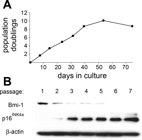
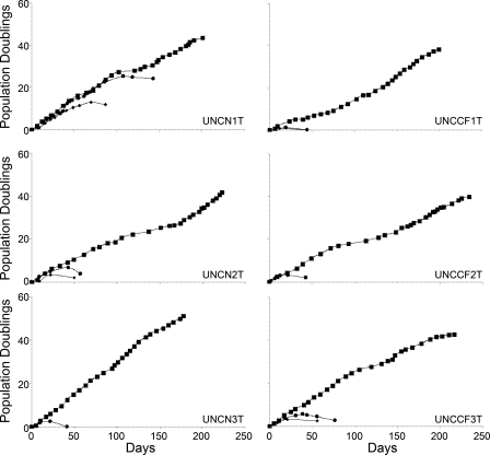
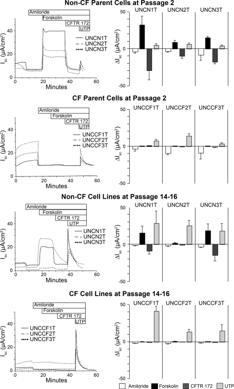
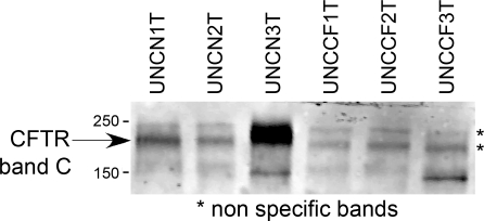
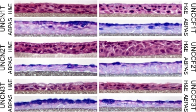
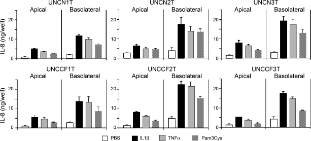
Immortalization of human bronchial epithelial (hBE) cells often entails loss of differentiation. Bmi-1 is a protooncogene that maintains stem cells, and its expression creates cell lines that recapitulate normal cell structure and function. We introduced Bmi-1 and the catalytic subunit of telomerase (hTERT) into three non-cystic fibrosis (CF) and three DeltaF508 homozygous CF primary bronchial cell preparations. This treatment extended cell life span, although not as profoundly as viral oncogenes, and at passages 14 and 15, the new cell lines had a diploid karyotype. Ussing chamber analysis revealed variable transepithelial resistances, ranging from 200 to 1,200 Omega.cm(2). In the non-CF cell lines, short-circuit currents were stimulated by forskolin and inhibited by CFTR(inh)-172 at levels mostly comparable to early passage primary cells. CF cell lines exhibited no forskolin-stimulated current and minimal CFTR(inh)-172 response. Amiloride-inhibitable and UTP-stimulated currents were present, but at lower and higher amplitudes than in primary cells, respectively. The cells exhibited a pseudostratified morphology, with prominent apical membrane polarization, few apoptotic bodies, numerous mucous secretory cells, and occasional ciliated cells. CF and non-CF cell lines produced similar levels of IL-8 at baseline and equally increased IL-8 secretion in response to IL-1beta, TNF-alpha, and the Toll-like receptor 2 agonist Pam3Cys. Although they have lower growth potential and more fastidious growth requirements than viral oncogene transformed cells, Bmi-1/hTERT airway epithelial cell lines will be useful for several avenues of investigation and will help fill gaps currently hindering CF research and therapeutic development.
Figures
References
-
- Alkema MJ, van der Lugt NM, Bobeldijk RC, Berns A, van Lohuizen M. Transformation of axial skeleton due to overexpression of bmi-1 in transgenic mice. Nature 374: 724–727, 1995. - PubMed
-
- Bea S, Tort F, Pinyol M, Puig X, Hernandez L, Hernandez S, Fernandez PL, van Lohuizen M, Colomer D, Campo E. BMI-1 gene amplification and overexpression in hematological malignancies occur mainly in mantle cell lymphomas. Cancer Res 61: 2409–2412, 2001. - PubMed
-
- Becker MN, Sauer MS, Muhlebach MS, Hirsh AJ, Wu Q, Verghese MW, Randell SH. Cytokine secretion by cystic fibrosis airway epithelial cells. Am J Respir Crit Care Med 169: 645–653, 2004. - PubMed
-
- Benali R, Tournier JM, Chevillard M, Zahm JM, Klossek JM, Hinnrasky J, Gaillard D, Maquart FX, Puchelle E. Tubule formation by human surface respiratory epithelial cells cultured in a three-dimensional collagen lattice. Am J Physiol Lung Cell Mol Physiol 264: L183–L192, 1993. - PubMed
-
- Boers JE, Ambergen AW, Thunnissen FB. Number and proliferation of basal and parabasal cells in normal human airway epithelium. Am J Respir Crit Care Med 157: 2000–2006, 1998. - PubMed
Publication types
MeSH terms
Substances
Grants and funding
LinkOut - more resources
Full Text Sources
Other Literature Sources
Medical
Molecular Biology Databases
Research Materials