. 2009 Feb;75(2):363-73.
doi: 10.1124/mol.108.047845. Epub 2008 Oct 31.

Preferential physical and functional interaction of pregnane X receptor with the SMRTalpha isoform

Affiliations

Preferential physical and functional interaction of pregnane X receptor with the SMRTalpha isoform

Chia-Wei Li et al. Mol Pharmacol. 2009 Feb.

Abstract

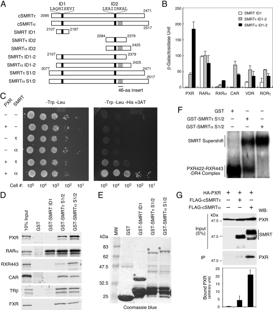
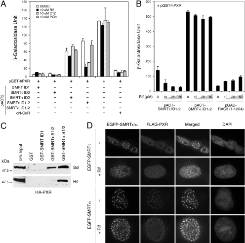
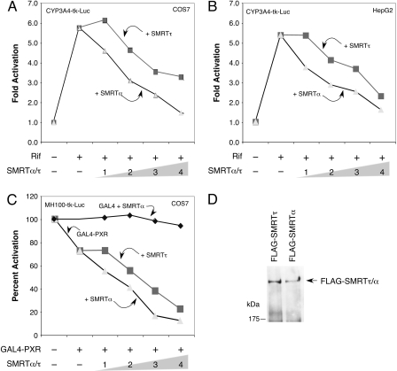
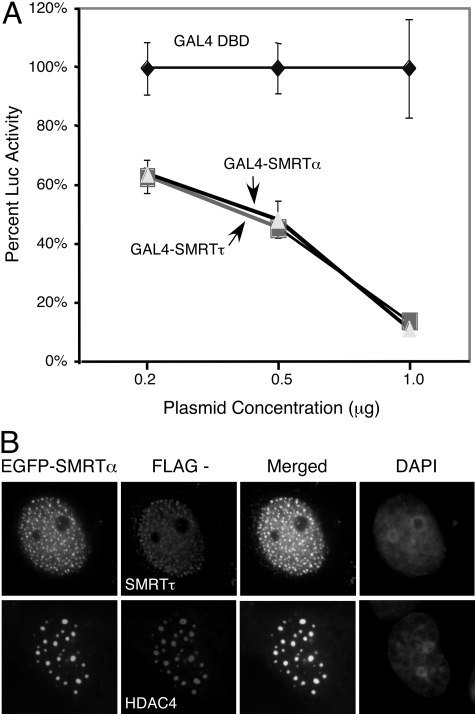
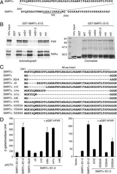
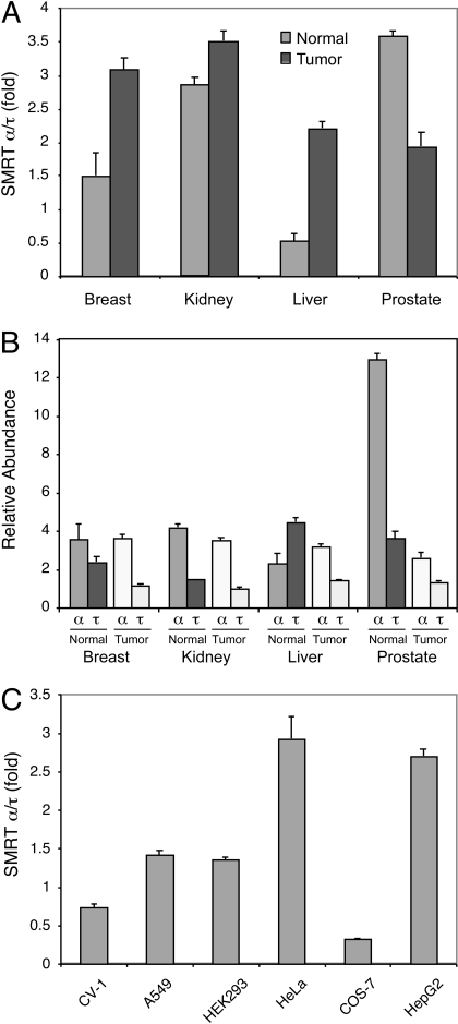
The silencing mediator for retinoid and thyroid hormone receptors (SMRT) serves as a platform for transcriptional repression elicited by several steroid/nuclear receptors and transcription factors. SMRT exists in two major splicing isoforms, alpha and tau, with SMRTalpha containing only an extra 46-amino acid sequence inserted immediately downstream from the C-terminal corepressor motif. Little is known about potential functional differences between these two isoforms. Here we show that the pregnane X receptor (PXR) interacts more strongly with SMRTalpha than with SMRTtau both in vitro and in vivo. It is interesting that the PXR-SMRTalpha interaction is also resistant to PXR ligand-induced dissociation, in contrast to the PXR-SMRTtau interaction. SMRTalpha consistently inhibits PXR activity more efficiently than does SMRTtau in transfection assays, although they possess comparable intrinsic repression activity and association with histone deacetylase. We further show that the mechanism for the enhanced PXR-SMRTalpha interaction involves both the 46-amino acid insert and the C-terminal corepressor motif. In particular, the first five amino acids of the SMRTalpha insert are essential and sufficient for the enhanced binding of SMRTalpha to PXR. Furthermore, we demonstrate that Tyr2354 and Asp2355 residues of the SMRTalpha insert are most critical for the enhanced interaction. In addition, expression data show that SMRTalpha is more abundantly expressed in most human tissues and cancer cell lines, and together these data suggest that SMRTalpha may play a more important role than SMRTtau in the negative regulation of PXR.


Figures

Fig. 1.
Fig. 1.
PXR interacts preferentially with SMRTα. A, schematic diagrams of various SMRTτ and SMRTα constructs used in this study. The proximal ID1 and distal ID2 corepressor motifs are indicated by black bars, with the motif core sequences shown at the top. The SMRTα-specific 46-aa insert is shown in gray. The amino acid positions of individual fragments are labeled numerically. B, PXR interacts preferentially with SMRTα in a yeast two-hybrid assay. The pGBT-hPXR, pAS-hRARα, pGBT-hRXRα, pGBT-hCAR, pGBT-hVDR, and pGBT-mRORγ were individually transformed into Y190 cells in combination with pACT-SMRT ID1 (aa 2107-2187), pACT-SMRTτ ID1-2 (aa 2107-2379), or pACT-SMRTα ID1-2 (aa 2107-2425). Three colonies from each plate were picked and grown in -Trp-Leu liquid media for 24 h. The expression of β-galactosidase was measured by liquid o-nitrophenyl β-d-galactopyranoside assay after normalization with cell numbers, and the average β-galactosidase units were calculated and plotted. C, survival assay of yeast cells cotransformed with pGBT-hPXR and pACT-SMRTτ ID1-2 or SMRTα ID1-2 constructs. Indicated numbers of transformed cells were spotted onto -Trp-Leu or -Trp-Leu-His + 3AT (50 mM) selection plates and incubated at 30°C for 2 days. The pGBT9 and pACT2 vectors were used as controls where indicated (-). D, interactions of SMRT isoforms with various NRs in GST pull-down assays. In vitro-translated 35S-labeled hPXR, hRARα, hRXRα443, hCAR, hTRβ, and hFXR were incubated with GST, GST-SMRT ID1, GST-SMRTτ S1/2 (aa 2077-2471), or GST-SMRTα S1/2 (aa 2077-2517) at 4°C overnight. After extensive washing with binding buffer, the bound proteins were collected by centrifugation and analyzed by SDS-polyacrylamide gel electrophoresis and autoradiography. E, Coomassie blue staining of GST and GST-SMRT fusion proteins used in this study. The GST proteins were purified from bacteria BL21 cells and analyzed by SDS-PAGE and Coomassie staining. Notable size difference of the intact GST-SMRT ID1, GST-SMRTτ S1/2, and GST-SMRTα S1/2 are marked by asterisks (*). F, preferential interaction of PXR-RXR heterodimers with SMRTα on a DR4 PXR response element. Gel-shift assay was conducted with in vitro-translated hPXR422, hRXRα443, and a 32P-labeled DR4 element. Equal amounts (6 μg) of purified GST, GST-SMRTτ S1/2, or GST-SMRTα S1/2 were added to individual reaction. The DNA-protein complexes were separated onto a native polyacrylamide gel and detected by autoradiography. G, coimmunoprecipitation of PXR with cSMRTτ/α from mammalian cell extracts. Approximately 1 mg of protein extracts obtained from HEK293 cells coexpressing HA-PXR and FLAG-cSMRTτ (aa 2095-2471) or FLAG-cSMRTα (aa 2095-2517) were immunoprecipitated with monoclonal anti-FLAG antibody-conjugated agarose beads. Approximately 5% of the extract used in each immunoprecipitation reaction (input) was analyzed by Western blot to show the relative amount of HA-PXR and cSMRTτ/α in the cell extracts. The total amount of immunoprecipitates (IP) was analyzed by Western blot with rabbit anti-HA antibody to detect the coimmunoprecipitated HA-PXR. The precipitated HA-PXR in each reaction was quantitated by densitometry, and results are plotted and shown at the bottom.
Fig. 2.
Fig. 2.
SMRTα-PXR interaction is resistant to ligand-induced dissociation. A, effects of PXR ligands on SMRT-PXR interactions in a yeast two-hybrid assay. Yeast colonies cotransformed with pGBT-hPXR and the indicated pACT-SMRT constructs or pACT-cN-CoR were treated with the hPXR-specific ligands Rif (10 μM) and CTZ (10 μM), or with the mPXR-specific ligand PCN (10 μM) for 48 h. The empty pACT2 vector was used as a control where indicated (-). The SMRTα ID2 and SMRTα ID1-2 show stronger, ligand-resistant interactions with PXR in comparison with SMRTτ. B, rifampicin concentration-dependent dissociation of SMRTτ/α-PXR interaction in a yeast two-hybrid assay. Yeast transformants containing pGBT-hPXR and pACT-SMRTτ ID1-2, pACT-SMRTα ID1-2, or pGAD-RAC3 (1-1204) were grown in -Trp-Leu media for 24 h. Aliquots of each sample were treated with increasing concentrations of Rif (10, 25, and 50 μM) and incubated for another 36 h. Rifampicin had little effect on the interaction of PXR with SMRTα ID1-2, whereas it reduced its interaction with SMRTτ ID1-2 and enhanced interaction with the coactivator RAC3. C, rifampicin had little effect on the formation of SMRTα-PXR complex. HA-PXR was overexpressed in HEK293 cells in the absence (Sol) or presence of Rif (10 μM). Approximately 50 μg of cell extracts was incubated with 5 μg of purified GST, GST-SMRT ID1, GST-SMRTτ S1/2, or GST-SMRTα S1/2 for 16 h at 4°C. The bound HA-PXR proteins were collected by centrifugation and analyzed by SDS-PAGE and Western blot using anti-HA antibody. D, SMRTα colocalizes with PXR in mammalian cells in the presence of rifampicin. COS-7 cells were transfected with pEGFP-hSMRTτ (full-length) or pEGFP-hSMRTα (full-length) together with FLAG-hPXR. Cells were recovered in Dulbecco's modified Eagle's medium media containing 10 μM rifampicin or DMSO for 12 h. Cells were fixed, and colocalization between SMRT and PXR was detected by immunostaining with anti-FLAG antibody and the EGFP signals. Rifampicin causes clear dissociation of PXR from SMRTτ nuclear foci, whereas it has little effect or no impact on the SMRTα-PXR colocalization.
Fig. 3.
Fig. 3.
Transcriptional activity of PXR is preferentially suppressed by SMRTα. COS-7 (A) and human liver HepG2 (B) cells were transiently transfected with pCMXHA-hPXR and pCYP3A4-tk-luc reporter together with a β-galactosidase expression vector as an internal control. Increasing amounts of pCMX-FLAG-SMRTα (full-length) or pCMX-FLAG-SMRTτ (full-length) were cotransfected as indicated (in micrograms). After transfection, cells were refed with fresh media containing 10 μM Rif where indicated (+) and recovered for 16 h. Relative -fold activations of the reporter in comparison with the control sample without treatment or SMRT cotransfection were determined from three independent experiments. SMRTα inhibits PXR transactivation stronger than SMRTτ in both cell types in a dose-dependent manner. C, transcriptional repression by GAL4-PXR is preferentially enhanced by SMRTα. COS-7 cells were transfected with GAL4-hPXR with increasing amounts (in micrograms) of pCMX-FLAG-SMRTτ/α (full-length) construct along with the GAL4-dependent MH100-tk-luc reporter and a β-galactosidase control vector. SMRTα did not affect GAL4 activity, whereas it preferentially enhanced the transcriptional repression activity of GAL-PXR. D, Western blot analysis showing comparable expression levels of FLAG-SMRTτ/α full-length proteins in the transfected cells.
Fig. 4.
Fig. 4.
SMRTα and SMRTτ possess comparable intrinsic repression activities. A, transcriptional repressions by GAL4-SMRTα (full-length) and GAL4-SMRTτ (full-length) are comparable. HEK293 cells were transfected with increasing amounts of GAL4 DBD or GAL4 DBD fusions of either full-length SMRTα (GAL4-SMRTα) or full-length SMRTτ (GAL4-SMRTτ), together with a GAL4-dependent MH100-tk-luc reporter. The relative percentages of luciferase activity in comparison with GAL4 DBD alone (set as 100%) were determined from three independent experiments. B, SMRTα colocalizes with SMRTτ and HDAC4. COS-7 cells were transiently transfected with EGFP-SMRTα (full-length) in combination with FLAG-SMRTτ (full-length) or FLAG-HDAC4. Transfected cells were fixed and immunostained with anti-FLAG monoclonal antibody and rhodamine-conjugated secondary antibody and visualized in comparison with the EGFP-SMRTα signals. Cell nuclei were stained with 4,6-diamidino-2-phenylindole.
Fig. 5.
Fig. 5.
Molecular determinants of SMRTα preference for PXR binding. A, sequence of the SMRTα-specific 46 amino acids is shown at the top with the flanking SMRTτ sequences shown at the bottom. This SMRTα-specific sequence is inserted after glycine at position 2352 after the distal ID2 corepressor motif (underlined). The numbers indicate amino acid positions. B, the ID2 motif mutation in SMRTα disrupts PXR interaction. The wild-type (WT) GST-SMRTα S1/2 and its site-directed mutants, mID1 (V2142A/I2143A), mID2 (I2345A/I2346A), mID1-2 (V2142A/I2143A, I2345A/I2346A), m3 (S2285E/K2286E/K2287E), and m4 (L2467A/I2468A), were tested for binding with 35S-labeled hPXR and hRARα in a GST pull-down assay. Right, Coomassie blue-stained GST proteins. The mID2-containing mutants (mID2 and mID1-2) show decreased binding to PXR, whereas only mID1-containing mutations (mID1 and mID1-2) affect their interactions with RARα. C, sequence comparison of the SMRTα-specific 46-aa insert and its mutants. The solid line on top marks the 46-aa sequence. Dashed lines represent deletions. The alanine substitutions in the SMRTτ 3G/A and the KYD, K2353A, Y2354A, and D2355A mutants of SMRTα are italicized. The plasmid pACT-SMRTα ID1-2 was used as a template to construct these mutants used in the following assays. D, yeast two-hybrid assays showing interactions of PXR with SMRTα-specific 46-aa-related mutants. Yeast Y190 cells were cotransformed individually with pGBT-hPXR and indicated pACT-SMRTτ/α ID1-2 wild-type and indicated mutant constructs.
Fig. 6.
Fig. 6.
Expression of SMRT isoforms in human tissues and cancer cells. cDNAs from normal (N) and tumor (T) tissues of the breast, kidney, liver, and prostate and from various established cell lines were amplified using primer sets specific for SMRTα or SMRTτ as described under Materials and Methods. Real-time PCR reactions were performed in an ABI PRISM 7900HT Sequence Detection System. Relative quantification of SMRTα versus SMRTτ was normalized to the internal control β-actin and calculated according to the 2-ΔΔCt method. A, SMRTα/τ expression ratios in human normal and tumor tissues. B, relative abundance of SMRTα and SMRTτ among tested human normal and tumor tissues. C, SMRTα/τ expression ratios in established cell lines.

Similar articles

Cited by

References

    1. Blumberg B, Sabbagh W Jr, Juguilon H, Bolado J Jr, van Meter CM, Ong ES, and Evans RM (1998) SXR, a novel steroid and xenobiotic-sensing nuclear receptor. Genes Dev 12 3195-3205. - PMC - PubMed
    1. Chen JD and Evans RM (1995) A transcriptional co-repressor that interacts with nuclear hormone receptors. Nature 377 454-457. - PubMed
    1. Falkner KC, Pinaire JA, Xiao GH, Geoghegan TE, and Prough RA (2001) Regulation of the rat glutathione S-transferase A2 gene by glucocorticoids: involvement of both the glucocorticoid and pregnane X receptors. Mol Pharmacol 60 611-619. - PubMed
    1. Gao YD, Olson SH, Balkovec JM, Zhu Y, Royo I, Yabut J, Evers R, Tan EY, Tang W, Hartley DP, et al. (2007) Attenuating pregnane X receptor (PXR) activation: a molecular modelling approach. Xenobiotica 37 124-138. - PubMed
    1. Geick A, Eichelbaum M, and Burk O (2001) Nuclear receptor response elements mediate induction of intestinal MDR1 by rifampin. J Biol Chem 276 14581-14587. - PubMed

Publication types

LinkOut - more resources