. 2009 Jan;149(1):549-60.
doi: 10.1104/pp.108.129866. Epub 2008 Oct 31.

Sulfur transfer through an arbuscular mycorrhiza

Affiliations

Sulfur transfer through an arbuscular mycorrhiza

James W Allen et al. Plant Physiol. 2009 Jan.

Abstract

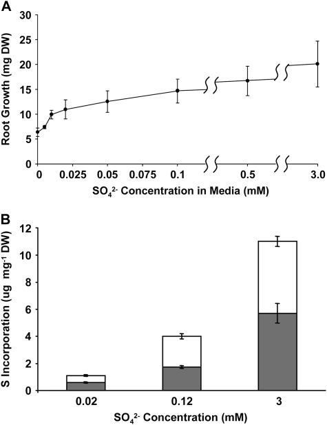
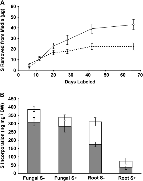
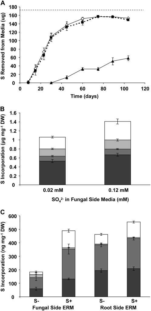
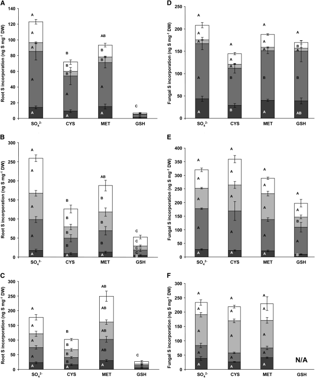
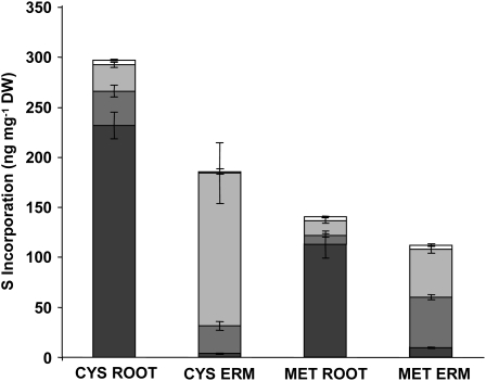
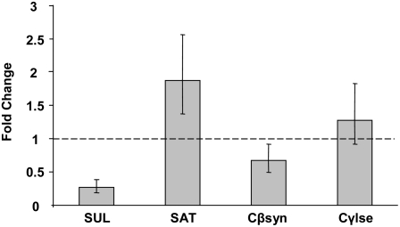
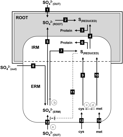
Despite the importance of sulfur (S) for plant nutrition, the role of the arbuscular mycorrhizal (AM) symbiosis in S uptake has received little attention. To address this issue, 35S-labeling experiments were performed on mycorrhizas of transformed carrot (Daucus carota) roots and Glomus intraradices grown monoxenically on bicompartmental petri dishes. The uptake and transfer of 35SO4(2-) by the fungus and resulting 35S partitioning into different metabolic pools in the host roots was analyzed when altering the sulfate concentration available to roots and supplying the fungal compartment with cysteine (Cys), methionine (Met), or glutathione. Additionally, the uptake, transfer, and partitioning of 35S from the reduced S sources [35S]Cys and [35S]Met was determined. Sulfate was taken up by the fungus and transferred to mycorrhizal roots, increasing root S contents by 25% in a moderate (not growth-limiting) concentration of sulfate. High sulfate levels in the mycorrhizal root compartment halved the uptake of 35SO4(2-) from the fungal compartment. The addition of 1 mm Met, Cys, or glutathione to the fungal compartment reduced the transfer of sulfate by 26%, 45%, and 80%, respectively, over 1 month. Similar quantities of 35S were transferred to mycorrhizal roots whether 35SO4(2-), [35S]Cys, or [35S]Met was supplied in the fungal compartment. Fungal transcripts for putative S assimilatory genes were identified, indicating the presence of the trans-sulfuration pathway. The suppression of fungal sulfate transfer in the presence of Cys coincided with a reduction in putative sulfate permease and not sulfate adenylyltransferase transcripts, suggesting a role for fungal transcriptional regulation in S transfer to the host. A testable model is proposed describing root S acquisition through the AM symbiosis.


Figures

Figure 1.
Figure 1.
The effect of sulfate concentration on the growth and S incorporation of nonmycorrhizal transformed carrot roots. A, Root growth measured as dry weight (DW) increase at different concentrations of Na2SO4 in 20 mL of modified liquid M media (St-Arnaud et al., 1996). B, The incorporation of S from solid media containing 0.02, 0.12, or 3 mm Na2SO4 labeled with 11.5 μCi of Na2[35SO4] was measured in roots after 4 weeks of labeled growth. Labeled root compounds were extracted with cold MeOH:water (70:30; white bars) before the cellular residue was solubilized in tissue solubilizer (gray bars). The fractions were analyzed by liquid scintillation counting. Error is expressed as sem.
Figure 2.
Figure 2.
Increased sulfate availability in the root compartments of split plates reduces transfer of S through the distal fungal ERM. A, The uptake of S by ERM from plates labeled with 30 μCi of Na2[35SO4] in 0.12 mm Na2SO4 in the fungal compartments was measured as the removal of radioactivity from the media when the root compartments were supplied with moderate S (0.12 mm Na2SO4; solid line) or high S (3 mm Na2SO4; broken line). B, Measurements of S incorporated into the roots (Root S±) and distal fungal (Fungal S±) ERM from the above experiment after 66 d of labeling; S+ refers to high sulfate levels (3 mm) in root compartments and S− to moderate levels (0.12 mm). Radioactivity was measured by scintillation counting after a crude separation by cold MeOH:water (70:30) extraction (white bars) followed by solubilization of the remaining tissue (gray bars). The aqueous alcohol extraction dissolves sulfate, sulfite, S amino acids, and potentially also unidentified sulfated esters, while the remainder includes proteins, sulfolipids, and other unknown products. Values are presented as mean ± SEM (n ≥ 5). DW, Dry weight.
Figure 3.
Figure 3.
Transfer of 35SO42− through the fungus versus direct root uptake. A, The uptake of S over 104 d from root and fungal compartment media. The root (proximal) compartments of split plates were labeled with 17.3 μCi of Na2[35SO4] in 0.2 mm Na2SO4 (171 μg S). One-half of the fungal (distal) compartments were labeled with 17.3 μCi of Na2[35SO4] in 0.2 mm Na2SO4 (171 μg S, white diamonds, solid line), while the others contained 0.02 mm sulfate without 35S (black squares, broken line). Distal uptake was monitored from labeled plates (black triangles, solid line). A straight broken line denotes the total initial S content of the proximal compartment media. B, Incorporation of 35S by roots when the fungal side was supplied with low unlabeled (0.02 mm) or moderate 35S-labeled (0.2 mm) sulfate was measured after 110 d of labeling by chemical fractionation into sulfate- (white bars), amino acid- (light gray bars), and protein- (medium gray bars) containing pools, and solubilized remaining tissue (dark gray bars) pools measured by liquid scintillation counting. C, Incorporation of 35S into different pools of ERM collected from the root or fungal side compartments of split plates when the fungal compartments contained 0.02 mm unlabeled sulfate (S−) or 0.2 mm of 35S-labeled (S+); as above, the root side compartments of all plates contained 0.2 mm 35S-labeled sulfate. Fractions are depicted as in A. Values represent means ± SEM (n ≥ 4). DW, Dry weight.
Figure 4.
Figure 4.
The effect of common S metabolite repressors Cys, Met, and GSH on the transfer and allocation of 35S in an arbuscular mycorrhiza. The incorporation of 35S into roots and fungal ERM was analyzed after labeling the distal compartments with 37.4 μCi of Na2[35SO4] in 0.12 mm Na2SO4 (91 μg S) alone (SO42−) or with 1 mm Cys, 1 mm Met, or 1 mm GSH. Root (A–C) and fungal (D–F) incorporation was analyzed after 2 (A and D), 4 (B and E), and 6 (C and F) weeks. Fungal ERM was collected from the distal compartments of the same plates as those from which root tissue was collected from the proximal compartments. Tissue was analyzed by chemical fractionation into pools containing sulfate (white bars), amino acids (light gray bars), proteins (medium gray bars), and the tissue solubilized biomass remaining after extraction (dark gray bars). Fractions were analyzed by liquid scintillation counting. Letters within the bars represent ANOVA single-factor analyses between bars of that type. Letters above the complete bar graphs are the analyses of total S uptake. Values are reported as mean ± SEM (n ≥ 5). DW, Dry weight.
Figure 5.
Figure 5.
Transfer of reduced S through a mycorrhizal symbiosis. Mycorrhizal roots were grown in 0.5 mm Na2SO4 and the fungal partner allowed to grow into distal compartments containing either 1 mm Cys labeled with 25.7 μCi [35S]Cys or 1 mm Met labeled with 54.2 μCi of [35S]Met as the sole S source. Roots and fungal distal ERM were collected after 4 weeks of labeling and the radioactive counts split into different biochemical fractions. Fractions included SO42− (white bars), amino acids (light gray bars), proteins (medium gray bars), and the remaining solubilized cellular material (dark gray bars) and were obtained through scintillation counting. Values are reported as mean ± SEM (n = 9). DW, Dry weight.
Figure 6.
Figure 6.
Fungal gene expression changes in response to the addition of 1 mm Cys to ERM. Expression relative to control plates of sequences with homology to high affinity sulfate permease (SUL), S-adenosyl transferase (SAT), cystathionine β-synthase (Cβsyn), and cystathionine γ-lyase (Cγlse) was analyzed by quantitative real-time PCR using a ribosomal protein sequence as a control as previously reported (Govindarajulu et al., 2005; see “Materials and Methods”). RNA was extracted from ERM tissue collected after 24 hr incubation with 0.12 mm SO42− and with or without 1 mm Cys added to the distal compartments of five split plates per sample. Values are reported as mean ± SEM (n = 3 biological replicates each consisting of tissue from five to seven plates).
Figure 7.
Figure 7.
Model depicting S transfer through an endomycorrhizal symbiosis. Roots and fungal mycelium both import SO42− from external sources (1 and 12), and fungal uptake can supply isolated mycelium (8). The transfer of SO42− through the mycorrhizal symbiosis is inversely related to root uptake (1 and 5). Cys and Met are imported by the fungus (13 and 14), resulting in a reduction of fungal uptake of SO42− (11) and the transfer of a reduced form of S to the root (4). The reduction of SO42− (7 and 2) and uptake of reduced S (13 and 14) lead to incorporation in the protein pools in both roots (3) and fungus (6). Putative steps in the assimilation pathway based on sequence data are depicted as gray arrows. Steps involving putative S assimilation genes are labeled in gray letters as follows: a, high affinity sulfate permease; b, sulfate adenylyltransferase; c, γ-cystathionine lyase; d, β-cystathionine synthetase. IRM, Intraradical mycelium.

References

    1. Banerjee R, Evande R, Kabil O, Ojha S, Taoka S (2003) Reaction mechanism and regulation of cystathionine beta-synthase. Biochim Biophys Acta 1647 30–35 - PubMed
    1. Baumgardner RE, Lavery TF, Rogers CM, Isil SS (2002) Estimates of the atmospheric deposition of sulfur and nitrogen species: Clean Air Status and Trends Network, 1990-2000. Environ Sci Technol 36 2614–2629 - PubMed
    1. Bhat KKS, Nye PH, Baldwin JP (1976) Diffusion of phosphate to plant roots in soil. 4. Concentration distance profile in rhizosphere of roots with root hairs in a low-p soil. Plant Soil 44 63–72
    1. Bradfield G, Somerfield P, Meyn T, Holby M, Babcock D, Bradley D, Segel IH (1970) Regulation of sulfate transport in filamentous fungi. Plant Physiol 46 720–727 - PMC - PubMed
    1. Breton A, Surdin-Kerjan Y (1977) Sulfate uptake in Saccharomyces cerevisiae: biochemical and genetic study. J Bacteriol 132 224–232 - PMC - PubMed

Publication types

Associated data

LinkOut - more resources