. 2009 Jan;296(1):H94-H105.
doi: 10.1152/ajpheart.00567.2008. Epub 2008 Oct 31.

Injection of isolated mitochondria during early reperfusion for cardioprotection

Affiliations

Injection of isolated mitochondria during early reperfusion for cardioprotection

James D McCully et al. Am J Physiol Heart Circ Physiol. 2009 Jan.

Abstract

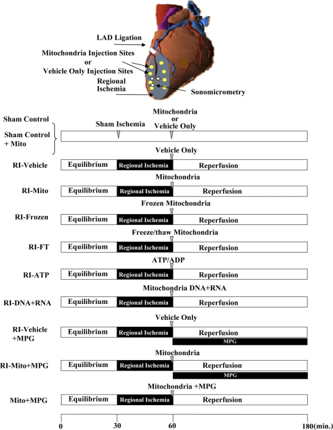
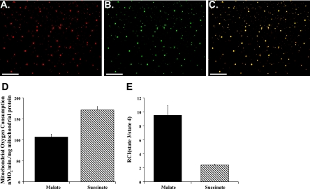
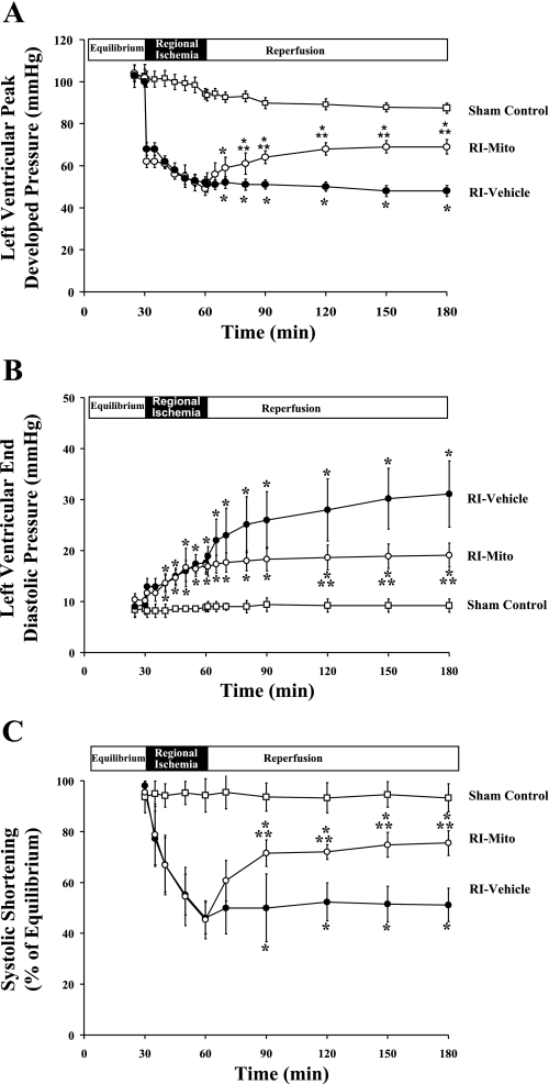
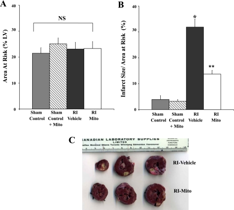
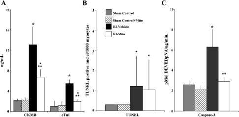
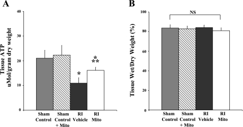
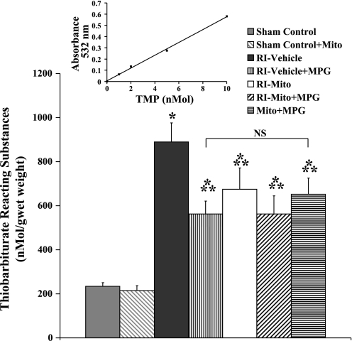
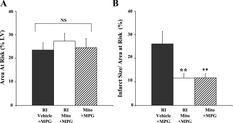
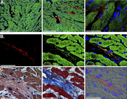
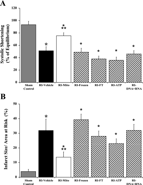
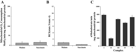
Previously, we demonstrated that ischemia induces mitochondrial damage and dysfunction that persist throughout reperfusion and impact negatively on postischemic functional recovery and cellular viability. We hypothesized that viable respiration-competent mitochondria, isolated from tissue unaffected by ischemia and then injected into the ischemic zone just before reperfusion, would enhance postischemic functional recovery and limit infarct size. New Zealand White rabbits (n = 52) were subjected to 30 min of equilibrium and 30 min of regional ischemia (RI) induced by snaring the left anterior descending coronary artery. At 29 min of RI, the RI zone was injected with vehicle (sham control and RI vehicle) or vehicle containing mitochondria (7.7 x 10(6) +/- 1.5 x 10(6)/ml) isolated from donor rabbit left ventricular tissue (RI-Mito). The snare was released at 30 min of RI, and the hearts were reperfused for 120 min. Our results show that left ventricular peak developed pressure and systolic shortening in RI-Mito hearts were significantly enhanced (P < 0.05 vs. RI-vehicle) to 75% and 83% of equilibrium value, respectively, at 120 min of reperfusion compared with 57% and 62%, respectively, in RI-vehicle hearts. Creatine kinase-MB, cardiac troponin I, and infarct size relative to area at risk were significantly decreased in RI-Mito compared with RI-vehicle hearts (P < 0.05). Confocal microscopy showed that injected mitochondria were present and viable after 120 min of reperfusion and were distributed from the epicardium to the subendocardium. These results demonstrate that viable respiration-competent mitochondria, isolated from tissue unaffected by ischemia and then injected into the ischemic zone just before reperfusion, significantly enhance postischemic functional recovery and cellular viability.


Figures

Fig. 1.
Fig. 1.
Top: Langendorff perfusion. White circles show positions of digital piezoelectric ultrasonic probes. Bottom: experimental protocol. Rabbit hearts were subjected to Langendorff perfusion consisting of 30 min of equilibrium, 30 min of regional ischemia [RI; achieved by occlusion of the left anterior descending coronary artery (LAD)], and 120 min of reperfusion. After 29 min of sham or RI, sham control and RI-vehicle hearts were injected with vehicle, sham control + Mito and RI-Mito hearts with vehicle containing isolated mitochondria (7.7 × 10⁶ ± 1.5 × 10⁶/ml), RI-frozen hearts with isolated mitochondria (7.7 × 10⁶ ± 1.5 × 10⁶/ml) that had been frozen overnight at −20°C, RI-FT with mitochondrial components from mitochondria (7.7 × 10⁶ ± 1.5 × 10⁶/ml) that had been frozen and then thawed, RI-ATP hearts with vehicle containing 1 mM ATP + 1 mM ADP, RI-DNA + RNA hearts with vehicle containing mitochondrial DNA + RNA isolated from 7.7 × 10⁶ ± 1.5 × 10⁶/ml mitochondria, and Mito + MPG hearts with isolated mitochondria (7.7 × 10⁶ ± 1.5 × 10⁶/ml) containing the reactive oxygen species scavenger N-(2 mecaptopropionyl)glycine (MPG, 300 μmol/l). At 30 min of RI, RI-vehicle + MPG and RI-Mito + MPG hearts were reperfused for 120 min with MPG (300 μmol/l) in Krebs-Ringer solution for 120 min.
Fig. 2.
Fig. 2.
Isolated mitochondrial viability and oxygen consumption. A–C: confocal-microscopic images of isolated mitochondria. A: isolated mitochondria labeled with MitoTracker Orange CMTMRos, which labels mitochondria maintaining membrane potential. B: mitochondria counterstained with mitochondria-specific MitoFluor Green. C: overlay of A and B. Original magnification ×400. D and E: state 3 (active) oxygen consumption (ADP-stimulated respiration) and respiratory control index (RCI; state 3/state 4) for malate-induced (complex I) and succinate-induced (complex II) energized mitochondria. Values are means ± SE of 6 analyses.
Fig. 3.
Fig. 3.
Global and regional myocardial function during equilibrium, RI, and reperfusion: left ventricular (LV) peak developed pressure (LVPDP; A), LV end-diastolic pressure (B), and systolic shortening (SS; C) during equilibrium, RI, and reperfusion in sham control, RI-vehicle, and RI-Mito hearts. Values are means ± SE. *P < 0.05 vs. sham control. **P < 0.05 vs. RI-vehicle.
Fig. 4.
Fig. 4.
Myocardial injury after 30 min of RI and 120 min of reperfusion: myocardial area at risk (AAR; A) and infarct size (IS) relative to AAR (IS/AAR, B) in sham control, sham control + Mito, RI-vehicle, and RI-Mito hearts after 30 min of RI and 120 min of reperfusion. Sham control hearts were subjected to sham RI only. There was no significant difference in IS between sham control and sham control + Mito. *P < 0.05 vs. sham control and sham control + Mito. **P < 0.05 vs. RI-vehicle. C: representative photograph of rabbit heart slices from RI-vehicle and RI-Mito hearts stained with 1% triphenyltetrazolium chloride. AAR is outlined by white line. Myocardial infarct (yellow) was significantly increased in RI-vehicle compared with RI-Mito hearts. Scale is shown in cm.
Fig. 5.
Fig. 5.
Markers of myocardial injury after 30 min of RI and 120 min of reperfusion. A: creatine kinase (CK-MB) and rabbit cardiac troponin I (cTnI) in myocardial effluents after 30 min of RI in sham control, sham control + Mito, RI-vehicle, and RI-Mito hearts. B and C: TdT-mediated dUTP nick-end-label (TUNEL)-positive nuclei per 1,000 myocytes and caspase-3-like activity after 30 min of RI and 120 min of reperfusion. pNA, peptide nucleic acid. Values are means ± SE. There were no significant differences in CK-MB, cTnI, TUNEL, or caspase-3 activity between sham control and sham control + Mito. *P < 0.05 vs. sham control and sham control + Mito. **P < 0.05 vs. RI-vehicle.
Fig. 6.
Fig. 6.
Tissue ATP content (A) and tissue wet-to-dry weight ratio (B) in AAR after 30 min of RI and 120 min of reperfusion. There was no significant difference in tissue ATP content in AAR between sham control and sham control + Mito. *P < 0.05 vs. sham control and sham control + Mito. **P < 0.05 vs. RI-vehicle. NS, no statistical significance between or within groups.
Fig. 7.
Fig. 7.
Thiobarbituric acid-reactive substances (TBARS) in AAR after 30 min of RI and 120 min reperfusion. Inset: representative standard curve. TMP, 1,1,3,3-tetramethoxypropane. There was no significant difference in TBARS in AAR between sham control and sham control + Mito. *P < 0.05 vs. sham control and sham control + Mito. **P < 0.05 vs. RI-vehicle.
Fig. 8.
Fig. 8.
Myocardial injury after 30 min of RI and 120 min of reperfusion: AAR (A) and IS/AAR (B). **P < 0.05 vs. RI-vehicle + MPG.
Fig. 9.
Fig. 9.
Mitochondrial viability and biodistribution after 120 min of reperfusion. A: representative confocal-microscopic images of tissue sections (5–7 μm) from AAR of RI-vehicle and RI-Mito hearts after 120 min of reperfusion. Left: sham control tissue section from AAR injected with MitoTracker Orange CMTMRos-labeled injection vehicle. No labeling of mitochondria was observed in RI-vehicle hearts. Middle: tissue section from AAR of RI-Mito heart injected with vehicle containing mitochondria (7.7 × 10⁶ ± 1.5 × 10⁶/ml). Injected mitochondria labeled with MitoTracker Orange CMTMRos were viable after 120 min reperfusion. Right: higher-power view of stained mitochondria. Note labeled mitochondria >2–3 mm from site of injection in subendocardium. Red, injected mitochondria; green, immunofluorescently stained microfilament protein α-actinin-2. Scale bars, 500 μm (left and middle) and 50 μm (right). *, Injection sites. B: RI-Mito heart tissue section from AAR shown in A. Left: injected mitochondria labeled with MitoTracker Orange. Middle: same tissue section stained with MitoFluor Green. Right: combined image. Injected mitochondria were viable and present within the myocardium after 120 min of reperfusion; <0.05% of MitoTracker Orange CMTMRos-labeled mitochondria were nonviable. Scale bars, 50 μm. C: adjacent serial sections stained with hematoxylin-eosin or Masson's trichrome (left and middle) and stained mitochondria overlaid on a differential interference contrast (DIC II) image (right). In fluorescently stained panels, nuclei were stained blue with 4′,6-diamidino-2-phenylindole. Scale bars, 50 μm.
Fig. 10.
Fig. 10.
SS (A) and IS/AAR (B) after 30 min of RI and 120 min of reperfusion in RI-frozen (n = 3), RI-FT (n = 3), RI-ATP (n = 3), RI-DNA + RNA (n = 3), sham control (n = 6), RI-vehicle (n = 7), and RI-Mito (n = 7) hearts. Values are means ± SE. *P < 0.05 vs. sham control. **P < 0.05 vs. all other RI groups.
Fig. 11.
Fig. 11.
Mitochondrial oxygen consumption (A) and RCI (B) in frozen mitochondria and complex I, II, III, IV, and V activity in mitochondria that had been frozen and then thawed (C).

References

    1. Birch-Machin MA, Briggs HL, Saborido AA, Bindoff LA, Turnbull DM. An evaluation of the measurement of the activities of complex I–IV in the respiratory chain of human skeletal muscle mitochondria. Biochem Med Metab Biol 51: 35–42, 1994. - PubMed
    1. Bolling SF, Bies LE, Bove EL. Effect of ATP synthesis promoters on postischemic myocardial recovery. J Surg Res 49: 205–211, 1990. - PubMed
    1. Dai W, Field LJ, Rubart M, Reuter S, Hale SL, Zweigerdt R, Graichen RE, Kay GL, Jyrala AJ, Colman A, Davidson BP, Pera M, Kloner RA. Survival and maturation of human embryonic stem cell-derived cardiomyocytes in rat hearts. J Mol Cell Cardiol 43: 504–516, 2007. - PMC - PubMed
    1. Dost T, Cohen MV, Downey JM. Redox signaling triggers protection during the reperfusion rather than the ischemic phase of preconditioning. Basic Res Cardiol 103: 378–384, 2008. - PMC - PubMed
    1. Feinberg H, Levitsky S, Lee SL. The quiescent heart: excitability, compliance, and vascular resistance. Am J Physiol Heart Circ Physiol 251: H1085–H1089, 1986. - PubMed

Publication types

MeSH terms