. 2008 Dec 26;283(52):36132-9.
doi: 10.1074/jbc.M808152200. Epub 2008 Oct 31.

FANCJ is a structure-specific DNA helicase associated with the maintenance of genomic G/C tracts

Affiliations

FANCJ is a structure-specific DNA helicase associated with the maintenance of genomic G/C tracts

Timothy B C London et al. J Biol Chem. .

Abstract

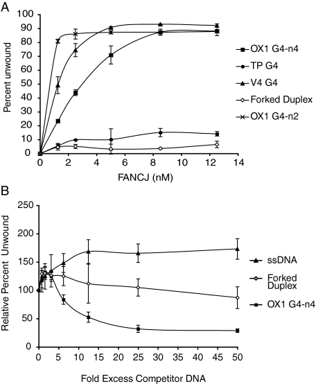
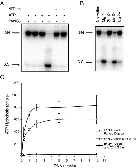
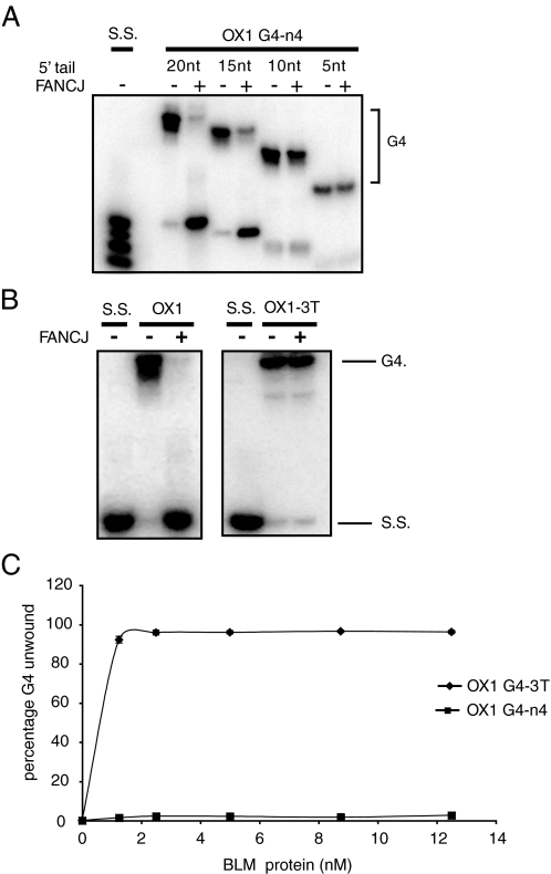
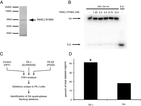
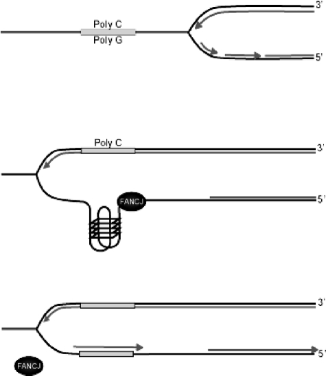
Fanconi anemia (FA) is a heritable human cancer-susceptibility disorder, delineating a genetically heterogenous pathway for the repair of replication-blocking lesions such as interstrand DNA cross-links. Here we demonstrate that one component of this pathway, FANCJ, is a structure-specific DNA helicase that dissociates guanine quadruplex DNA (G4 DNA) in vitro. Moreover, in contrast with previously identified G4 DNA helicases, such as the Bloom's helicase (BLM), FANCJ unwinds G4 substrates with 5'-3' polarity. In the FA-J human patient cell line EUFA0030 the loss of FANCJ G4 unwinding function correlates with the accumulation of large genomic deletions in the vicinity of sequences, which match the G4 DNA signature. Together these findings support a role for FANCJ in the maintenance of potentially unstable genomic G/C tracts during replication.


Figures

FIGURE 1.
FIGURE 1.
Purification of DNA substrates. DNA substrates were annealed and end-labeled as described under “Experimental Procedures.” Substrates were next purified after separation by preparative polyacrylamide gel electrophoresis. A, OX-1 (bimolecular (n-2) and tetramolecular (n-4) forms as indicated), TP-B, and V4 G4 DNA substrates are shown. Unannealed single-stranded DNA is also marked. B, G4 DNA substrates with shortened 5′ single-stranded tails are indicated. Excised are denoted by the boxes.
FIGURE 2.
FIGURE 2.
FANCJ is an ATP-dependent G4 unwinding enzyme. A, recombinant FANCJ protein was expressed and purified from Sf9 cells. Protein was resolved by SDS-PAGE and visualized using simply blue stain (Invitrogen). B, FANCJ unwinds G4 DNA substrates. 32P-End-labeled OX1 G4 DNA was incubated with increasing concentrations of FANCJ (as indicated) in the presence of ATP. Unwinding of G4 DNA is detected by the production of a single-stranded oligonucleotide DNA product (S.S.). Reaction products were analyzed by polyacrylamide gel electrophoresis and visualized using a PhosphorImager (GE Healthcare).
FIGURE 3.
FIGURE 3.
FANCJ preferentially unwinds G4 structure. A, FANCJ unwinds G4 DNA but not forked duplex DNA. The graph shows the efficiency at which FANCJ unwinds different DNA substrates (as indicated). Reactions were performed as described above. B, unwinding of OX1 DNA is not competed by preincubation with forked duplex DNA or single-stranded DNA competitors. Experiments were performed using 2 nm FANCJ as described under “Experimental Procedures.” G4 unwinding was normalized to reactions containing no competitor (100%).
FIGURE 4.
FIGURE 4.
G4 DNA unwinding requires ATP hydrolysis and magnesium ions. A, ATP requirement. Unwinding assays were performed as described in the legend Fig. 1, with 2 nm FANCJ but with no nucleotide, with 5 mm ATP or 5 mm ATPγS as indicated. B, cation requirement. G4 unwinding assay were performed as before with various cations (5 mm) as indicated. C, the ATPase function of FANCJ is stimulated by G4 and forked duplex DNA. ATP hydrolysis by FANCJ (10 pmol) was measured using thin layer chromatography and [γ-32P]ATP in the presence of different concentrations of forked duplex or OX1 G4 DNA as indicated. ATP hydrolysis of FANCJK52R protein in which the ATP binding site has been mutated was also measured.
FIGURE 5.
FIGURE 5.
FANCJ unwinds G4 DNA with 5′–3′ polarity. A, FANCJ-mediated unwinding of G4 DNA requires a single-stranded tail of ∼15 nt. FANCJ was incubated with 32P-end-labeled OX1 G4 DNA substrates that have ssDNA tails of different lengths (as indicated). Reactions were carried out as described in the legend to Fig. 1 and under “Experimental Procedures.” Whereas OX1 DNA substrates with tails of 15 or 20 nt were efficiently unwound, those with ssDNA tails of 5 or 10 nt were not unwound by FANCJ. Denatured substrates are shown as markers (S.S.) B, FANCJ unwinds G4 DNA only when the ssDNA tail is positioned 5′ to the G4 structure (OX1), indicating a 5′–3′ polarity. OX1 substrate with a 3′-ssDNA tail (OX1–3T) was not unwound. All reactions were performed under standard reaction conditions described under “Experimental Procedures.” C, BLM efficiently unwinds G4 DNA in the 3′ to 5′ direction. Reactions contained 2.5 nm OX1–3T and the indicated concentrations of BLM and were carried out for 30 min at 37 °C before products were resolved by SDS-PAGE and visualized using a PhosphorImager.
FIGURE 6.
FIGURE 6.
FA-J cells accumulate deletions in regions containing G4 DNA. A, mutant FANCJR798X protein was expressed and purified from Sf9 cells. Protein was resolved by SDS-PAGE and visualized using simply blue stain (Invitrogen). B, FANCJR798X does not unwind G4 DNA substrate in vitro. 32P-End-labeled OX1 G4 DNA was incubated with increasing concentrations of FANCJ (as indicated) in the presence of ATP. Migration of the expected single-stranded oligonucleotide DNA product is indicated (S.S.). Reaction products were analyzed by polyacrylamide gel electrophoresis and visualized using a PhosphorImager (GE Healthcare). C, schematic of array CGH analysis of control, FA-J, and FA-D2 cell lines and subsequent analysis of FA-J-specific deletion breakpoint regions for sequences predicted to form G4 quadruplexes. D, G4 quadruplex DNA flanks deletions in FA-J cells, in an orientation specific manner. 6 kb of sequence up- and downstream of each deletion observed by CGH as specific to FA-J cells was analyzed using the Quadparser algorithm to identify putative G4 DNA signatures. Deletions that associate with G4 DNA signatures were then classified by strand orientation. G4+ designates G4 DNA signatures that lies 3′ to a deleted region, and G4– are those regions that are associated with a G4 DNA signatures situated 5′ to the deleted region. Statistical analysis was performed using a Hidden Markov model to show significance (*, p = 0.04) for the correlation between G4+ DNA signatures and FA-J-specific deleted regions.
FIGURE 7.
FIGURE 7.
Model depicting the role of FANCJ in the maintenance of G-tracts. FANCJ resolves G4 DNA during replication. This model is a modification of that proposed by Cheung et al. (8) for DOG-1 in C. elegans. During replication in regions of the genome with long G/C tracts, the separation of DNA strands reveals stretches of ssDNA with the potential to form stable G4 DNA structures that may cause a block in DNA synthesis. In wild type cells FANCJ resolves this potential replication block by unwinding G4 DNA in a 5′–3′ direction, enabling synthesis to continue. In the absence of FANCJ/DOG-1 the G4 sequence is most likely excised and resected by nucleases and DNA rejoined by cellular DNA repair machinery giving rise to a deletion.

References

    1. Seal, S., Thompson, D., Renwick, A., Elliott, A., Kelly, P., Barfoot, R., Chagtai, T., Jayatilake, H., Ahmed, M., Spanova, K., North, B., McGuffog, L., Evans, D. G., Eccles, D., Easton, D. F., Stratton, M. R., and Rahman, N. (2006) Nat. Genet. 38 1239–1241 - PubMed
    1. Levitus, M., Waisfisz, Q., Godthelp, B. C., de Vries, Y., Hussain, S., Wiegant, W. W., Elghalbzouri-Maghrani, E., Steltenpool, J., Rooimans, M. A., Pals, G., Arwert, F., Mathew, C. G., Zdzienicka, M. Z., Hiom, K., De Winter, J. P., and Joenje, H. (2005) Nat. Genet. 37 934–935 - PubMed
    1. Litman, R., Peng, M., Jin, Z., Zhang, F., Zhang, J., Powell, S., Andreassen, P. R., and Cantor, S. B. (2005) Cancer Cell 8 255–265 - PubMed
    1. Levran, O., Attwooll, C., Henry, R. T., Milton, K. L., Neveling, K., Rio, P., Batish, S. D., Kalb, R., Velleuer, E., Barral, S., Ott, J., Petrini, J., Schindler, D., Hanenberg, H., and Auerbach, A. D. (2005) Nat. Genet. 37 931–933 - PubMed
    1. Joenje, H., and Patel, K. J. (2001) Nat. Rev. 2 446–457 - PubMed

Publication types

Substances