. 2008 Nov;90(11):2390-8.
doi: 10.2106/JBJS.H.00049.

Differentiation potential of multipotent progenitor cells derived from war-traumatized muscle tissue

Affiliations

Differentiation potential of multipotent progenitor cells derived from war-traumatized muscle tissue

Leon J Nesti et al. J Bone Joint Surg Am. 2008 Nov.

Abstract

Background: Recent military conflicts have resulted in numerous extremity injuries requiring complex orthopaedic reconstructive procedures, which begin with a thorough débridement of all contaminated and necrotic tissue in the zone of injury. The site of injury is also the site of healing, and we propose that débrided muscle tissue contains cells with robust reparative and regenerative potential.

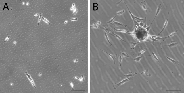
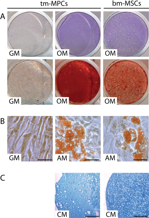
Methods: Débrided muscle from soldiers who had sustained traumatic open extremity injuries was collected during surgical débridement procedures at Walter Reed Army Medical Center. With modifications to a previously described stem-cell-isolation protocol, mesenchymal progenitor cells were harvested from traumatized muscle, enriched, expanded in culture, and exposed to induction media for osteogenesis, adipogenesis, and chondrogenesis.

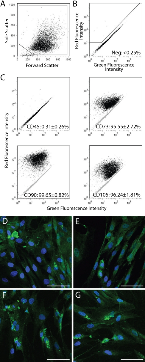
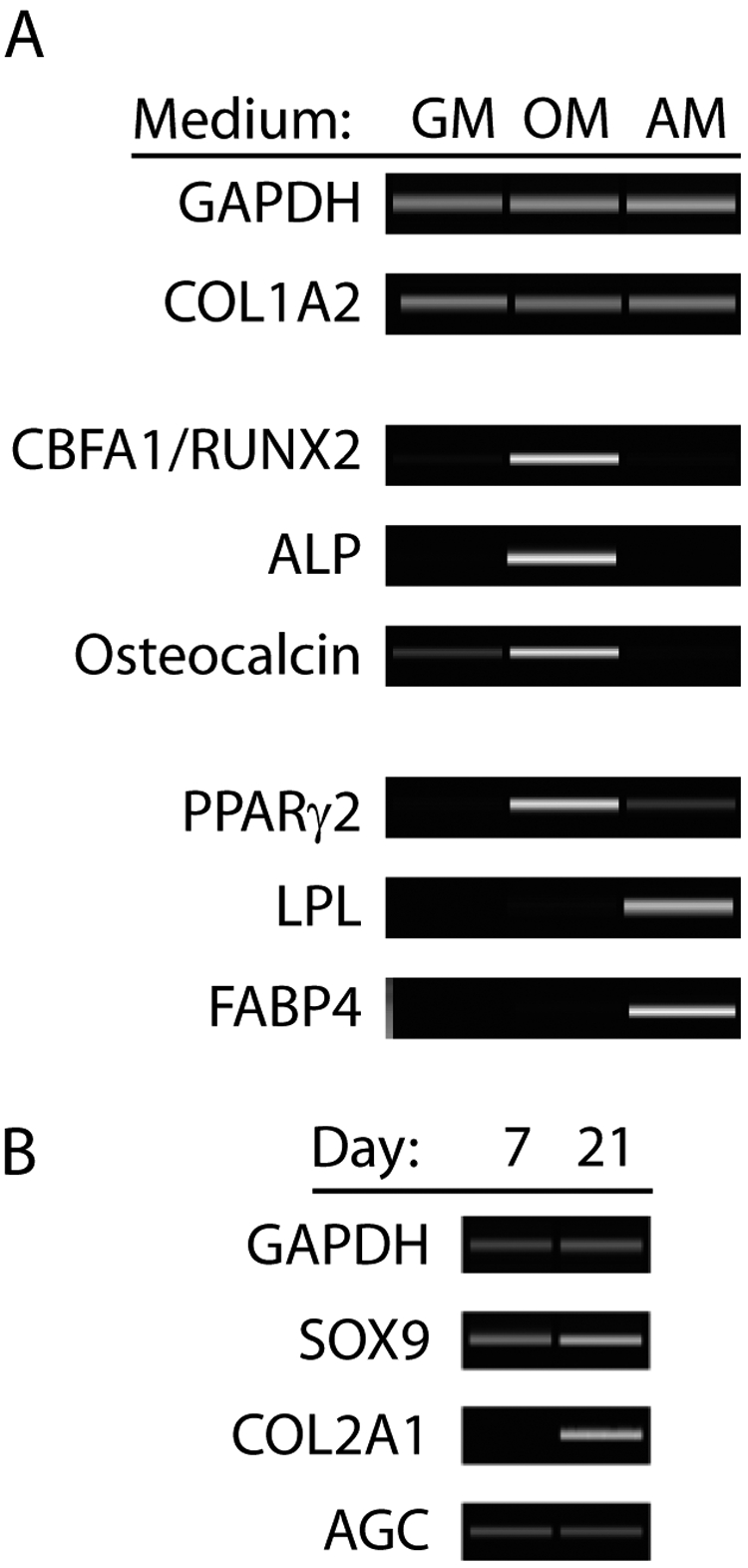
Results: The isolated mesenchymal progenitor cells stained positive for cell-surface markers (CD73, CD90, CD105), which are characteristic of adult human mesenchymal stem cells. Histological identification of lineage-specific markers demonstrated the potential of these cells to differentiate into multiple mesenchymal lineages. Reverse transcription-polymerase chain reaction analysis confirmed multilineage mesenchymal differentiation at the gene-expression level.

Conclusions: To our knowledge, the present report provides the first description of mesenchymal progenitor cell isolation from traumatized human muscle. These cells may play an integral role in tissue repair and regeneration and merit additional investigation as they could be useful in future cell-based tissue-engineering strategies.


Figures

Fig. 1
Fig. 1
Phase contrast microscopy showing multipotent cells obtained as adherent cells twenty-four hours after plating. Traumatized muscle-derived multiprogenitor cells (A) and bone marrow-derived mesenchymal stem cells (B) were plated, and, after two hours, the cultures were washed extensively with phosphate-buffered saline solution. Cells were visualized with phase contrast microscopy after twenty-four hours. The morphology of many of the muscle-derived cells is spindle-shaped and elongated, similar to that of bone marrow-derived mesenchymal stem cells. Bar = 100 μm.
Fig. 2
Fig. 2
A through G: Immunophenotyping of multiprogenitor cells derived from traumatized muscle. A: Whole multiprogenitor cells were identified and gated on the basis of their size (forward scatter) and granularity (side scatter). B: Multiprogenitor cells stained with a non-immunogenic isotype control were used to define the gate for positive phytoerythrin (red fluorescence) staining. C: Cells were stained with phytoerythrin-conjugated antibodies raised against CD45, CD73, CD90, and CD105. The average percentage of positive cells for each marker in ten multiprogenitor cell populations is listed in each panel. D through G: Multiprogenitor cells were stained with fluorescein isothiocyanate-conjugated antibodies raised against (D) CD29, (E) CD44, (F) CD105, and (G) CD146. Confocal laser scanning microscopy revealed positive green cell-surface staining for all four markers. Bar = 50 μm.
Fig. 3
Fig. 3
A, B, and C: Histological analysis of differentiated multiprogenitor cells derived from traumatized muscle. Traumatized muscle-derived mulitprogenitor cells (tm-MPCs) were cultured in growth medium (GM), osteogenic induction medium (OM), adipogenic induction medium (AM), or in three-dimensional pellet cultures with chondrogenic medium (CM). Bone marrow-derived mesenchymal stem cells (bm-MSCs) were used as positive controls for osteogenic, adipogenic, and chondrogenic differentiation. A: Multiprogenitor cells cultured in osteogenic induction medium exhibited enhanced alkaline phosphatase activity (Fast Blue BB; top) and mineralized matrix (alizarin red; bottom). B: Multiprogenitor cells cultured in adipogenic induction medium developed intracellular lipid droplets that stained positively with Oil Red O. Bar = 50 μm. C: Histological sections of the multiprogenitor cell pellet cultures showed positive matrix staining with alcian blue, suggesting the presence of sulfated proteoglycan-rich extracellular matrix. Bar = 500 μm.
Fig. 4
Fig. 4
A and B: Gene-expression analysis of differentiation of traumatized muscle-derived multiprogenitor cells. A: Traumatized muscle-derived multiprogenitor cells were cultured in growth medium (GM), osteogenic induction medium (OM), or adipogenic induction medium (AM) and then were lysed and RNA-extracted. Gene-expression profiles were then analyzed with use of reverse transcription polymerase chain reaction. Cells cultured in osteogenic induction medium showed upregulated expression of CBFA1/RUNX2, alkaline phosphatase (ALP), and osteocalcin, characteristic of osteoblastic phenotype. Cells cultured in adipogenic induction medium exhibited upregulated expression of PPARγ2, lipoprotein lipase (LPL), and fatty acid binding protein 4 (FABP4), characteristic of adipocytic phenotype. B: Pellet cultures of multiprogenitor cells in chondrogenic medium expressed the chondrogenic genes SOX9, COL2A1, and aggrecan (AGC). The expression of COL2A1 was upregulated from Day 7 to Day 21, suggesting chondrocyte maturation. Glyceraldehyde 3-phosphate dehydrogenase (GAPDH) expression was analyzed as a control for RNA loading.

References

    1. Jussila J. Measurement of kinetic energy dissipation with gelatine fissure formation with special reference to gelatine validation. Forensic Sci Int. 2005;150:53-62. - PubMed
    1. Jussila J, Kjellstrom BT, Leppaniemi A. Ballistic variables and tissue devitalisation in penetrating injury—establishing relationship through meta-analysis of a number of pig tests. Injury. 2005;36:282-92. - PubMed
    1. Williams AJ, Hartings JA, Lu XC, Rolli ML, Tortella FC. Penetrating ballistic-like brain injury in the rat: differential time courses of hemorrhage, cell death, inflammation, and remote degeneration. J Neurotrauma. 2006;23:1828-46. - PubMed
    1. Caterson EJ, Nesti LJ, Danielson KG, Tuan RS. Human marrow-derived mesenchymal progenitor cells: isolation, culture expansion, and analysis of differentiation. Mol Biotechnol. 2002;20:245-56. - PubMed
    1. Noth U, Osyczka AM, Tuli R, Hickok NJ, Danielson KG, Tuan RS. Multilineage mesenchymal differentiation potential of human trabecular bone-derived cells. J Orthop Res. 2002;20:1060-9. - PubMed

Publication types