

Macrophage infiltration into adipose tissue: initiation, propagation and remodeling
- PMID: 18978945
- PMCID: PMC2575346
- DOI: 10.2217/17460875.3.5.545
Macrophage infiltration into adipose tissue: initiation, propagation and remodeling
Abstract
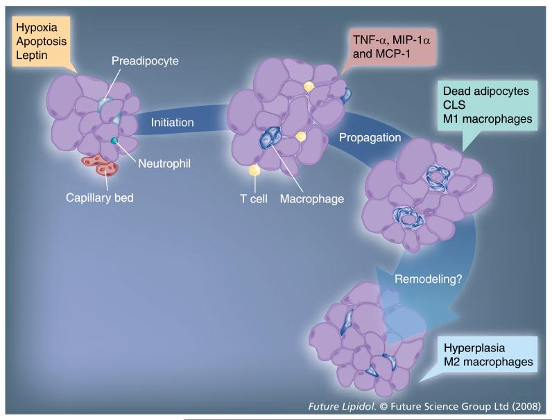
It has long been known that adipose tissue in obesity is in a heightened state of inflammation. Recently, our understanding of this has been transformed by the knowledge that immune cells such as macrophages and T cells can infiltrate adipose tissue and are responsible for the majority of inflammatory cytokine production. These seminal findings have opened up a new area in biology that is garnering the interest of scientists involved in research relating to cell motility, inflammation, obesity, physiology, diabetes and cardiovascular disease. Some important general questions relevant to this field are: how are macrophages recruited to adipose tissue in obesity? What are the physiological consequences of macrophage-adipocyte interactions? Do these inflammatory macrophages contribute to pathophysiological conditions associated with obesity, such as insulin resistance, dyslipidemia, diabetes and cardiovascular disease? This review focuses on the first of these important questions.
Figures
References
-
-
Xu H, Barnes GT, Yang Q, et al. Chronic inflammation in fat plays a crucial role in the development of obesity-related insulin resistance. J. Clin. Invest. 2003;112:1821–1830.•• Describes the observation that macrophages infiltrate adipose tissue in obesity. In addition, these authors demonstrate that the presence of adipose tissue macrophages temporally precedes systemic hyperinsulinemia.
-
-
-
Weisberg SP, McCann D, Desai M, Rosenbaum M, Leibel RL, Ferrante AW., Jr Obesity is associated with macrophage accumulation in adipose tissue. J. Clin. Invest. 2003;112:1796–1808.•• Demonstrated increased macrophage infiltration into white adipose tissue in obese mice and humans. The authors demonstrate that the adipose tissue macrophages are responsible for the majority of inflammatory cytokine production and that these cells are derived from the bone marrow.
-
-
- Kintscher U, Hartge M, Hess K, et al. T-lymphocyte infiltration in visceral adipose tissue. A primary event in adipose tissue inflammation and the development of obesity-mediated insulin resistance Arterioscler. Thromb. Vasc. Biol DOI: 10.1161/ATVBAHA.108.165100 2008. (Epub ahead of print). - PubMed
-
- Rausch ME, Weisberg S, Vardhana P, Tortoriello DV. Obesity in C57BL/6J mice is characterized by adipose tissue hypoxia and cytotoxic T-cell infiltration. Int. J. Obes. (Lond.) 2008;32:451–463. - PubMed
-
- Wu H, Ghosh S, Perrard XD, et al. T-cell accumulation and regulated on activation, normal T cell expressed and secreted upregulation in adipose tissue in obesity. Circulation. 2007;115:1029–1038. - PubMed
Grants and funding
LinkOut - more resources
Full Text Sources
Medical