. 2008;3(11):e3629.
doi: 10.1371/journal.pone.0003629. Epub 2008 Nov 3.

ICAM-2 expression mediates a membrane-actin link, confers a nonmetastatic phenotype and reflects favorable tumor stage or histology in neuroblastoma

Affiliations

ICAM-2 expression mediates a membrane-actin link, confers a nonmetastatic phenotype and reflects favorable tumor stage or histology in neuroblastoma

Karina Jin Yoon et al. PLoS One. 2008.

Abstract

The actin cytoskeleton is a primary determinant of tumor cell motility and metastatic potential. Motility and metastasis are thought to be regulated, in large part, by the interaction of membrane proteins with cytoplasmic linker proteins and of these linker proteins, in turn, with actin. However, complete membrane-to-actin linkages have been difficult to identify. We used co-immunoprecipitation and competitive peptide assays to show that intercellular adhesion molecule-2 (ICAM-2)/alpha-actinin/actin may comprise such a linkage in neuroblastoma cells. ICAM-2 expression limited the motility of these cells and redistributed actin fibers in vitro, and suppressed development of disseminated tumors in an in vivo model of metastatic neuroblastoma. Consistent with these observations, immunohistochemical analysis demonstrated ICAM-2 expression in primary neuroblastoma tumors exhibiting features that are associated with limited metastatic disease and more favorable clinical outcome. In neuroblastoma cell lines, ICAM-2 expression did not affect AKT activation, tumorigenic potential or chemosensitivity, as has been reported for some types of transfected cells. The observed ICAM-2-mediated suppression of metastatic phenotype is a novel function for this protein, and the interaction of ICAM-2/alpha-actinin/actin represents the first complete membrane-linker protein-actin linkage to impact tumor cell motility in vitro and metastatic potential in an in vivo model. Current work focuses on identifying specific protein domains critical to the regulation of neuroblastoma cell motility and metastasis and on determining if these domains represent exploitable therapeutic targets.


Conflict of interest statement

Competing Interests: The authors have declared that no competing interests exist.

Figures

Figure 1
Figure 1. In neuroblastoma cells, membrane-bound ICAM-2 associates with α-actinin and actin.
A) Immunoblot of whole cell lysates (35 µg protein/lane) of six neuroblastoma cell lines, comparing endogenous levels of expression of ICAM-2. B) Immunoblots of SK-N-AS cells transfected with control plasmid (pIRESneo) or with plasmid encoding ICAM-2 (pIRES.ICAM2) show that transfectants expressed similar levels of α-actinin, ezrin, and actin, but SK-N-ASpIRES.ICAM2 cells expressed a higher level of ICAM-2 than SK-N-ASpIRESneo cells (25 µg protein/lane). C) ICAM-2 expressed in SK-N-ASpIRES.ICAM2 cells localized to cell membranes (examples indicated by red arrows). D) Co-immunoprecipitation co-precipitation/mass spectroscopic analysis (actin) showed that actin co-precipitated with ICAM-2 in ICAM-2 transfectants. Band #1 was >99% identical to human actin. E) Immunoprecipitation/immunoblot showing equal amounts of α-actinin precipitated from lysates of SK-N-ASpIRESneo and SK-N-ASpIRES.ICAM2 cells. F) Immunoprecipitation/immunoblots showing that ICAM-2 and actin co-precipitated with α-actinin in lysates from SK-N-ASpIRES.ICAM2 cells, but little if any ICAM-2 and actin co-precipitated with α-actinin in SK-N-ASpIRESneo cells.
Figure 2
Figure 2. ICAM-2 expression affected the subcellular distribution of actin fibers and cell motility in two types of in vitro assays.
A) Confocal fluorescence microscopic images of actin fibers visualized by FITC-phalloidan (green) staining demonstrate that ICAM-2 expression redistributed F-actin from a transverse to a juxtamembrane localization. Cell nuclei were visualized using DAPI (blue). B) Competitive peptides to inhibit ICAM-2/α-actinin binding or α-actinin/actin binding reversed the ICAM-2-mediated juxtamembrane distribution of actin fibers in neuroblastoma cells. C) In scratch assays, ICAM-2 transfectants migrated more slowly than control transfectants. The width of scratches at 0 hours for pIRESneo and pIRES.ICAM2 transfectants were 1127 and 1044 pixels, respectively. At 72 hours, no gap remained in wells containing SK-N-ASpIRESneo cells, while a scratch distance of ∼450 pixel width remained in wells containing SK-N-ASpIRES.ICAM2 cells. D) In modified Boyden chamber assays, ICAM-2 expression inhibited invasion of SK-N-AS cells through Matrigel to the distal side of porous membranes, as detected by Diff-Quick (IMEB, Inc., San Marcos, CA), and quantitated using an Alpha Imager software, as detailed in Materials and Methods.
Figure 3
Figure 3. The effect of ICAM-2 on actin distribution and cell motility depends on an intact intracellular domain.
A) SK-N-AS cells were transfected to express a truncated form of ICAM-2 with a deleted cytoplasmic domain (ICAM2ΔCD). Expression of full length ICAM-2 and ICAM2ΔCD was similar in respective transfected cell lines. B) ICAM-2, α-actinin, and actin co-precipitated only in transfectants expressing full length ICAM-2. C) ICAM2ΔCD had no effect on actin fiber distribution. (Compare with Fig. 2A). D) ICAM2ΔCD did not affect migration of SK-N-AS cells in scratch assays. Pixel width of scratches for pIRESneo and pIRES.ICAM2ΔCD transfectants at 0 hours and 27 hours were similar. No gap remained for either cell line 45 hours after scratch.
Figure 4
Figure 4. ICAM-2 expression inhibited the development of disseminated tumors but did not affect establishment or development of local subcutaneous tumors.
A) ICAM-2 did not affect the growth of neuroblastoma cells as subcutaneous tumors in SCID mice, a measure of tumorigenic potential. B) ICAM-2 prevented the development of disseminated tumors, following intravenous tail vein injection of neuroblastoma cells, a measure of metastatic potential. C) Mice injected intravenously with SK-N-ASpIRES.ICAM-2 cells survived significantly longer (P<0.001) than mice receiving pIRESneo or pIRES.ICAM2ΔCD transfected cells. D) The failure of ICAM2ΔCD expression to suppress development of disseminated tumors was not due to cessation of expression of the transfected cDNA, since expression of ICAM-2ΔCD protein persisted until time of death.
Figure 5
Figure 5. RNAi techniques indicated that the observed ICAM-2-induced phenotype was unlikely to be due to artificial overexpression of this protein.
A) SJNB-1A cells express a relatively high endogenous level of ICAM-2 (Fig. 1A). The expression of ICAM-2 was inhibited in a population of SJNB-1A cells transfected to stably express ICAM-2 shRNA. This inhibition was transient even in the presence of selection pressure by G418 in vitro. B) Consistent with the proposed hypothesis, the high endogenous level of ICAM-2 expression in SJNB-1A cells is co-incident with a juxtamembrane distribution of actin fibers (left panel); and when ICAM-2 expression is decreased, actin fibers are transverse (right panel). C) Inhibition of ICAM-2 expression increased the development of disseminated neuroblastoma tumors in SCID mice injected intravenously with SJNB-1A ICAM-2 shRNA cells.
Figure 6
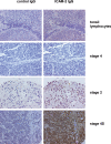
Figure 6. Immunohistochemical analyses ICAM-2 expression with a tissue microarray containing primary tumor specimens from 90 individual patients showed that a relatively high level of ICAM-2 expression was associated with primary tumor cells recognized to have limited metastatic potential.
ICAM-2 antibody binding was detected using a peroxidase-tagged secondary antibody and DAB substrate (brown), by standard methods. Specimens were counterstained with hematoxylin/eosin, to visualize morphology of tumor and stromal cells. Staining was scored as: percent of stained cells×staining intensity (0, 1+, 2+, or 3+). The scores for ICAM-2 expression of the Stage 4, Stage 2, and Stage 4S tumor specimens pictured were 0, 270, and 300, respectively. Normal lymphocytes (a positive control) received a score of 100 (100% of lymphocytes×a staining intensity of 1+). Each specimen is described in more detail in the text.

References

    1. Bretscher A. Regulation of cortical structure by the ezrin-radixin-moesin protein family. Curr Opin Cell Biol. 1999;11:109–116. - PubMed
    1. Sun C-X, Robb VA, Gutmann DH. Protein 4.1 tumor suppressors: getting a FERM grip on growth regulation. J Cell Sci. 2002;2002:1153991–3400. - PubMed
    1. Yamazaki D, Kurisu S, Takenawa T. Regulation of cancer cell motility through actin reorganization. Cancer Sci. 2005;96:379–386. - PMC - PubMed
    1. Honda K, Yamada T, Endo R, Ino Y, Gotoh M, et al. Actinin-4, a novel actin-bundling protein associated with cell mobility and cancer invasion. J Cell Biol. 1998;140:1383–1393. - PMC - PubMed
    1. Gluck U, Ben-Ze'ev A. Modulation of alpha-actinin levels affects cell motility and confers tumorigenicity on 3T3 cells. J Cell Sci. 1994;107:1773–1782. - PubMed

Publication types

MeSH terms