. 2009 Feb;26(2):395-403.
doi: 10.1007/s11095-008-9756-3. Epub 2008 Nov 1.

Intrascleral drug delivery to the eye using hollow microneedles

Affiliations

Intrascleral drug delivery to the eye using hollow microneedles

Jason Jiang et al. Pharm Res. 2009 Feb.

Abstract

Purpose: This study tested the hypothesis that hollow microneedles can infuse solutions containing soluble molecules, nanoparticles, and microparticles into sclera in a minimally invasive manner.

Methods: Individual hollow microneedles were inserted into, but not across, human cadaver sclera and aqueous solutions containing sulforhodamine or fluorescently tagged nanoparticles or microparticles were infused into sclera at constant pressure. The infused volume of fluid was measured and imaged histologically as a function of scleral thickness, infusion pressure, needle retraction depth and the presence of spreading enzymes (hyaluronidase and collagenase).

Results: Individual hollow microneedles were able to insert into sclera. Fluid infusion was extremely slow after microneedle insertion into the sclera without retraction, but partial retraction of the microneedle over a distance of 200-300 microm enabled infusion of 10-35 microl of fluid into the tissue. Scleral thickness and infusion pressure had insignificant effects on fluid delivery. Nanoparticle suspensions were also delivered into sclera, but microparticles were delivered only in the presence of hyaluronidase and collagenase spreading enzymes, which suggested the role of scleral glycosaminoglycans and collagen fibers as rate-limiting barriers.

Conclusion: This study shows that hollow microneedles can infuse solutions into the sclera for minimally invasive delivery of soluble molecules, nanoparticles and microparticles.


Figures
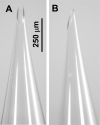
Figure 1
Figure 1
Representative hollow microneedle: (A) front view and (B) side view. The microneedle shown has an approximately elliptical tip opening with a long axis of ~100 μm and a short axis of ~40 μm with a bevel tip angle of 25°.
Figure 2
Figure 2
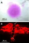
Representative human cadaver sclera after microneedle infusion of sulforhodamine solution. (A) Top view image of the scleral surface using bright-field microscopy shows infusion of sulforhodamine over an area of many square millimeters. (B) Histological section using fluorescence microscopy shows the site of microneedle insertion (indicated by arrows) and the distribution of injected sulforhodamine preferentially localized to the upper portion of the tissue. A microneedle was inserted 720 μm into the sclera and then retracted 60 μm every 3 min to a maximum retraction of 240 μm. Sulforhodamine solution was infused into the tissue at a pressure of 15 psi.
Figure 3
Figure 3
Effect of microneedle retraction on fluid delivery into anterior, medial and posterior regions of human cadaver sclera. In each experiment, a microneedle was inserted into the sclera at a distance of 720 – 1080 μm and sulforhodamine solution was infused at a pressure of 15 psi. Every 3 min, the microneedle was retracted 60 μm. The bar on the left of each pair of bars shows the infusion volume after insertion without retraction. The bar on the right of each pair shows the infusion volume after insertion and retraction. Along the x axis, the reported insertion distance is the average insertion distance of that experimental data set. The reported retraction distance is the average amount of microneedle retraction needed before appreciable fluid flow began. Data are expressed as mean values (n ≥ 17) with standard deviation bars.
Figure 4
Figure 4
Effect of pressure on fluid delivery into anterior, medial and posterior regions of human cadaver sclera. Individual microneedles were inserted 720 μm into the sclera and then retracted 60 μm every 3 min to a maximum retraction of 140 – 300 μm with an infusion pressure of 5 (■), 10 (formula image), 15 (formula image), 20 (formula image) and 25 (formula image) psi. Data are expressed as mean values (n ≥ 3) with standard deviation bars.
Figure 5
Figure 5
Representative histological image of human cadaver sclera after infusion of fluorescent nanoparticles (280 μm diameter). The microneedle was inserted 720 μm into the anterior region of sclera and then retracted 60 μm every 3 min to a maximum retraction of 240 μm. A 1.0 wt% nanoparticles suspension was infused into the tissue at a pressure of 15 psi over 15 min. The site of microneedle insertion is indicated by arrows.
Figure 6
Figure 6
Representative histological images of human cadaver sclera after infusion of fluorescent nanoparticles as a function of nanoparticle concentration and scleral position. Microneedles were inserted 720 – 1080 μm into anterior, medial and posterior regions of the sclera and then retracted 60 μm every 3 min to a maximum retraction of 240 – 360μm. In each experiment, a 20 μl suspension with a solids content of 0.5, 1, 5 or 10 wt% was infused into the tissue at a pressure of 15 psi. Dotted lines have been added to each image to more clearly indicate the upper and lower edges of the scleral tissue.
Figure 7
Figure 7
Representative histological images of human cadaver sclera after infusion of fluorescent microparticles as a function of exposure to the spreading enzymes (A) collagenase and (B) hyaluronidase. Tissues were either untreated (control), incubated in the enzyme for 1 h before microparticle infusion (pretreatment), or co-injected with a mixture of the enzyme microparticles (co-injection). Microneedles were inserted 720 – 960 μm into anterior, medial and posterior regions of the sclera and then retracted 60 μm every 3 min to a maximum retraction of 240 – 300μμm. In each experiment, a 20 μl suspension with a solids content of 1.3 wt% was infused into the tissue at a pressure of 15 psi. Dotted lines have been added to each image to more clearly indicate the upper and lower edges of the scleral tissue.

References

    1. Ghate D, Edelhauser HF. Ocular drug delivery. Expert Opin Drug Deliv. 2006;3:275–87. - PubMed
    1. Del Amo EM, Urtti A. Current and future ophthalmic drug delivery systems. A shift to the posterior segment. Drug Discov Today. 2008;13:135–43. - PubMed
    1. Maurice DM. Drug delivery to the posterior segment from drops. Surv Ophthalmol. 2002;47(Suppl 1):S41–52. - PubMed
    1. Jager RD, Aiello LP, Patel SC, Cunningham ET., Jr. Risks of intravitreous injection: a comprehensive review. Retina. 2004;24:676–98. - PubMed
    1. Geroski DH, Edelhauser HF. Transscleral drug delivery for posterior segment disease. Adv Drug Deliv Rev. 2001;52:37–48. - PubMed

Publication types