. 2009 Feb;322(1-2):15-23.
doi: 10.1007/s11010-008-9935-x. Epub 2008 Nov 4.

The proteoglycan osteoglycin/mimecan is correlated with arteriogenesis

Affiliations

The proteoglycan osteoglycin/mimecan is correlated with arteriogenesis

Andreas Kampmann et al. Mol Cell Biochem. 2009 Feb.

Abstract

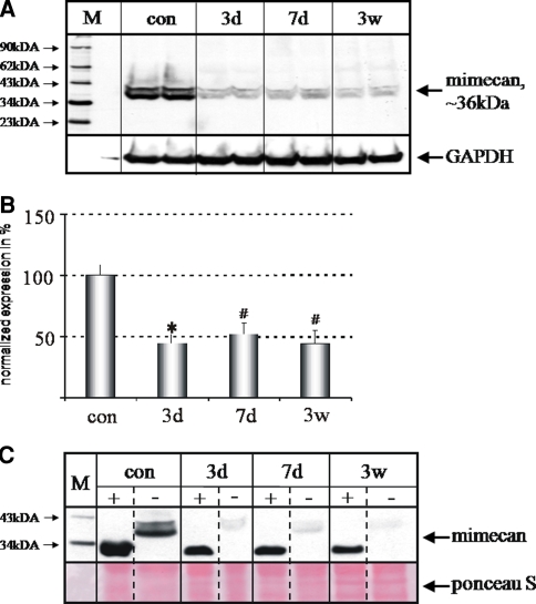
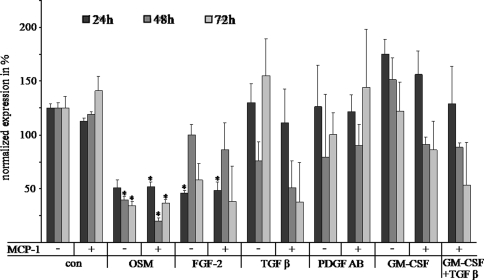
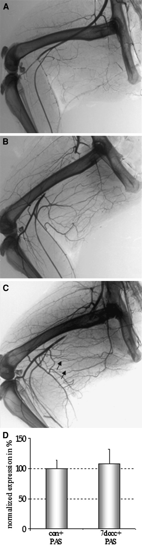
Arteriogenesis or collateral growth is able to compensate for the stenosis of major arteries. Using differential display RT-PCR on growing and quiescent collateral arteries in a rabbit femoral artery ligation model, we cloned the rabbit full-length cDNA of osteoglycin/mimecan. Osteoglycin was present in the adventitia of collateral arteries as a glycosylated protein without keratan sulfate side chains, mainly produced by smooth muscle cells (SMCs) and perivascular fibroblasts. Northern blot, Western blot, and immunohistochemistry confirmed a collateral artery-specific downregulation of osteoglycin from 6 h to 3 weeks after the onset of arteriogenesis. Treatment of primary SMCs with the arteriogenic protein fibroblast growth factor-2 (FGF-2) resulted in a similar reduction of osteoglycin expression as observed in vivo. Application of the FGF-2 inhibitor polyanethole sulfonic acid (PAS) blocked the downregulation of osteoglycin and interfered with arteriogenesis. From our study we conclude that downregulation of osteoglycin is a fundamental requirement for proper arteriogenesis.


Figures

Fig. 1
Fig. 1
Sequence of rabbit osteoglycin. The rabbit osteoglycin cDNA consisted of 2,493 bp and contained an open reading frame coding for a protein of 298aa. The protein starts with a signal peptide of 19 amino acids (underlined). The three consensus sequences for N-linked glycosylation are printed in red. The leucine-rich repeats of the S-type are marked in blue, that of the T-type in green
Fig. 2
Fig. 2
Osteoglycin mRNA expression at various time points after occlusion of the femoral artery. ABar graphs representing Northern blot results on osteoglycin expression in growing collateral arteries from 3 h to 3 weeks after femoral artery ligation (for 3–24 h: = 4 for each time point; for 3d to 3w: = 8 for each time point; control: = 8). B Representative Northern blot showing osteoglycin expression (top) in quiescent (con) and growing collateral arteries at days 3, 7 as well as 3 weeks after femoral artery occlusion. The blot was rehybridized with an 18S rRNA specific probe (bottom). Results are expressed as mean ± SEM (* P < 05 versus con; con control)
Fig. 3
Fig. 3
Western blot analyses of osteoglycin. A Representative Western blot showing the level of osteoglycin protein in growing collateral arteries at days 3, 7 and 3 weeks after femoral artery occlusion as well as in control vessels. B Quantification of the Western blot analyses. n = 7 for control and for d3, n = 6 for d7, and n = 5 for 3 weeks. Results are expressed as mean ± SEM (* P < 05 versus con). C Protein preparations before (−) and after (+) deglycosylation (n = 4 for each time point). Ponceau S staining was used in all Western blots to demonstrate equal loading of the protein samples. (M Cruz marker molecular weight standard, con control, 3d 3 days after occlusion, 7d 7 days after occlusion, 3w 3 weeks after occlusion)
Fig. 4
Fig. 4
Localization of osteoglycin mRNA (A, C, D), protein (B, E, F), and the proliferation marker ki-67 (G) in collateral arteries and surrounding tissue. A, C, D In situ hybridization. A The hybridization with the sense probe was used as a negative control. C, D Positive signals for osteoglycin were detected in the SMCs of the collateral media (arrows) and in fibroblasts (arrowheads). B, E, F Immunohistochemistry. B Negative control for immunoperoxidase reaction. E Quiescent collateral arteries showed a thin media and continuous internal elastic lamina (arrow). Note the prominent staining for osteoglycin in the adventitia (black arrowheads). F, G (consecutive sections): After 7 days of femoral artery occlusion growing collateral arteries show fragmented elastic lamina (arrows in F), prominent neointima formation (NI in F), and SMC proliferation (arrows in G; ki67 staining). Note the strong reduction of osteoglycin protein in the adventitia of growing collateral arteries (compare black arrowheads in E and F), whereas in the fascia of the muscle the signal intensity remains the same (red arrowheads)
Fig. 5
Fig. 5
Stimulation of SMCs with distinct cytokines and growth factors. Bar graphs representing Northern blot results on osteoglycin expression in primary rabbit SMCs after 24, 48, and 72 h of stimulation with distinct growth factors and cytokines. The factors and concentrations used are indicated in the “Material and Methods” section. Results are expressed as mean ± SEM (* P < 0.05 versus con; con control, OsM Oncostatin M). All experiments were performed in triplicate
Fig. 6
Fig. 6
Application of PAS. Postmortem angiograms of rabbit hind limbs. A After 1 week of infusion of PAS without ligation of the femoral artery. B After 1 week of femoral artery ligation with concomitant infusion of PAS. C After 1 week of femoral artery ligation without infusion of PAS. Upon femoral artery ligation, only rabbits not treated with PAS (C) showed collateral arteries with corkscrew formation (arrows), a typical sign for growing collaterals. DBar graphs representing Northern blot results on osteoglycin expression after 1 week of infusion of PAS in resting collateral arteries (con + PAS) and in growing collateral arteries 7 days after ligation of the femoral artery (7d occ + PAS; = 4). Results are expressed as mean ± SEM. No statistical difference was found

Similar articles

Cited by

References

    1. WHO (1999) WHO Library Cataloguing in Publication Data. The world health report 1999: Making a difference I. Title: Making a difference ISBN 92 4 156194 7 (NLM Classification: WA 540.1) ISSN 1020-3311
    1. None
    2. Schaper W, Piek JJ, Munoz-Chapuli R et al (1999) Collateral circulation of the heart. In: Ware JA, Simons M (eds) Angiogenesis and cardiovascular disease. Oxford University Press, New York, pp 159–198
    1. {'text': '', 'ref_index': 1, 'ids': [{'type': 'DOI', 'value': '10.1161/01.RES.0000242560.77512.dd', 'is_inner': False, 'url': 'https://doi.org/10.1161/01.res.0000242560.77512.dd'}, {'type': 'PubMed', 'value': '16931799', 'is_inner': True, 'url': 'https://pubmed.ncbi.nlm.nih.gov/16931799/'}]}
    2. Eitenmuller I, Volger O, Kluge A et al (2006) The range of adaptation by collateral vessels after femoral artery occlusion. Circ Res 99:656–662. doi:10.1161/01.RES.0000242560.77512.dd - PubMed
    1. {'text': '', 'ref_index': 1, 'ids': [{'type': 'PubMed', 'value': '12692088', 'is_inner': True, 'url': 'https://pubmed.ncbi.nlm.nih.gov/12692088/'}]}
    2. Deindl E, Ziegelhoffer T, Kanse SM et al (2003) Receptor-independent role of the urokinase-type plasminogen activator during arteriogenesis. FASEB J 17:1174–1176 - PubMed
    1. {'text': '', 'ref_index': 1, 'ids': [{'type': 'PubMed', 'value': '9321814', 'is_inner': True, 'url': 'https://pubmed.ncbi.nlm.nih.gov/9321814/'}]}
    2. Ito WD, Arras M, Winkler B et al (1997) Angiogenesis but not collateral growth is associated with ischemia after femoral artery occlusion. Am J Physiol 273:H1255–H1265 - PubMed

LinkOut - more resources