. 2008 Nov 25;47(47):12515-22.
doi: 10.1021/bi8008328.

Impeded electron transfer from a pathogenic FMN domain mutant of methionine synthase reductase and its responsiveness to flavin supplementation

Affiliations

Impeded electron transfer from a pathogenic FMN domain mutant of methionine synthase reductase and its responsiveness to flavin supplementation

Carmen G Gherasim et al. Biochemistry. .

Abstract

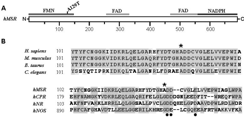
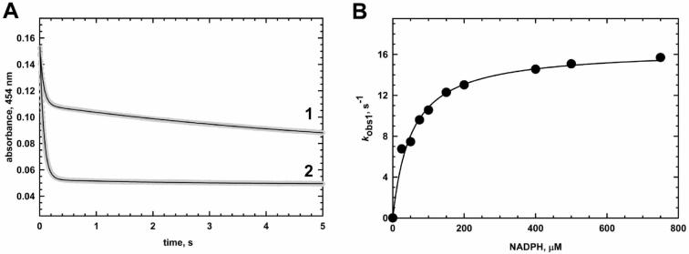
Methionine synthase reductase (MSR) is a diflavin oxidoreductase that transfers electrons from NADPH to oxidized cobalamin and plays a vital role in repairing inactive cobalamin-dependent methionine synthase. MSR deficiency is a recessive genetic disorder affecting folate and methionine metabolism and is characterized by elevated levels of plasma homocysteine. In this study, we have examined the molecular basis of MSR dysfunction associated with a patient mutation, A129T, which is housed in the FMN binding domain and is adjacent to a cluster of conserved acidic residues found in diflavin oxidoreductases. We show that the substitution of alanine with threonine destabilizes FMN binding without affecting the NADPH coenzyme specificity or affinity, indicating that the mutation's effects may be confined to the FMN module. The A129T MSR mutant transfers electrons to ferricyanide as efficiently as wild type MSR but the rate of cytochrome c, 2,6-dichloroindophenol, and menadione reduction is decreased 10-15 fold. The mutant is depleted in FMN and reactivates methionine synthase with 8% of the efficiency of wild type MSR. Reconstitution of A129T MSR with FMN partially restores its ability to reduce cytochrome c and to reactivate methionine synthase. Hydrogen-deuterium exchange mass spectrometric studies localize changes in backbone amide exchange rates to peptides in the FMN-binding domain. Together, our results reveal that the primary biochemical penalty associated with the A129T MSR mutant is its lower FMN content, provide insights into the distinct roles of the FAD and FMN centers in human MSR for delivering electrons to various electron acceptors, and suggest that patients harboring the A129T mutation may be responsive to riboflavin therapy.


Figures

Figure 1
Figure 1
(A) Position of the A129T mutation in MSR relative to the FAD/NADPH and FMN binding domains. (B) Multiple sequence alignment of MSR homologs from other organisms and other diflavin oxidoreductases, in the region encompassing the mutated residue. The sequence alignment was prepared using BioEdit software using the sequence of MSR for the following species: Homo sapiens (NP_002445), Mus musculus (NP_766068), Bos taurus (AAY86763), Caenorhabditis elegans (Q17574) and cytochrome P450 reductase (NP_000932), novel reductase 1 (NP_055249) and neuronal nitric oxide synthase (NP_000611) respectively, all from Homo sapiens. The A129 residue, mutated in this study is represented by *, and the putative acidic cluster residues that are proximal to A129 in MSR are denoted by black dots.
Figure 2
Figure 2
Activation of methionine synthase by A129T and wild-type MSR. The activity of methionine synthase was determined as described previously (10) at the indicated A129T (○) or wild-type (●) MSR:methionine synthase (MS) ratios.
Figure 3
Figure 3
Kinetics of flavin reduction in wild type and A129T MSR determined by stopped-flow spectroscopy. (A) Single-wavelength absorption experiments for the reduction of 7 μM wild-type (1) and 11.27 μM A129T MSR (2) with 500 μM NADPH were followed at 454 nm under anaerobic conditions in 50 mM potassium phosphate, pH 7.0 at 25°C. Biphasic absorption traces for the reduction of MSR proteins were obtained by fitting the data in panels A with a double exponential equation as described under Methods. The black line running through each experimental trace (shown in gray) represents the best fit using equation 3. Panel B shows the dependence of kobs1 for the A129T MSR reaction on NADPH concentration.
Figure 4
Figure 4
Effect of exogenous FMN on MSR activity. (A) Cytochrome c reductase activity of wild-type (●) and A129T (○) MSR in the absence or presence of various FMN concentrations were measured by incubating the respective proteins in the assay mixture containing 50 mM Tris pH 7.5, 60 μM cytochrome c and 100 μM NADPH and 0-60 μM FMN in a final volume of 1 ml. Reactions were initiated by addition of NADPH and the initial velocities were monitored at 37°C; (B) Methionine synthase activity determined in the presence or absence of exogenous FMN when wild-type (●) or A129T (○) MSR where used as reactivating protein partner.
Figure 5
Figure 5
Differences in the kinetics of deuterium incorporation in A129T MSR (○) versus wild-type MSR (●). Time courses are shown for peptide 36-55 (A), peptide 56-75 (B) and peptide 86-102 (C).
Figure 6
Figure 6
(A). Map of peptides that exhibit differences in H/D exchange kinetics in the A129T mutant compared to wild-type MSR. Peptides identified by H/D exchange studies map to the FMN binding domain of MSR and are shown in green (36-55), magenta (56-75) and red (86-102) and the A129 residue is shown in surface representation. The modeled structure for the FMN binding domain was created using the alignment mode of the SWISS-MODEL program (www.swissmodel.expasy.org/SWISS-MODEL.html) with cytochrome P450 reductase as template. (B) Location of the A129T mutation in the modeled structure of the FMN-binding domain of MSR. Superimposition of cytochrome P450 reductase (PDB file 1AMO) and a modeled structure of the FMN domain of human MSR showing the location of A129 relative to the FMN cofactor. The putative acidic cluster residues in MSR and the FMN cofactor are shown in ball and stick representation

Similar articles

Cited by

References

    1. Mills JL, McPartlin JM, Kirke PN, Lee YJ, Conley MR, Weir DG, Scott JM. Homocysteine metabolism in pregnancies complicated by neural-tube defects. Lancet. 1995;345:149–151. - PubMed
    1. Clarke R, Smith AD, Jobst KA, Refsum H, Sutton L, Ueland PM. Folate, vitamin B12, and serum total homocysteine levels in confirmed Alzheimer disease. Arch Neurol. 1998;55:1449–1455. - PubMed
    1. Refsum H, Ueland PM, Nygard O, Vollset SE. Homocysteine and Cardiovascular Disease. Annu. Rev. Medicine. 1998;49:31–62. - PubMed
    1. Rosenblatt DS. Inborn errors of folate and cobalamin metabolism. Cambridge University Press; Cambridge: 2001.
    1. Finkelstein JD. Inborn errors of sulfur-containing amino acid metabolism. J Nutr. 2006;136:1750S–1754S. - PubMed

Publication types

MeSH terms

Associated data