Review
. 2008 Nov 1;14(21):6744-50.
doi: 10.1158/1078-0432.CCR-08-1032.

Improving gemcitabine-mediated radiosensitization using molecularly targeted therapy: a review

Affiliations
Review

Improving gemcitabine-mediated radiosensitization using molecularly targeted therapy: a review

Meredith A Morgan et al. Clin Cancer Res. .

Abstract

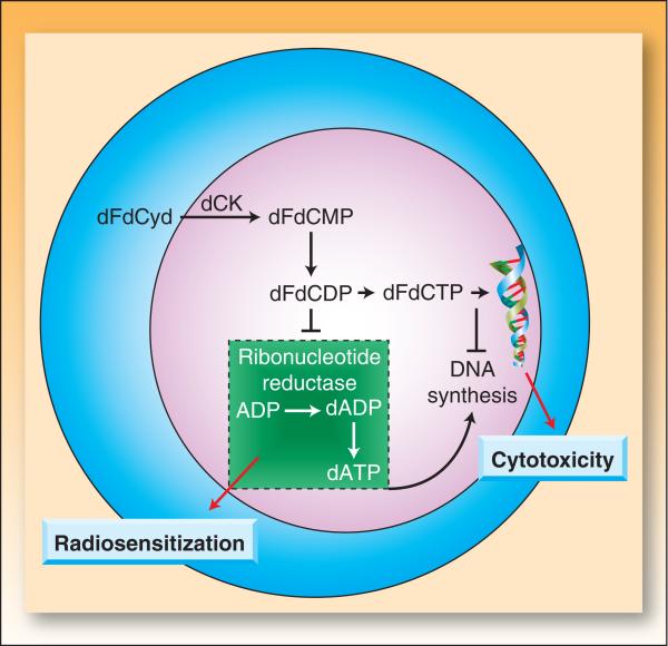
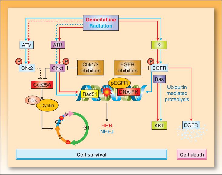
In the last three decades, gemcitabine has progressed from the status of a laboratory cytotoxic drug to a standard clinical chemotherapeutic agent and a potent radiation sensitizer. In an effort to improve the efficacy of gemcitabine, additional chemotherapeutic agents have been combined with gemcitabine (both with and without radiation) but with toxicity proving to be a major limitation. Therefore, the integration of molecularly targeted agents, which potentially produce less toxicity than standard chemotherapy, with gemcitabine radiation is a promising strategy for improving chemoradiation. Two of the most promising targets, described in this review, for improving the efficacy of gemcitabine radiation are epidermal growth factor receptor and checkpoint kinase 1.


Figures

Figure 1
Figure 1. Gemcitabine mechanisms of action
Following cellular incorporation, gemcitabine (dFdCyd) undergoes a series of sequential phosphorylations mediated by deoxycytidine kinase. dFdCDP is a direct inhibitor of ribonucleotide reductase which results in inhibition of deoxyribonucleotide triphosphate synthesis, specifically deoxyadenosine triphosphate (dATP). Depletion of dATP pools is crucial for radiosensitization (19). dFdCTP is incorporated into DNA during synthesis and contributes to cytotoxicity (88-90).
Figure 2
Figure 2. The effect of caspase 9 on gemcitabine-mediated radiosensitization
Wild type or caspase 9 dominant negative (DN) MCF-7 cells were exposed to equicytotoxic concentrations of gemcitabine for 24 hours prior to treatment with 0 − 8 Gy radiation. Survival was determined by a clonogenic survival assay as previously described (91). Data are expressed as the radiation enhancement ratio which was calculated as the ratio of the mean inactivation dose for drug treated cells to non-drug treated cells (ER = 1). Data are from the mean of 3 independent experiments ± standard error.
Figure 3
Figure 3. The effects of gemcitabine and radiation on cell cycle checkpoints and EGFR signaling
Radiation-induced double strand breaks or gemcitabine-induced replication stress trigger the activation of ataxia telangiectasia mutated (ATM) and ATM and Rad3-related (ATR) kinases, respectively (92). Active ATM/ATR phosphorylate and activate Chk1 and Chk2 (93-95) which phosphorylate Cdc25 phosphatases, leading to their inactivation through degradation (Cdc25A) or cytoplasmic sequestration (Cdc25C) (93, 96). In the absence of Cdc25 phosphatase activity, cyclin dependent kinases (Cdk1 and Cdk2) remain bound by inhibitory phosphorylations, resulting in arrest of the cell cycle at G1/S, intra-S, or G2/M. Treatment of cells with gemcitabine prior to radiation results in radiosensitization that can be attributed to a number of events (Box 1), including dATP depletion and S-phase arrest. Inhibition of Chk1 sensitizes cells to gemcitabine and radiation by a number of potential mechanisms including abrogation of cell cycle arrest, premature mitotic entry, and inhibition of Rad 51 focus formation resulting in impaired homologous recombination repair (HRR). EGFR is phosphorylated in response to radiation or gemcitabine by an unknown mechanism(s) (97). Radiation triggers translocation of EGFR into the nucleus (58, 60). This process coincides with transport of Ku70/80 and protein phosphatase 1 into the nucleus, resulting in increases in DNA-PK, repair of DNA-strand breaks (NHEJ; nonhomologous endjoining), and cell survival. Activation of EGFR in response to gemcitabine can also result in activation of the survival signal AKT (46). Activating Ras mutations can result in activation of Ras-dependent pathways, such as PI3K/AKT, even in the presence of EGFR inhibitors. EGFR inhibitors prevent gemcitabine and/or radiation-mediated EGFR signaling and are thought to impair cell survival signals and DNA repair. EGFR inhibition blocks nuclear transport of EGFR and DNA-PK activity (60, 98). In some instances, phosphorylation of EGFR by gemcitabine promotes ubiquitination of the receptor leading to degradation along a proteosome/lysosome pathway (63). EGFR degradation results in down-regulation of the survival signal pAKT, leading to apoptosis. Blocking EGFR degradation at various steps of this pathway reduces gemcitabine-mediated cytotoxicity. Whether an EGFR-activating insult leads to cell survival or cell death may ultimately be determined by the severity and duration of the stress. The colored arrows indicate the effects mediated by gemcitabine (red) versus radiation (blue). Dotted lines indicate less pronounced effects.
Figure 4
Figure 4. The effects of Chk1 inhibition on radiation and chemoradiation sensitivity
MiaPaca-2 cells were treated with 30 nM PD-321852 for 24 hrs pre- and post-ionizing radiation (0 −10 Gy) (A) or for 2 hours with gemcitabine (50nM) and then with AZD7762 (100nM) for 1 hour pre- and 24 hours post-irradiaiton (B). Cells were then plated at cloning densities and grown for 10 days to determine the surviving fraction, which represents the fraction of cells surviving radiation treatment relative to un-irradiated controls. Cell survival curves were then fitted using the linear quadratic equation, and the mean inactivation dose was calculated according to the method of Fertil et al. (99). The radiation enhancement ratio was calculated by dividing the mean inactivation dose under control conditions by the mean inactivation dose of Chk1 inhibitor-treated cells.

Similar articles

Cited by

References

    1. Safran H, Dipetrillo T, Iannitti D, et al. Gemcitabine, paclitaxel, and radiation for locally advanced pancreatic cancer: a Phase I trial. International journal of radiation oncology, biology, physics. 2002;54:137–41. - PubMed
    1. Blackstock AW, Melin SA, Butler JM, et al. Irinotecan/gemcitabine followed by twice-weekly gemcitabine/radiation in locally advanced pancreatic cancer. Oncology (Williston Park) 2002;16:25–8. - PubMed
    1. Kachnic LA, Shaw JE, Manning MA, Lauve AD, Neifeld JP. Gemcitabine following radiotherapy with concurrent 5-fluorouracil for nonmetastatic adenocarcinoma of the pancreas. International journal of cancer. 2001;96:132–9. - PubMed
    1. Symon Z, Davis M, McGinn CJ, Zalupski MM, Lawrence TS. Concurrent chemoradiotherapy with gemcitabine and cisplatin for pancreatic cancer: from the laboratory to the clinic. International journal of radiation oncology, biology, physics. 2002;53:140–5. - PubMed
    1. Morgan MA, Meirovitz A, Davis MA, Kollar LE, Hassan MC, Lawrence TS. Radiotherapy combined with gemcitabine and oxaliplatin in pancreatic cancer cells. Translational Oncology. 2008;1:36–43. - PMC - PubMed

MeSH terms