

Cell proliferation, cell cycle abnormalities, and cancer outcome in patients with Barrett's esophagus: a long-term prospective study
- PMID: 18980994
- PMCID: PMC2587072
- DOI: 10.1158/1078-0432.CCR-07-5063
Cell proliferation, cell cycle abnormalities, and cancer outcome in patients with Barrett's esophagus: a long-term prospective study
Abstract
Purpose: Elevated cellular proliferation and cell cycle abnormalities, which have been associated with premalignant lesions, may be caused by inactivation of tumor suppressor genes. We measured proliferative and cell cycle fractions of biopsies from a cohort of patients with Barrett's esophagus to better understand the role of proliferation in early neoplastic progression and the association between cell cycle dysregulation and tumor suppressor gene inactivation.
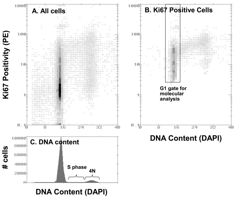
Experimental design: Cell proliferative fractions (determined by Ki67/DNA multiparameter flow cytometry) and cell cycle fractions (DNA content flow cytometry) were measured in 853 diploid biopsies from 362 patients with Barrett's esophagus. The inactivation status of CDKN2A and TP53 was assessed in a subset of these biopsies in a cross-sectional study. A prospective study followed 276 of the patients without detectable aneuploidy for an average of 6.3 years with esophageal adenocarcinoma as an end point.
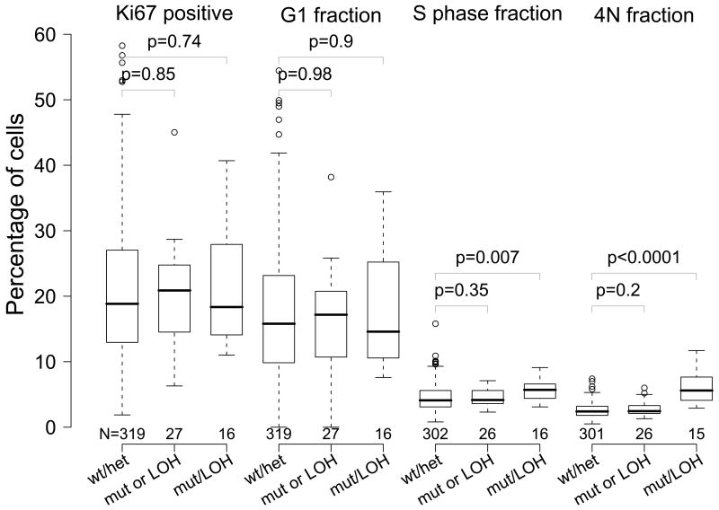
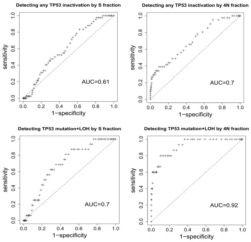
Results: Diploid S and 4N (G(2)/tetraploid) fractions were significantly higher in biopsies with TP53 mutation and loss of heterozygosity. CDKN2A inactivation was not associated with higher Ki67-positive, diploid S, G(1), or 4N fractions. High Ki67-positive and G(1)-phase fractions were not associated with the future development of esophageal adenocarcinoma (P=0.13 and P=0.15, respectively), whereas high diploid S-phase and 4N fractions were (P=0.03 and P<0.0001, respectively).
Conclusions: High Ki67-positive proliferative fractions were not associated with inactivation of CDKN2A and TP53 or future development of cancer in our cohort of patients with Barrett's esophagus. Biallelic inactivation of TP53 was associated with elevated 4N fractions, which have been associated with the future development of esophageal adenocarcinoma.
Figures
References
-
- Preston-Martin S, Pike MC, Ross RK, Jones PA, Henderson BE. Increased cell division as a cause of human cancer. Cancer Res. 1990;50:7415–21. - PubMed
-
- Gerdes J. Ki-67 and other proliferation markers useful for immunohistological diagnostic and prognostic evaluations in human malignancies. Semin Cancer Biol. 1990;1:199–206. - PubMed
-
- Hartwell LH, Culotti J, Pringle JR, Reid BJ. Genetic control of the cell division cycle in yeast. Science. 1974;183:46–51. - PubMed
Publication types
MeSH terms
Substances
Grants and funding
LinkOut - more resources
Full Text Sources
Other Literature Sources
Medical
Research Materials
Miscellaneous
