. 2008 Dec;36(22):6959-76.
doi: 10.1093/nar/gkn771. Epub 2008 Nov 3.

Proteome-wide identification of poly(ADP-ribose) binding proteins and poly(ADP-ribose)-associated protein complexes

Affiliations

Proteome-wide identification of poly(ADP-ribose) binding proteins and poly(ADP-ribose)-associated protein complexes

Jean-Philippe Gagné et al. Nucleic Acids Res. 2008 Dec.

Abstract

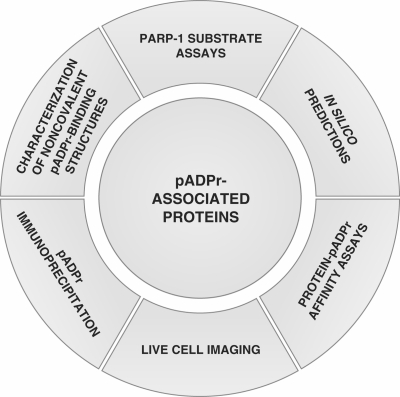
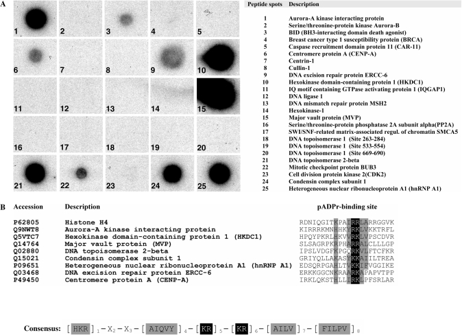
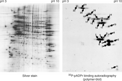
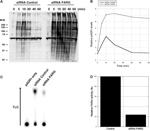
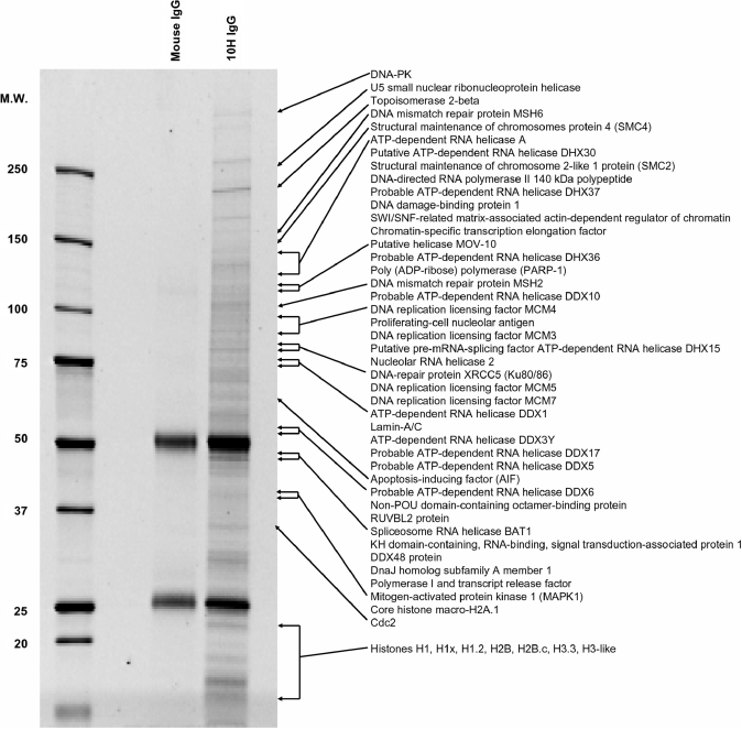
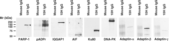
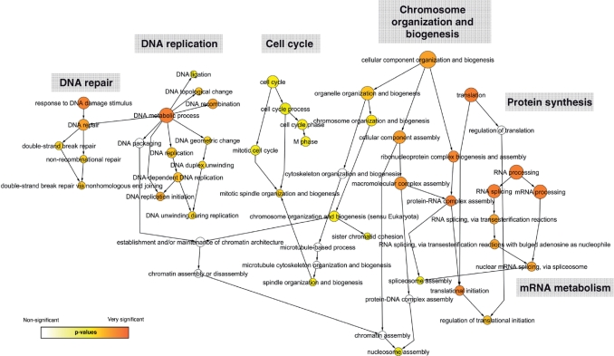
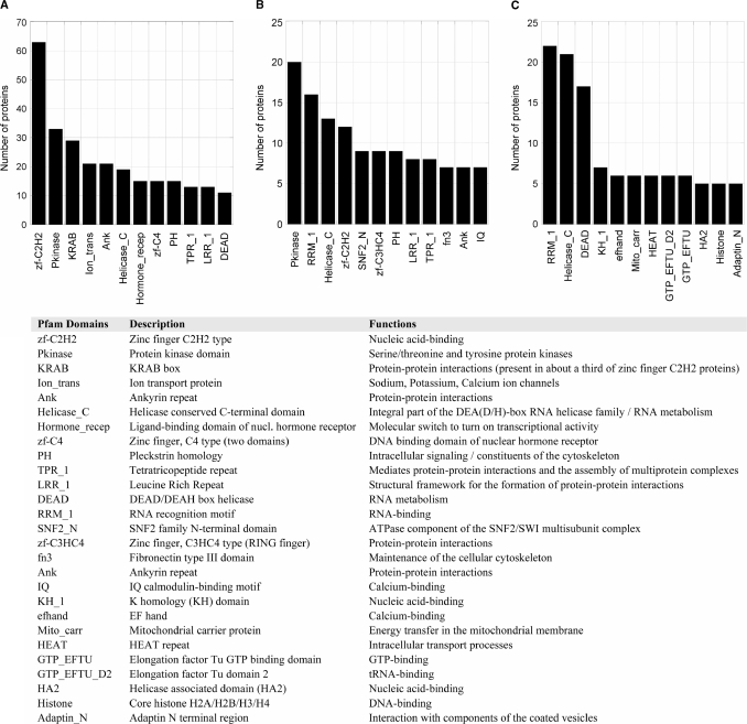
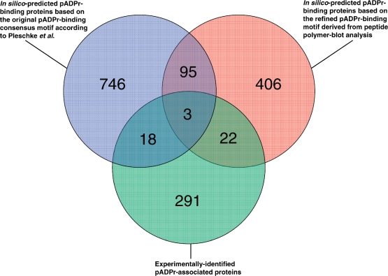
Poly(ADP-ribose) (pADPr) is a polymer assembled from the enzymatic polymerization of the ADP-ribosyl moiety of NAD by poly(ADP-ribose) polymerases (PARPs). The dynamic turnover of pADPr within the cell is essential for a number of cellular processes including progression through the cell cycle, DNA repair and the maintenance of genomic integrity, and apoptosis. In spite of the considerable advances in the knowledge of the physiological conditions modulated by poly(ADP-ribosyl)ation reactions, and notwithstanding the fact that pADPr can play a role of mediator in a wide spectrum of biological processes, few pADPr binding proteins have been identified so far. In this study, refined in silico prediction of pADPr binding proteins and large-scale mass spectrometry-based proteome analysis of pADPr binding proteins were used to establish a comprehensive repertoire of pADPr-associated proteins. Visualization and modeling of these pADPr-associated proteins in networks not only reflect the widespread involvement of poly(ADP-ribosyl)ation in several pathways but also identify protein targets that could shed new light on the regulatory functions of pADPr in normal physiological conditions as well as after exposure to genotoxic stimuli.


Figures

Figure 1.
Figure 1.
Current experimental strategies for the identification of pADPr-associated proteins. Six types of experimental approaches have been used to date to identify poly(ADP-ribosyl)ated and pADPr binding proteins (pADPr-associated proteins). As illustrated clockwise starting from the top: (i) identification of in vitro and in vivo poly(ADP-ribosyl) transferase activity of PARP-1 onto acceptor proteins (covalent modifications) using immunological or radioactivity-based detection methods; (ii) identification of noncovalent pADPr binding regions within protein amino acid sequences based on similarity with a consensus pADPr binding motif; (iii) affinity-based assays for the identification, validation and quantitative evaluation of noncovalent pADPr binding [surface plasmon resonance (SPR), electrophoretic mobility shift assays (EMSA), peptide and protein polymer blot analysis, mono- and bidimensional gel electrophoresis coupled with polymer blot analysis followed by MS]; (iv) in vivo evaluation of recruitment and accumulation of pADPr binding proteins using micro irradiation-induced DNA damage assays in live cell experiments; (v) immunoprecipitation assays using specific anti-pADPr antibodies to pull-down pADPr-associated proteins; and (vi) the characterization of PARP-associated protein domains as functional noncovalent pADPr binding modules (e.g. the Macro domain A1pp or the zinc-finger domain PBZ).
Figure 2.
Figure 2.
Polymer blot analysis. (A) Synthetic peptides corresponding to putative pADPr binding proteins as revealed by in silico prediction based on the consensus sequence proposed by Pleschke et al. (3) were dot-blotted onto nitrocellulose-coated glass slides, screened with 32P-pADPr and autoradiographied (see Supplementary Table S1 for the complete list of predictions). Amino acid sequences used for peptide synthesis are provided in Supplementary Table S4. (B) A refined pADPr binding consensus is generated from the sequence alignment of validated pADPr binding peptides. Experimentally verified pADPr binding regions from pADPr dot blot analysis were aligned to derive a refined pADPr binding motif and to computationally screen protein sequence database with the goal of achieving higher reliability and minimizing false positive identifications (see Supplementary Table S2 for a listing of proteins that match the refined pADPr binding consensus).
Figure 3.
Figure 3.
Two-dimensional polymer blot analysis. HeLa cell extracts were separated by tube-gel isoelectric focusing, resolved by SDS–PAGE and transferred onto PVDF membrane. The left panel shows the bidimensional protein separation as revealed by silver staining. The right-hand panel shows a corresponding two-dimensional-gel transferred onto a PVDF membrane for polymer blot analysis. Incubation with 32P-pADPr followed by autoradiography reveals a binding-protein pattern. Numbered spots were excised for identification by peptide mass fingerprinting (see Table 2 for complete spot identifications).
Figure 4.
Figure 4.
pADPr levels are increased and sustained in SK-N-SH cells treated with PARG siRNA following alkylation-induced DNA damage. (A) Time course western blot analysis of pADPr accumulation in SK-N-SH cells following 100 μM MNNG treatment in both control and PARG siRNA treated cells. Crude protein extracts were loaded and subjected to SDS–PAGE and immunoblotting. Blots were revealed with anti-pADPr polyclonal antibody 96-10 as described in Materials and methods section. (B) Western blot quantification of pADPr levels from control (full line) and PARG siRNA-treated cells (dashed line) following 100 μM MNNG treatment. (C) Evaluation of PARG knockdown using PARG TLC activity assays. Silencing was conducted over 6 days, passaging cells every 48 h to achieve maximum PARG knockdown. Whole-cell extracts were prepared from cultured cells that had been treated with either control or PARG siRNA. PARG activities were detected by TLC analysis of reaction mixtures containing these cell extracts and 32P-labeled pADPr as a substrate. Substrate remained at the origin of the TLC plate, while ADP-ribose products migrated towards the top of the TLC plate following development by 0.3 M LiCl and 0.9 M ethanoic (acetic) acid. (D) Relative PARG residual activity in SK-N-SH cells after 6-day silencing as determined from the TLC quantification of PARG reaction products.
Figure 5.
Figure 5.
SDS–PAGE analysis of pADPr-associated proteins from MNNG-treated and PARG-silenced SK-N-SH cells after immunoprecipitation with anti-pADPr antibodies. pADPr-associated proteins were immunoprecipitated using anti-pADPr mouse monoclonal antibody clone 10H bound to Protein G coated magnetic beads. Immunoprecipitates were resolved by 4–12% SDS–PAGE and stained with SYPRO Ruby fluorescent dye. Normal mouse IgGs were used to assess nonspecific binding. Selected proteins identified by LC-MS/MS, mostly involved in DNA/RNA transactions, are shown (see Supplementary Table S3 for complete protein listing).
Figure 6.
Figure 6.
Validation of selected pADPr-associated proteins identified by LC-MS/MS using western blot analysis of pADPr immunoprecipitates. The specificity of the pADPr immunoprecipitation using anti-pADPr 10H monoclonal antibodies was evaluated by immunoblot analysis as described in Materials and methods section. The same proteins were not precipitated by normal mouse IgGs, confirming the specificity of the pull-down.
Figure 7.
Figure 7.
Graphical network representation of pADPr-associated proteins identified by LC-MS/MS in anti-pADPr immunoprecipitates. Functional categorization of pADPr-associated proteins was performed using GO annotations. Over-represented categories were statistically identified using BiNGO and visualized with Cytoscape. The size of the terms (circles) is representative of the proportional protein abundance in the dataset and the color shading indicates the degree of statistical significance (darker shades indicate stronger significance).
Figure 8.
Figure 8.
Distribution of protein domains and families in computationally predicted and experimentally validated pADPr binding proteins. The graph shows the number of occurrences for the 12 most common Pfam domains in (A) putative pADPr binding proteins identified through a 90% similarity sequence search based on the original consensus pADPr binding motif (Supplementary Table S1), (B) a sub-dataset of putative pADPr binding proteins corresponding to the refined pADPr binding module derived from peptides polymer blot analysis (Supplementary Table S2) and (C) pADPr-associated proteins identified by MS in anti-pADPr immunoprecipitation assays in SK-N-SH cells following alkylation-induced DNA damage (Supplementary Table S3).
Figure 9.
Figure 9.
Venn diagram illustrating the overlap between the three pADPr binding candidates datasets described in this study. Overlapping circles shows the distribution of in silico predicted proteins revealed through the use of the consensus pADPr binding motif proposed by Pleschke et al. (3) and the refined pADPr binding motif derived from sequence alignment of experimentally validated pADPr binding regions from peptide polymer blot analysis. The relationship between the two in silico prediction approaches and experimental identification of pADPr-associated proteins in pADPr immunoprecipitates is illustrated as a third intercepting circle.

References

    1. Nishizuka Y, Ueda K, Honjo T, Hayaishi O. Enzymic adenosine diphosphate ribosylation of histone and poly adenosine diphosphate ribose synthesis in rat liver nuclei. J. Biol. Chem. 1968;243:3765–3767. - PubMed
    1. D’Amours D, Desnoyers S, D'Silva I, Poirier GG. Poly(ADP-ribosyl)ation reactions in the regulation of nuclear functions. Biochem. J. 1999;342(Pt 2):249–268. - PMC - PubMed
    1. Pleschke JM, Kleczkowska HE, Strohm M, Althaus FR. Poly(ADP-ribose) binds to specific domains in DNA damage checkpoint proteins. J. Biol. Chem. 2000;275:40974–40980. - PubMed
    1. Karras GI, Kustatscher G, Buhecha HR, Allen MD, Pugieux C, Sait F, Bycroft M, Ladurner AG. The macro domain is an ADP-ribose binding module. EMBO J. 2005;24:1911–1920. - PMC - PubMed
    1. Egloff MP, Malet H, Putics A, Heinonen M, Dutartre H, Frangeul A, Gruez A, Campanacci V, Cambillau C, Ziebuhr J, et al. Structural and functional basis for ADP-ribose and poly(ADP-ribose) binding by viral macro domains. J. Virol. 2006;80:8493–8502. - PMC - PubMed

Publication types