. 2008 Nov 15;181(10):6747-56.
doi: 10.4049/jimmunol.181.10.6747.

Aging impairs IFN regulatory factor 7 up-regulation in plasmacytoid dendritic cells during TLR9 activation

Affiliations

Aging impairs IFN regulatory factor 7 up-regulation in plasmacytoid dendritic cells during TLR9 activation

Heather W Stout-Delgado et al. J Immunol. .

Abstract

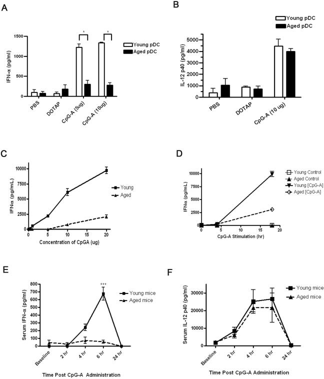
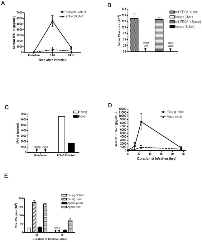
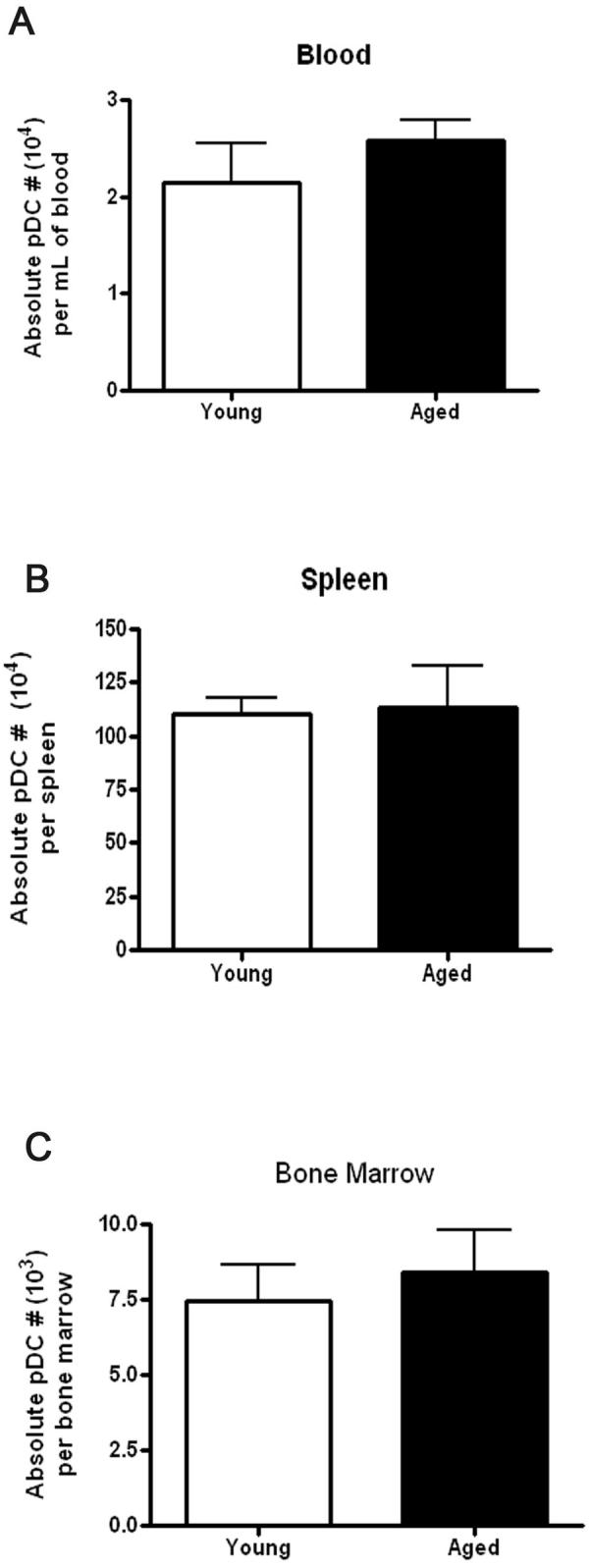
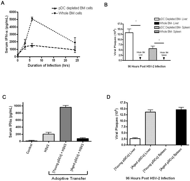
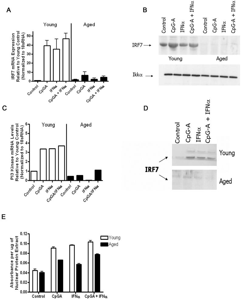
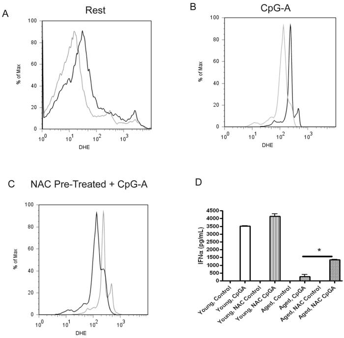
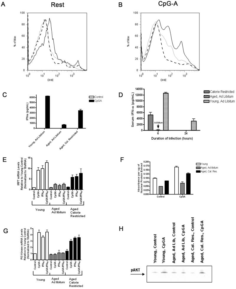
Plasmacytoid dendritic cells (pDCs) are innate sensors that produce IFN-alpha in response to viral infections. Determining how aging alters the cellular and molecular function of these cells may provide an explanation of increased susceptibility of older people to viral infections. Hence, we examined whether aging critically impairs pDC function during infection with HSV-2, a viral pathogen that activates TLR9. We found that impaired IFN-alpha production by aged murine pDCs led to impaired viral clearance with aging. Upon TLR9 activation, aged pDCs displayed defective up-regulation of IFN-regulatory factor 7, a key adaptor in the type I IFN pathway, as compared with younger counterparts. Aged pDCs had more oxidative stress, and reducing oxidative stress in aged pDCs partly recovered the age-induced IFN-alpha defect during TLR9 activation. In sum, aging impairs the type I IFN pathway in pDCs, and this alteration may contribute to the increased susceptibility of older people to certain viral infections.


Figures

Figure 1
Figure 1
IFN-α production is impaired in CpG type A-activated aged pDCs compared to young pDCs in vitro and to systemic administration of CpG type A in vivo. A-B, The pDCs were isolated from the bone marrow of young and aged C57BL/6 mice and incubated for 18h with CpG-ODN at 5 or 10 μg/ml. IFN-α (A) and IL-12p40 (B) production was measured. Control cells were stimulated with either PBS or DOTAP. C, Aged pDCs exhibited defective IFN-α in dose response experiments at 24h post CpG stimulation. IL-12p40 responses were preserved in dose response experiments in aged pDCs as compared to young counterparts (data not shown). D, IFN-α responses by aged pDCs were inferior to young pDCs in time course experiments. *p<0.05 (Student's t-test). The results in A-D are representative of at least four independent experiments with similar findings. N=3 per group for each experiment. E-F, Young and aged C57BL/6 mice were administered type A CpG i.v., and then serum IFN-α (E) and IL-12p40 (F) levels were assessed by ELISA. ***p<0.0001. The results are representative of at least three independent experiments with similar results. N=3 per group for each experiment.
Figure 2
Figure 2
Aged hosts produce less IFN-α during HSV-2 infection as compared with young hosts. A, IFN-α levels during systemic HSV-2 infection were measured in control and anti-PDCA-1 (pDC depleting mAB)-treated young mice. B, Viral load was measured in young mice that were treated with anti-PDCA-1 or control antibody. C, Aged pDCs exhibit an impaired IFN-α response to in vitro HSV-2 infection compared to young pDCs. N = 3/group in at least two independent experiments. D, Young and aged C57BL6 mice were systemically infected with HSV-2 and then serum IFN-α levels were measured. These results are representative of at least four independent experiments (N=3 per group) with similar results. E, Spleen and liver samples were isolated from virally infected mice during HSV-2 infection, and viral load was measured by plaque assay. These results are representative of at least two independent experiments with similar results (N=3 per group).
Figure 3
Figure 3
Aged pDCs manifest an impaired IFN-α response to TLR7 activation with RNA40, and aged mice manifest an impaired IFN-α response to MCMV infection and a reduced ability to clear this virus. A, Aged pDCs manifest an impaired IFN-α response during 18h of TLR7 activation with 50 μg/ml RNA40 (N=3/group). Groups were significantly different with a p-value of <0.05. Control cells were treated with PBS. B-C, Aged mice infected with MCMV exhibited reduced IFN-α response (B) and viral load (C) as compared to young mice (N=3/group). Aged and young infected groups were significantly different with p-value of <0.05. Parameters were measured after 36h of viral infection. Control groups received PBS.
Figure 4
Figure 4
Aged mice maintain similar numbers of pDC within the blood, spleen and bone marrow as compared to young mice. The pDCs from blood, bone marrow, and spleen from young and aged mice were enumerated using flow cytometry (PDCA-1+ staining; N>6/group).
Figure 5
Figure 5
Transfer of young whole bone marrow cells into aged hosts augments IFN-α levels and viral clearance in HSV-2-infected aged hosts in a pDC-dependent fashion. Aged C57BL/6 mice were adoptively transferred i.v. with 5×10⁶ whole or pDC-depleted bone marrow cells isolated from young C57BL/6 mice. A, Mice were infected with HSV-2 at 72h post-transfer, and serum IFN-α levels were measured by ELISA. B, Liver and spleen samples were isolated at 96h post-HSV-2 infection, and viremia was measured. *p<0.05 (Student's t-test). C, Aged mice were adoptively transferred with either 1×10⁴ young or aged pDCs from syngeneic donors 72 hours prior to infection with HSV-2. IFN-α responses were measured at 24h post-infection. Non-infected (denoted as control) and non-adoptively transferred infected controls (denoted as HSV-2) are shown. D, Viral load was measured in the liver and spleen from aged mice that were adoptively transferred with aged or young pDCs (N=3/group). Results are representative of data from at least two independent experiments with similar results.
Figure 6
Figure 6
Aged pDCs manifest an impaired ability to upregulate IRF-7 or PI3-kinase during either TLR9 or IFN-α receptor stimulation. A, Aged or young pDCs were stimulated with CpG-A, IFN-α, or both, and then the upregulation of IRF-7 mRNA levels from each experimental group were measured, normalized to 18sRNA, and compared to young controls. B, The same groups as in (A) except that IRF-7 or Iκκα protein levels were measured by western blot. Results are representative of data from at least two independent experiments with similar results (N=3/group). C, The experimental groups are the same as in (A), except that PI3-kinase levels were measured by real-time PCR. D, IRF-7 nuclear translocation was analyzed by western blotting. Young and aged nuclear protein fractions were electrophoresed on two separate SDS-PAGE gels that were run concurrently under the same conditions. Results are representative of data from at least two independent experiments with similar results (N=3/group). E, Nuclear IRF-7 activity was measured in young and aged pDCs by ELISA.
Figure 7
Figure 7
Aging leads to increased oxidative stress in pDCs at rest and during CpG-A activation. A-B, Aged pDCs exhibit evidence of increased oxidative stress (measured by DHE fluorescence) at rest (A) and during CpG-A (B) stimulation. Grey line represents young cells, and the black line indicates aged cells. Data represents at least two independent experiments with similar results (N=3/group). C, Pre-treating aged pDCs with NAC (black line) reduces the ROS profile during CpG-A activation compared to aged pDCs pre-treated with PBS (grey line). Data represents at least two independent experiments with similar results (N=3/group). D, NAC treatment augments IFN-α responses in aged pDCs during CpG-A stimulation. *p<0.05 Student's t-test. Young and aged pDCS were pre-treated for 1 h at 37 °C with or without 1 μM NAC diluted in normal cell culture media. Cells were cultured with 10 μg of CpG-A at 37 °C for 18h. NAC control refers to cells that were pre-treated with NAC but not activated with CpGA. Control cells that were neither pre-treated with NAC nor activated with CpG-A are denoted as control. Data represents at least two independent experiments with similar results (N=3/group).
Figure 8
Figure 8
CR partly recovers the age-induced defective type I IFN signaling response in pDC. A-B, The pDCs harvested from CR aged mice exhibit reduced oxidative stress (grey line) at rest and during 18h of activation with CpG-A as compared to pDCs harvested from AL fed aged controls (black line). Young AL fed controls are shown (dashed line). Data represent at least four independent experiments with similar results (N=3/group). C-D, The pDCs harvested from CR aged mice displayed augmented IFN-α responses during in vitro activation with 18h CpG-A or in vivo infection with HSV-2 as compared to pDCs from AL-fed aged and young controls. Data represent at least two independent experiments with similar results (N=3/group). E, The pDCs from CR aged mice exhibit increased IRF-7 gene expression during either CpG-A or IFN-α stimulation as compared to pDCs from AL-fed aged mice. Gene expression from pDCs harvested from young AL-fed controls is shown. Data represent at least three independent experiments with similar results (N=3/group). F, The pDCs from CR aged mice exhibit increased IRF-7 activity, expressed as absorbance per μg nuclear extract, during CpG-A activation compared to pDCs from age-matched AL-fed mice. The pDCs from young AL-fed mice are shown. G, The pDCs from CR aged mice exhibit increased PI3-kinase upregulation during either CpG-A or IFN-α stimulation as compared to pDCs from ALfed aged mice. Gene expression from pDCs harvested from young AL-fed controls is shown. Data represents at least three independent experiments with similar results (N=3/group). H, The pDCs from CR aged mice exhibit increased phosphorylation of AKT during CpG-A activation compared to pDCs from age-matched AL-fed mice. The pDCs from young AL-fed mice are shown.

Similar articles

Cited by

References

    1. Pawelec G, Barnett Y, Forsey R, Frasca D, Globerson A, McLeod J, Caruso C, Franceschi C, Fulop T, Gupta S, Mariani E, Mocchegiani E, Solana R. T cells and aging, January 2002 update. Front Biosci. 2002;7:d1056–1183. - PubMed
    1. Linton P-J, Dorschkind K. Age related changes in lymphocyte development and function. Nat Immunol. 2004;5:133–139. - PubMed
    1. Yung R. Changes in immune function with age. Rheum. Dis. Clin. N. Am. 2000;26:455–473. - PubMed
    1. Weng NP. Aging of the Immune System: How Much Can the Adaptive Immune System Adapt? Immunity. 2006;24:495. - PMC - PubMed
    1. Haynes L, SL S. Why Aging T cells Fail: Implications for vaccination. Immunity. 2006;24:663–666. - PMC - PubMed

Publication types