. 2008 Nov 15;181(10):6779-88.
doi: 10.4049/jimmunol.181.10.6779.

Regulatory roles for NKT cell ligands in environmentally induced autoimmunity

Affiliations

Regulatory roles for NKT cell ligands in environmentally induced autoimmunity

Jaya Vas et al. J Immunol. .

Abstract

The development of autoimmune diseases is frequently linked to exposure to environmental factors such as chemicals, drugs, or infections. In the experimental model of metal-induced autoimmunity, administration of subtoxic doses of mercury (a common environmental pollutant) to genetically susceptible mice induces an autoimmune syndrome with rapid anti-nucleolar Ab production and immune system activation. Regulatory components of the innate immune system such as NKT cells and TLRs can also modulate the autoimmune process. We examined the interplay among environmental chemicals and NKT cells in the regulation of autoimmunity. Additionally, we studied NKT and TLR ligands in a tolerance model in which preadministration of a low dose of mercury in the steady state renders animals tolerant to metal-induced autoimmunity. We also studied the effect of Sphingomonas capsulata, a bacterial strain that carries both NKT cell and TLR ligands, on metal-induced autoimmunity. Overall, NKT cell activation by synthetic ligands enhanced the manifestations of metal-induced autoimmunity. Exposure to S. capsulata exacerbated autoimmunity elicited by mercury. Although the synthetic NKT cell ligands that we used are reportedly similar in their ability to activate NKT cells, they displayed pronounced differences when coinjected with environmental agents or TLR ligands. Individual NKT ligands differed in their ability to prevent or break tolerance induced by low-dose mercury treatment. Likewise, different NKT ligands either dramatically potentiated or inhibited the ability of TLR9 agonistic oligonucleotides to disrupt tolerance to mercury. Our data suggest that these differences could be mediated by the modification of cytokine profiles and regulatory T cell numbers.


Conflict of interest statement

Disclosures

The authors have no financial conflict of interest.

Figures

FIGURE 1
FIGURE 1
Synthetic variants of α-GalCer used in this study.
FIGURE 2
FIGURE 2
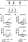
NKT cell activation increases susceptibility to mercury-induced autoimmunity. C57BL/6.SJL mice received HgCl2 (30 µg) on days 0, 2, and 4 with or without 2 µg of 4-deoxy α-GalCer (A) or PBS 57 (B) on days 0, 3, and 7. Animals were thereafter evaluated for ANoA. ANoA were detected by immunofluorescence on HEp-2 cells using isotype-specific FITC conjugates. Results are expressed as titers (inverse of the highest serum dilution that yielded nucleolar fluorescence) ± SD. **p< 0.001; ***p< 0.0001 vs HgCl2-treated group.
FIGURE 3
FIGURE 3
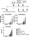
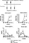
Differential effects of NKT cell activation on mercury-induced autoimmunity. A.SW mice received HgCl2 (30 µg) on days 0, 2, and 4 with or without 2 µg of PBS 57 or 4-deoxy α-GalCer on days 0 and 2. Animals were thereafter evaluated for ANoA and serum Ig levels. IgG1 and IgG2a ANoA were detected by immunofluorescence on HEp-2 cells using isotype-specific FITC conjugates. Results are expressed as titers (inverse of the highest serum dilution that yielded nucleolar fluorescence) ± SD. Serum IgE levels were measured by ELISA, as described in Materials and Methods, and are expressed in µg/ml ± SD. *p< 0.05; **p< 0.001; ***p< 0.0001 vs HgCl2-treated group. Note: The IgG1 ANoA curves are superimposed for the PBS 57 plus HgCl2 and the HgCl2-alone group.
FIGURE 4
FIGURE 4
Mercury potentiates PBS 57-induced NKT cell expansion. A.SW mice received HgCl2 (30 µg) or vehicle on days 0, 2, and 4 with or without 2 µg of PBS 57 (A) or 4-deoxy α-GalCer (B) on days 0 and 2. On day 5, splenocytes were stained with empty or PBS 57-loaded allophycocyanin-conjugated CD1d tetramers and PE-conjugated anti-B220 to negatively gate B cells. Absolute numbers and percentages of NKT cells are expressed as mean ± SD. #,p < 0.05 vs HgCl2-treated group; *, p < 0.05 vs PBS 57-treated group.
FIGURE 5
FIGURE 5
NKT cell activation by PBS 57, but not 4-deoxy α-GalCer, prevents tolerance establishment. A.SW mice received 3 µg of PBS 57, 4-deoxy α-GalCer, or vehicle 4 h before administration of a low dose (3 µg) of HgCl2 on day −7. All mice then received three injections of 30 µg of HgCl2 on days 0, 2, and 4. Animals were thereafter evaluated for ANoA and serum IgE levels. ANoA were detected by immunofluorescence on HEp-2 cells using isotype-specific FITC conjugates. Results are expressed as titers (inverse of the highest serum dilution that yielded nucleolar fluorescence) ± SD. Serum IgE levels were measured by ELISA, as described in Materials and Methods, and are expressed in µg/ml ± SD. *, p < 0.05; ***, p < 0.0001 vs vehicle-treated group.
FIGURE 6
FIGURE 6
NKT cell ligands can either synergize with or antagonize TLR9-induced breakage of tolerance. A.SW mice received a single low dose (3 µg) of HgCl2 on day −7. Animals then received either NKT cell ligand (2 µg), CpG 1826 (100 µg), or combinations of the two at the indicated timepoints. All groups received 30 µg of HgCl2 on days 0, 2, and 4. Animals were thereafter evaluated for ANoA and serum Ig levels. For clarity, results are shown at a single timepoint, week 3 for autoantibodies and week 2 for serum IgE. ANoA were detected by immunofluorescence on HEp-2 cells using isotype-specific FITC conjugates. Results are expressed as titers (inverse of the highest serum dilution that yielded nucleolar fluorescence) ± SD. Serum IgE levels were measured by ELISA, as described in Materials and Methods, and are expressed in µg/ml ± SD. *p< 0.05 vs CpG 1826-treated group.
FIGURE 7
FIGURE 7
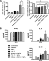
Mice receiving PBS 57 and mercury show increased regulatory T cells and Th2 cytokine production. A.SW mice received HgCl2 or vehicle on days 0, 2, and 4. Additionally, some groups received injections of 4 µg of PBS 57, 4-deoxy α-GalCer, or vehicle (on days 0 and 2). A, On day 8, mice were sacrificed and splenocytes were stained with Abs to surface CD4 and CD25 and for intracellular Foxp3. Both percentages and absolute numbers of regulatory T cells are shown. B, Splenocytes from same animals were also stimulated with PMA/ionomycin. Culture supernatants collected at 48 and 96 h were analyzed for IFN-γ, IL-4, and IL-10 levels by sandwich ELISA, as described in Materials and Methods. Cytokine levels are expressed in pg/ml ± SD. # p< 0.05; ##, p< 0.001 vs indicated group; *p< 0.05 vs PBS 57-treated group.
FIGURE 8
FIGURE 8
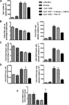
PBS 57 restores balance between T and Teffector cell populations in the spleen. A.SW mice received a single low dose (3 µg) of HgCl2 on day −7. Animals then received either NKT cell ligand (2 µg) with CpG 1826 (100 µg) at the indicated timepoints. All groups received 30 µg of HgCl2 on days 0, 2, and 4. At day 8, mice were sacrificed and splenocytes were stained with Abs to surface CD4 and CD25 and for intracellular Foxp3. A, Absolute numbers of cells per spleen following RBC lysis. B, Percentage (right) and absolute numbers (left) of splenic CD4+ CD25+ cells. C, Percentage (right) and absolute numbers (left) of TregsD, Percentage (right) and absolute numbers (left) of TeffectorsE, Ratio of Tregs:Teffectors. *, p < 0.05 vs PBS 57 plus CpG 1826-treated and vehicle-treated groups.
FIGURE 9
FIGURE 9
Effect of NKT cell and TLR ligands on cytokine production. A.SW mice received a single low dose (3 µg) of HgCl2 on day −7. Animals then received either NKT cell ligand (2 µg) with CpG 1826 (100 µg) at the indicated timepoints. All groups received 30 µg of HgCl2 on days 0, 2, and 4. On day 8, mice were sacrificed and splenocytes were stimulated with PMA/ionomycin or anti-CD3 plus anti-CD28 mAbs. Culture supernatants collected at indicated timepoints were analyzed for IFN-7 (following anti-CD3 plus anti-CD28 stimulation) or IL-4 and IL-10 (following PMA/ionomycin stimulation) by sandwich ELISA, as described in Materials and Methods. Cytokine levels are expressed in pg/ml ± SD. *p< 0.05 vs PBS 57 plus CpG 1826-treated and CpG 1826-treated groups; #, p< 0.05 vs CpG 1826-treated group.
FIGURE 10
FIGURE 10
Administration of heat-killed microbes exacerbates mercury-induced autoimmunity. A.SW mice received HgCl2 (30 µg) or vehicle on days 0, 2, and 4 with or without 100 µ of heat-killed S. capsulata on days 0 and 2. Animals were thereafter evaluated for ANoA and serum Ig levels. ANoA were detected by immunofluorescence on HEp-2 cells using isotype-specific FITC conjugates. Results are expressed as titers (inverse of the highest serum dilution that yielded nucleolar fluorescence) ± SD. Serum Ig levels were measured by ELISA, as described in Materials and Methods, and are expressed in mg/ml ± SD (IgG2a) or µg/ml ± SD (IgE). *p< 0.05; ***p< 0.0001 vs HgCl2-treated group.

References

    1. Lawrence DA, McCabe MJ., Jr. Immunomodulation by metals. Int. Immunopharmacol. 2002;2:293–302. - PubMed
    1. Cardenas A, Roels H, Bernard AM, Barbon R, Buchet JP, Rosello J, Hotter G, Mutti A, Franchini I. Markers of early renal changes induced by industrial pollutants. I. Application to workers exposed to mercury vapors. Br. J. Ind. Med. 1998;50:17–27. - PMC - PubMed
    1. Enestrom S, Hultman P. Does amalgam affect the immune system? A controversial issue. Int. Arch. Allergy Immunol. 1995;106:180–203. - PubMed
    1. Goyer RA. Environmentally related diseases of the urinary tract. Med. Clin. N. Am. 1990;74:377–389. - PubMed
    1. Moszczynski P, Slowinski S, Rutkowski J, Bem S, Jakus-Stoga D. Lymphocytes, T and NK cells, in men occupationally exposed to mercury vapours. Int. J. Occup. Med. Environ. Health. 1995;8:49–56. - PubMed

Publication types

MeSH terms