. 2008 Nov 15;181(10):7194-204.
doi: 10.4049/jimmunol.181.10.7194.

Macrophage antigen complex-1 mediates reactive microgliosis and progressive dopaminergic neurodegeneration in the MPTP model of Parkinson's disease

Affiliations

Macrophage antigen complex-1 mediates reactive microgliosis and progressive dopaminergic neurodegeneration in the MPTP model of Parkinson's disease

Xiaoming Hu et al. J Immunol. .

Abstract

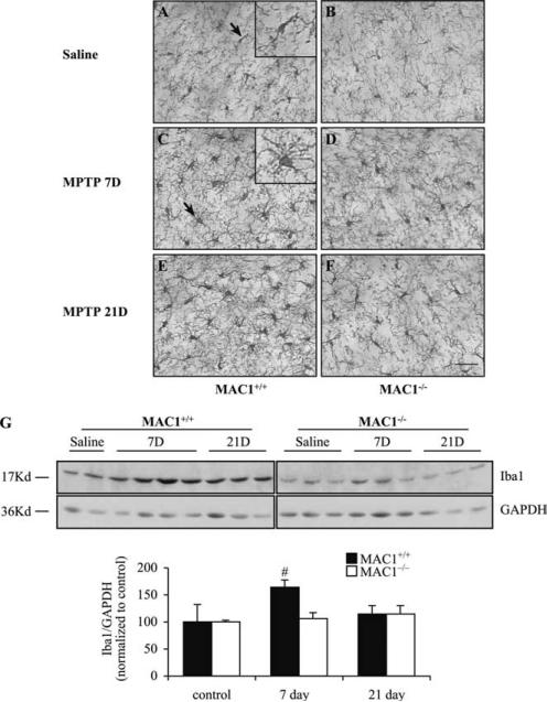
Neuronal death is known to trigger reactive microgliosis. However, little is known regarding the manner by which microglia are activated by injured neurons and how microgliosis participates in neurodegeneration. In this study we delineate the critical role of macrophage Ag complex-1 (MAC1), a member of the beta(2) integrin family, in mediating reactive microgliosis and promoting dopaminergic (DAergic) neurodegeneration in the 1-methyl-4-phenyl-1,2,3,6-tetrahydropyridine (MPTP) model of Parkinson's disease. MAC1 deficiency greatly attenuated the DAergic neurodegeneration induced by MPTP or 1-methyl-4-phenyl-pyridium iodide (MPP(+)) exposure both in vivo and in vitro, respectively. Reconstituted experiments created by adding microglia from MAC1(-/-) or MAC1(+/+) mice back to MAC1(+/+) neuron-enriched cultures showed that microglia with functional MAC1 expression was mandatory for microglia-enhanced neurotoxicity. Both in vivo and in vitro morphological and Western blot studies demonstrated that MPTP/MPP(+) produced less microglia activation in MAC1(-/-) mice than MAC1(+/+) mice. Further mechanistic studies revealed that a MPP(+)-mediated increase in superoxide production was reduced in MAC1(-/-) neuron-glia cultures compared with MAC1(+/+) cultures. The stunted production of superoxide in MAC1(-/-) microglia is likely linked to the lack of translocation of the cytosolic NADPH oxidase (PHOX) subunit (p47(phox)) to the membrane. In addition, the production of PGE(2) markedly decreased in neuron plus MAC1(-/-) microglia cocultures vs neuron plus MAC1(+/+) microglia cocultures. Taken together, these results demonstrate that MAC1 plays a critical role in MPTP/MPP(+)-induced reactive microgliosis and further support the hypothesis that reactive microgliosis is an essential step in the self-perpetuating cycle leading to progressive DAergic neurodegeneration observed in Parkinson's disease.


Figures

FIGURE 1
FIGURE 1
MPP+-induced DAergic neurotoxicity was attenuated in mesencephalic neuron-glia cultures from MAC1–/– mice compared with MAC1+/+ mice. Mouse (MAC1+/+ and MAC1–/–) mesencephalic neuron-glia cultures in 24-well plates were treated with vehicle or different concentration of MPP+ (0.125 μM, 0.25 μM and 0.5 μM) for 7 days. A, Representative microscopic images were shown for TH-positive neurons treated with vehicle or different concentrations of MPP+. Scale bar: 30 μm. B, The effect of MPP+ on the number of DAergic neurons was assessed by counting the number of TH-positive cells remaining in the neuron-glia cultures after vehicle or MPP+ treatments. C, MPP+-induced DAergic neurotoxicity was quantified by the [3H]DA uptake assay, which measures the capacity of DAergic neurons to take up 3H-labeled DA. Results were expressed as a percentage of the vehicle-treated controls and were the mean ± SEM from four independent experiments in duplicate. #, p < 0.05 compared with corresponding vehicle-treated controls. *, p < 0.05 compared with MAC1+/+ cultures after same treatments.
FIGURE 2
FIGURE 2
Expression of MAC1 on microglia is important for MPP+-induced neurotoxicity. A, Mesencephalic neuron-enriched cultures from MAC1+/+ or MAC1–/– mice were compared for their susceptibility to MPP+-induced DAergic neurotoxicity. B, Neuron-enriched cultures from MAC1+/+ mice with microglia (MAC1+/+ or MAC1–/–) added back were compared for their susceptibility to MPP+-induced neurotoxicity. Microglia (1 × 10⁵/well) were added back to neuron-enriched cultures 24 h before treatment. Cultures were treated with vehicle or 0.25 μM MPP+ for 7 days, and MPP+-induced neurotoxicity was quantified by counting TH-positive neurons. The data were expressed as the percentage of corresponding vehicle-treated control cultures and were the mean ± SEM from three independent experiments in triplicate. #, p < 0.05 relative to neuron-enriched cultures with or without MAC1–/– microglia added back. C, Representative microscopic images were shown for TH-positive neurons treated with vehicle or MPP+. Scale bar: 50 μm.
FIGURE 3
FIGURE 3
Analysis of MPP+-induced reactive microgliosis in vitro. Mesencephalic neuron-glia cultures from MAC1+/+ and MAC1–/– mice were treated with vehicle or MPP+ (0.25 μM). A, Cultures were fixed at 1, 4, and 7 days (D) after treatments. Microglia was visualized by immunostaining of the F4/80 Ag, a microglia marker. The images presented are representative of three independent experiments. Scale bar: 50 μm. B, Western blot analysis of microglial activation. Cell lysates of cultures from MAC1+/+ and MAC1–/– mice were prepared 4 days after MPP+ treatment. Immunoblot analysis was performed to assess Iba1 Ag. GAPDH was used as loading control (Ctrl). The ratio of densitometry values of Iba1 and GAPDH was analyzed and normalized to each respective control. The experiment has been performed three times. Results were presented as the mean ± SEM. *, p < 0.05 relative to corresponding vehicle-treated control cultures.
FIGURE 4
FIGURE 4
MAC1 mediates the activation of PHOX and production of superoxide. A, Mesencephalic neuron-glia cultures from MAC1+/+ and MAC1–/– mice were treated with vehicle or MPP+ (0.25 μM) for 4 or 7 days. B, Microglia-enriched cultures from MAC1+/+ and MAC1–/– mice were treated with vehicle, PMA (25 nM), LPS (10 ng/ml), or MPP+ (0.25 μM). Extracellular superoxide generation was measured by the SOD-inhibitable reduction of tetrazolium salt, WST-1. Data were mean ± SEM from three independent experiments in triplicates. #, p < 0.05 relative to corresponding vehicle-treated control cultures. *, p < 0.05 relative to MAC1+/+ cultures after same treatments. C, Enriched microglial cells from MAC1+/+ and MAC1–/– mice were treated with vehicle or LPS for 10 min. Cells were incubated with a rabbit polyclonal Ab against p47phox and then with a FITC-conjugated goat anti-rabbit Ab. Focal planes spaced at 0.4 μm intervals were imaged. The signal of p47phox (FITC-p47phox; green) and the merged view of cell morphology and p47phox (DIC plus FITC-p47phox) were shown. Scale bar: 20 μm. D, Western blot assays for p47phox levels in membrane and cytosolic fractions of microglia from MAC1+/+ and MAC1–/– mice 10 min after vehicle or LPS treatment. Densitometry analysis was performed with values of p47phox normalized to each respective loading control (GAPDH for cytosolic fraction, gp91 for membrane fraction) and further normalized to MAC1+/+ controls. Experiments were performed at least three times.
FIGURE 5
FIGURE 5
Involvement of MAC1 in MPP+-induced PGE2 production. Neuron-enriched cultures from MAC1+/+ mice with or without microglia (MAC1+/+ or MAC1–/–) added back were compared for their ability to release PGE2 into the culture medium. Microglia (1 × 10⁵/well) were added back to MAC1+/+ neuron-enriched cultures 24 h before treatment. Neuron-microglia cocultures were treated with vehicle or 0.25 μM MPP+. Culture media were collected 4 days after treatment. Data were mean ± SEM from three independent experiments in triplicates. #, p < 0.05 relative to corresponding vehicle treated control cultures. *, p < 0.05 relative to cocultures of neuron and MAC1–/– microglia after same treatment.
FIGURE 6
FIGURE 6
MPTP produced less DAergic neurotoxicity in MAC1–/– mice than MAC1+/+ mice. MAC1+/+ and MAC1–/– mice were injected s.c. with MPTP (15 mg/kg/day) or saline for 6 consecutive days. MPTP-mediated damage of DAergic neurons in SNpc was evaluated 21 days after the last injection by immunostaining with an Ab against TH. A–D, Immunostaining of TH-positive neurons in saline-treated (MAC1+/+ (A); MAC1–/– (C)) and MPTP-treated (MAC1+/+ (B); MAC1–/– (D)) mice. Scale bar: 50 μm. E, Quantification of SNpc TH-positive neurons in the mice in A–D. Results were presented as the mean ± SEM. Five to nine animals were used in each group. #, p < 0.05 compared with corresponding saline-treated controls. *, p < 0.05 compared with MAC1–/– mice after same treatment. F, HPLC determination of striatal MPP+ levels in MAC1+/+ and MAC1–/– mice 90 min after s.c. MPTP injection. Results were presented as the mean ± SEM. Three animals were used in each group.
FIGURE 7
FIGURE 7
Analysis of MPTP-induced reactive microgliosis in vivo. MAC1+/+ and MAC1–/– mice were sacrificed 7 days and 21 days following saline or MPTP s.c. injection. A–F, Brain sections were immuno-stained with an Iba1 Ab specific for microglia. Magnified images of resting (arrow in A) and activated microglia (arrow in C) were shown in the insets. The images were representative of three to four animals per group. Scale bar: 50 μm (inset: 25 μm). G, Western blot results with detection of Iba1 in midbrain tissue extracts from MAC1+/+ and MAC1–/– mice. GAPDH was used as loading control. The ratio of densitometry values of Iba1 and GAPDH was analyzed and normalized to each respective control. Results were presented as the mean ± SEM. #, p < 0.05 relative to corresponding vehicle-treated control.

References

    1. Leenders KL, Oertel WH. Parkinson's disease: clinical signs and symptoms, neural mechanisms, positron emission tomography, and therapeutic interventions. Neural Plast. 2001;8:99–110. - PMC - PubMed
    1. Kreutzberg GW. Microglia: a sensor for pathological events in the CNS. Trends Neurosci. 1996;19:312–318. - PubMed
    1. Gao HM, Hong JS. Why neurodegenerative diseases are progressive: uncontrolled inflammation drives disease progression. Trends Immunol. 2008;29:357–365. - PMC - PubMed
    1. McGeer PL, Itagaki S, Boyes BE, McGeer EG. Reactive microglia are positive for HLA-DR in the substantia nigra of Parkinson's and Alzheimer's disease brains. Neurology. 1988;38:1285–1291. - PubMed
    1. Benveniste EN, Nguyen VT, O'Keefe GM. Immunological aspects of microglia: relevance to Alzheimer's disease. Neurochem. Int. 2001;39:381–391. - PubMed

Publication types